
Agglutination Test: Types (Direct, Passive, Reverse Passive) with Examples

On this page
When specific antibodies (agglutinins) bind antigens that are on the surface of a particle, whether a bacterial cell, a red blood cell, or an artificial latex bead, they cross-link those particles into visible clumps. That visible clumping is agglutination, and a test built on it is an agglutination test. Its great practical advantage is that the result is read by eye, with no instrument, which is why agglutination underlies everything from bedside blood grouping to rapid antigen detection in a district laboratory.
This article organizes the many agglutination methods by their underlying design, because once you see the three basic arrangements, direct, passive, and reverse passive, every named test on the list becomes a variation you can predict rather than memorize.
The agglutination reaction is one class of antigen-antibody reaction. For how it sits alongside precipitation, complement fixation, and the labeled methods, and for the prozone phenomenon that affects all agglutination tests, see Antigen-antibody reactions.
The three basic arrangements
Every agglutination test is built on one of three designs, defined by what is naturally particulate and what has to be attached to a carrier.
Direct agglutination. The antigen is naturally on the surface of the particle (a bacterial cell, a red blood cell), and antibody added to it causes direct clumping. Nothing is artificially coated. ABO blood grouping and bacterial slide serotyping are direct agglutination.
Indirect (passive) agglutination. The antigen of interest is soluble, so it is coated onto a carrier particle (latex bead, treated red cell). Antibody in the patient's serum then agglutinates the coated particles. Because the carrier is a passive vehicle for the antigen, this detects antibody in the patient. Example: latex particles coated with rubella antigen to detect anti-rubella antibodies.
Reverse passive agglutination. The mirror image: antibody is coated onto the carrier particle, so the test detects antigen in the patient's specimen. Example: latex beads coated with anti-cryptococcal antibody to detect cryptococcal antigen in CSF. Coagglutination is a reverse passive method.
The antihuman globulin (Coombs) test uses an anti-human antibody to bridge and agglutinate red cells already coated with incomplete antibody. See Coombs test.
The single most useful discrimination: passive detects antibody (antigen is on the bead); reverse passive detects antigen (antibody is on the bead).
Formats: slide vs tube
Slide (rapid) agglutination. A drop of antiserum is mixed with particulate antigen on a slide or card. Fast (seconds to a minute), used for bacterial identification, serotyping of Salmonella and Shigella, and blood grouping. The Rose Bengal plate test for brucellosis is a slide agglutination example, using a buffered, stained Brucella antigen for rapid screening. See Rose Bengal plate test.
Because it evaporates, it must be read promptly, and clumping appearing after a minute (from drying) is disregarded. A saline control alongside checks the antigen is not autoagglutinable.
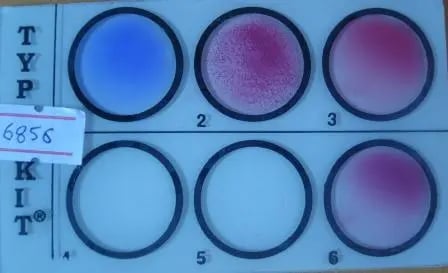 Image: Widal Test is one of the most common slide agglutination test method
Tube agglutination. The quantitative method. A fixed volume of antigen suspension is added to serial dilutions of serum; the reciprocal of the highest dilution still showing agglutination is the titer. Used for the serological diagnosis of typhoid (Widal), brucellosis, and typhus (Weil-Felix). Tube tests for brucellosis in particular can be complicated by the prozone phenomenon, so several dilutions must be tested to avoid a false-negative.
Particle agglutination methods
Latex agglutination. Antibody or antigen is bound to polystyrene latex beads (0.8–1 µm). The bead size makes the clumping easy to read by eye, and each bead carries many binding sites, amplifying the signal. Latex agglutination detects bacterial polysaccharide antigens at levels as low as about 1 ng/mL, so conditions (pH, ionic strength) must be standardized. Rheumatoid factor is a known cause of false positives; specimens may be pretreated (boiling or EDTA) to counter this. Reactions are graded 1+ to 4+, with 2+ the usual minimum positive. A control latex (coated with non-immune antibody from the same species) is run alongside; reactivity with both test and control latex means the result is nonspecific and uninterpretable. Clinical uses: Cryptococcus neoformans antigen in CSF or serum, group B Streptococcus, Clostridioides difficile toxins A and B, and rotavirus.
Coagglutination. A reverse passive method that uses killed Staphylococcus aureus (Cowan I strain) as the carrier. The staphylococcal cell wall is rich in protein A, which binds the Fc (base) of the coating antibody, leaving both Fab arms free to capture antigen. Highly specific, but generally less sensitive than latex for small amounts of antigen, so it is used more for identifying cultured organisms (Lancefield grouping of streptococci, S. pneumoniae, Neisseria meningitidis, N. gonorrhoeae, Haemophilus influenzae) than for direct antigen detection in specimens.
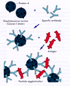 Fig: Coagglutination Method
Hemagglutination (as a carrier method). When red blood cells are the carrier, the reaction is hemagglutination. In direct hemagglutination the RBC's own surface antigen is the target (ABO grouping). In passive hemagglutination the RBC is coated with an unrelated soluble antigen so that the test detects antibody to that antigen (historic treponemal tests such as MHA-TP and TPHA use this design). Note: the inhibition of viral hemagglutination is a distinct application covered in the Hemagglutination inhibition (HAI) test.
Agglutination inhibition
A competitive format where the absence of visible clumping is the positive result. A patient sample and a carrier-bound version of the analyte compete for a limited amount of antibody. If the analyte is present in the sample, it soaks up the antibody and blocks agglutination of the carrier. Used to detect small soluble analytes: the classic latex agglutination inhibition urine test for hCG (pregnancy) and for drugs of abuse. This inverse logic (no clumping = positive) is the single most confusing point in agglutination testing and is worth stating explicitly.
How to Remember
- Which is on the bead tells you what you detect. If antigen is coated on the carrier, the test catches the patient's antibody (passive). If antibody is coated on the carrier, the test catches the patient's antigen (reverse passive). Whatever is on the bead is the "bait," and you are fishing for its partner.
- Inhibition flips the meaning of clumping. In a normal agglutination test, clumping is positive. In an agglutination inhibition test, clumping is negative and no clumping is positive, because the analyte you are hunting works by blocking the clumping. Whenever you see "inhibition," expect the result to read backwards.
Key exam facts in one table
| Point | What to remember |
|---|---|
| Agglutination | Antibody cross-links particle-bound antigen into visible clumps, read by eye |
| Direct | Antigen is naturally on the particle (ABO grouping, bacterial serotyping) |
| Indirect (passive) | Soluble antigen coated on carrier; detects patient's antibody |
| Reverse passive | Antibody coated on carrier; detects patient's antigen |
| On-the-bead rule | Antigen on bead = detect antibody; antibody on bead = detect antigen |
| Slide format | Rapid (seconds), read promptly, needs saline autoagglutination control |
| Tube format | Quantitative; titer = reciprocal of highest dilution still agglutinating |
| Latex | Beads 0.8–1 µm; detects polysaccharide antigen to ~1 ng/mL; RF causes false positives |
| Coagglutination | Reverse passive; S. aureus Cowan I protein A binds antibody Fc, Fab free for antigen |
| Hemagglutination | RBC as carrier; direct (ABO) or passive (coated RBC, e.g. TPHA) |
| Agglutination inhibition | No clumping = positive (analyte blocks agglutination); hCG, drugs of abuse |
| Prozone | Antibody excess blocks clumping → false negative; test several dilutions |
Where students get confused
"Passive and reverse passive are just two names for the same thing." They are opposites. Passive coats antigen on the bead to detect antibody. Reverse passive coats antibody on the bead to detect antigen. Fix the "what is on the bead" question first and the rest follows.
"Clumping always means positive." Not in an agglutination inhibition test. There, the analyte you are looking for prevents clumping, so a smooth, un-clumped result is the positive one. This trips up nearly everyone reading a latex inhibition pregnancy or drug test.
"Coagglutination and latex agglutination are interchangeable." Both are reverse passive particle methods, but coagglutination uses S. aureus protein A as the carrier and is more specific but less sensitive for small antigen amounts, so it is used mainly to identify cultured organisms, not to detect scarce antigen directly in a specimen. Latex is the more sensitive antigen-detection tool.
"A stronger antibody titer always gives stronger clumping." Antibody excess can block agglutination (the prozone phenomenon), giving a false-negative at low dilutions that becomes positive as you dilute out. This is why quantitative agglutination is read across a dilution series, not at a single concentration.
References
- Levinson, W. (2020). Review of Medical Microbiology and Immunology (16th ed.). McGraw-Hill.
- Tille, P. M. (2022). Bailey & Scott's Diagnostic Microbiology (15th ed.). Elsevier.
- Procop, G. W., et al. (2017). Koneman's Color Atlas and Textbook of Diagnostic Microbiology (7th ed.). Wolters Kluwer.
- Johnson, J., Duffy, K., New, L., Holliman, R. E., Chessum, B. S., & Fleck, D. G. (1989). Direct agglutination test and other assays for measuring antibodies to Toxoplasma gondii. Journal of Clinical Pathology, 42(5), 536–541.
Frequently Asked Questions
What is an agglutination test?
What is the difference between passive and reverse passive agglutination?
Why does a positive agglutination inhibition test show no clumping?
What is coagglutination?
Why can a high antibody level give a false-negative agglutination result?

Tankeshwar Acharya, MSc (Medical Microbiology)
Tankeshwar Acharya is an Assistant Professor in the Department of Microbiology at Patan Academy of Health Sciences (PAHS), Nepal, where he has been teaching and practicing clinical microbiology for over 14 years. He is the founder of Microbe Online, one of the leading free microbiology education resources on the web, covering bacteriology, mycology, parasitology, immunology, and clinical laboratory diagnostics written from direct experience in both the classroom and the diagnostic laboratory.
Comments
No comments yet. Be the first to share your thoughts.
Leave a comment
All comments are reviewed before they appear.