
Blood Agar: Composition, Preparation, and How to Read Hemolysis

On this page
The green colony that might be a pathogen, or might be normal throat flora
A throat swab from a child with a sore throat is streaked onto blood agar. The next morning the plate is covered in growth, most of it the mixed flora that lives in everyone's throat. The task is not to identify everything on the plate. It is to answer one question: is there anything here that needs treatment?
Blood agar answers it by showing how each colony treats red blood cells. The commensal streptococci that make up most of the plate are alpha-hemolytic, surrounded by a soft green halo. But among them, a small colony with a wide, clear, colorless zone stands out. That clear zone is beta-hemolysis, complete destruction of the red cells, and in a throat culture it is the signal that raises group A Streptococcus pyogenes, the cause of strep throat, rheumatic fever, and post-streptococcal kidney disease.
This is the everyday value of blood agar. It grows nearly everything in the specimen, and in the same step it sorts what grew by how it interacts with blood, turning a crowded plate into a short list of things worth a second look, often before any biochemical test is run. Reading those zones correctly, telling a wide beta zone from a narrow one, true green alpha from a deceptive clear-center variant, is one of the first real diagnostic skills a microbiologist learns, and one of the most used. The rest of this article is about how to read them.
Blood agar is an enriched bacterial growth medium. Fastidious organisms, such as streptococci, do not grow well on ordinary growth media but grow on blood agar.
Blood agar is a growth medium made from a nutrient-rich base enriched with 5% sheep blood. Common bases include tryptic soy agar and Columbia agar base; the representative formulation in the table below is a Columbia-type base.
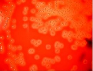 Figure: Beta hemolysis in sheep blood agar.
Blood agar consists of a base containing a protein source (e.g. tryptones), soybean protein digest, sodium chloride (NaCl), agar, and 5% sheep blood.
Blood contains inhibitors for certain bacteria such as Neisseria and Haemophilus genera, so the blood agar must be heated to inactivate these inhibitors and to release essential growth factors (X factor/hemin and V factor/NAD). Heating of blood agar converts it into chocolate agar (heated blood turns a chocolate color) and supports the growth of these bacteria.
Blood agar is the single most important and universally used primary plating medium in clinical microbiology. It is included in virtually every specimen workup (from throat swabs and wound cultures to blood cultures and CSF) because it supports the growth of nearly all clinically significant bacteria while simultaneously providing hemolysis patterns that give immediate presumptive identification clues within 18–24 hours of incubation.
The key diagnostic value of blood agar lies not just in growing organisms but in revealing how each organism interacts with red blood cells; a genetically determined characteristic that directly reflects bacterial virulence factors and narrows identification rapidly before any biochemical testing is performed.
Composition of Blood Agar
Ingredients | Gram/liter |
Beef heart peptone | 10 gm |
Tryptose | 10 gm |
Sodium chloride | 5 gm |
Agar | 15 gm |
Sheep blood | 5% |
Final pH at 25°C 7.3 ± 0.2 |
|
Protein sources may differ among manufacturers and may be pancreatic digest of casein, papaic (papain) digest of soybean meal, neutralized peptone, yeast extract, or a combination of them. Please check the paper insert in the purchased media.
Choice of the Blood
Sheep blood is the first choice to prepare BA plates, followed by horse, rabbit, or goat blood.
Human blood, particularly expired citrated donor blood, should not be used because this may contain substances inhibitory to the growth of some pathogens. Residual antibiotics in host blood and antibodies like ASO or anti-M protein could interfere with the growth of S. pyogenes. Citrate inhibits the growth of beta-hemolytic streptococci. Infected human blood may also contain infectious agents.
Preparation of Blood Agar
Preparation of blood agar from dehydrated blood agar base
- Prepare the Blood Agar base as instructed by the manufacturer.
- Sterilize by autoclaving at 121°C for 15 minutes.
- Transfer thus prepared BA base to a 50°C water bath.
- When the agar base is cooled to 50°C, add sterile sheep blood aseptically and mix well gently. Avoid the formation of air bubbles. You must have warmed the blood to room temperature at the time of dispensing to the molten agar base.
- Dispense 15 ml amounts to sterile Petri plates aseptically
- Label the medium with the date of preparation and give it a batch number (if necessary).
- Store the plates at 2-8°C, preferably in sealed plastic bags to prevent loss of moisture. The shelf life of thus prepared BA is up to four weeks.
Note: If you are planning to prepare a batch of blood agar plates, prepare few blood agar plates first to ensure that blood is sterile.
Quality control of Blood Agar
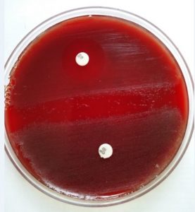 Figure: Optochin and bacitracin sensitivity of the isolates in Blood agar
- The final pH of the blood agar is 7.3 ± 0.2 at 25°C (approximately 7.1 to 7.5).
- Inoculate the plates with 5-hour broth cultures of Streptococcus pyogenes and S. pneumoniae. Inoculate also a plate with H. influenzae and streak with S. aureus (i.e. Satellitism Test).
- Incubate the plates in a carbon dioxide-enriched atmosphere at 35-37°C overnight.
- Check for the growth characteristics of each species
- S. pyogenes: Beta-hemolysis
- S. pneumoniae: Alpha-hemolysis
- H. influenzae: Satellitism
Inoculation and Incubation
- Allow the plate to reach room temperature and dry the agar surface before use. Inoculate as soon as possible after the specimen is collected.
- Inoculate directly from the specimen. Blood agar takes almost every routine specimen type: throat and wound swabs, sputum, urine, cerebrospinal fluid, and blood culture subcultures.
- If culturing from a swab, roll the swab over a small area at the edge of the plate first, then streak out from that area for isolation.
- Streak for well-isolated colonies over four quadrants using a sterile loop. Stab the loop into the agar in a few places. Streptolysin O is oxygen-labile, so stabbing lets the subsurface, reduced-oxygen environment reveal the full beta zone that a surface-only streak can understate.
- Incubate at 35 to 37°C for 18 to 24 hours. A carbon-dioxide-enriched atmosphere improves recovery of streptococci and makes the beta zone of group A Streptococcus easier to read.
- If growth is light or hemolysis is unclear at 24 hours, reincubate and read again at 48 hours. Some organisms, such as the umbilicated colony of Streptococcus pneumoniae, develop their characteristic appearance only by 48 to 72 hours.
- Read hemolysis by holding the plate up to a light source with the light coming from behind, and inspect subsurface or stab areas, not just the surface colony.
Uses of Blood Agar
Blood agar has two major uses:
- Primary isolation of nearly all clinically significant bacteria from most specimen types
- Isolation, identification, and susceptibility testing of streptococci. On blood agar, an optochin disc presumptively identifies S. pneumoniae (optochin-sensitive) among alpha-hemolytic colonies, while a bacitracin disc presumptively identifies group A S. pyogenes (bacitracin-sensitive) among beta-hemolytic colonies.
- Determine the type of hemolysis, if any.
Hemolysis
 Figure: Types of hemolysis (α, β and γ)
Certain bacterial species produce extracellular enzymes that lyse red blood cells in the blood agar (hemolysis). These hemolysins diffuse outward from the colonies and destroy red cells in the medium, either completely or partially. In complete (beta) hemolysis the hemoglobin is fully broken down to colorless products, leaving a clear zone. In partial (alpha) hemolysis the hemoglobin is only oxidized to green pigments, leaving a greenish zone.
Four hemolysis patterns are described on sheep blood agar: alpha (α), beta (β), gamma (γ), and alpha-prime (α′, wide-zone alpha). A separate and often-confused pattern is the double-zone (target) hemolysis of Clostridium perfringens, covered below.
Hemolysis is best observed by examining colonies grown under anaerobic conditions or inspecting sub-surface colonies. Hold the blood agar plate up to a light source and observe it with the light coming from behind (transmitted light) to read the type of hemolysis.
If either type of hemolysis is present, then one will observe a zone of hemolysis surrounding a growing colony.
 Figure: Various types of Hemolysis
Alpha (α) Hemolysis
Alpha hemolysis is the partial lysis of RBCs to produce a greenish-grey or brownish discoloration around the bacterial colony. In streptococci, alpha hemolysis is caused largely by hydrogen peroxide (H₂O₂) produced by the organism, which oxidizes hemoglobin to green products (verdohemoglobin and related biliverdin-type pigments) in the medium surrounding the colony. This is partial degradation, not the complete cell lysis seen in beta hemolysis, which is why the zone is green rather than clear.
Many of the alpha-hemolytic streptococci are part of the normal flora of humans but Streptococcus pneumoniae which is also alpha-hemolytic causes serious pneumonia and other deadly infectious diseases.
Viridans group of streptococci also gives alpha-hemolysis.
Beta (β) Hemolysis
Beta-hemolysis is the complete lysis of RBCs, resulting in a distinct, clear, colorless zone surrounding and under the colony. The RBC membrane is destroyed. Organisms of Group A beta-hemolytic streptococci-Streptococcus pyogenes and Group B, beta-hemolytic streptococci-Streptococcus agalactiae are beta-hemolytic.
Group A streptococci produce two hemolysins: streptolysin O (SLO), which is oxygen-labile and active only under reduced oxygen, and streptolysin S (SLS), which is oxygen-stable and responsible for the beta hemolysis seen around surface colonies. Because SLO contributes only under reduced oxygen, stabbing the loop into the agar or incubating in increased CO2 makes the full beta zone easier to read.
Other beta-hemolytic organisms are Staphylococcus aureus, Listeria monocytogenes, and Bacillus cereus.
Gamma (γ) or Non-hemolysis
Gamma-hemolysis indicates no hemolysis of RBCs. There is no change in the medium under and surrounding the colonies.
Alpha-Prime (Alpha') Hemolysis
Alpha-prime (α′) hemolysis, also called wide-zone alpha hemolysis, produces a small zone of intact or minimally affected red cells immediately around the colony, surrounded by a wider zone of partial (alpha, green) hemolysis further out. It is an alpha variant, most often described with certain streptococci, and can be mistaken for beta hemolysis if only the outer zone is read.
 Figure: Double zone hemolysis produced by Clostridium perfringens
Do not confuse this with the double-zone (target) hemolysis of Clostridium perfringens, which is a different pattern: an inner zone of complete (clear) beta hemolysis from theta-toxin, surrounded by an outer zone of partial (green) alpha hemolysis from alpha-toxin (lecithinase). The double zone is a strong presumptive identifier for C. perfringens in anaerobic cultures, but it is a beta-plus-alpha combination, not true alpha-prime.
Summary of hemolysis types:
| Type | Appearance | Mechanism | Classic examples |
|---|---|---|---|
| Alpha (α) | Green/brown discoloration around colony | Partial RBC lysis; hemoglobin → verdohemoglobin (green) | S. pneumoniae, viridans streptococci |
| Beta (β) | Complete clear zone around colony | Complete RBC lysis by hemolysins (streptolysin O/S, etc.) | S. pyogenes, S. agalactiae, S. aureus |
| Gamma (γ) | No change around colony | No hemolysis | Enterococcus faecalis, Klebsiella spp. |
| Alpha-prime (α') | Narrow rim of intact cells at colony + wider outer green zone | Wide-zone alpha variant | Some streptococci |
| Double zone (target) | Inner clear (beta) zone + outer green (alpha) zone | Theta-toxin (beta) + alpha-toxin/lecithinase (alpha) | Clostridium perfringens |
How to Remember
- Alpha = a little (partial), and green. Partial lysis, greenish discoloration. The organism only half-destroys the red cells, and the H₂O₂ it makes oxidizes hemoglobin to a green product. Think "alpha, a-little, algae-green."
- Beta = best/complete, and clear. Complete lysis, a clear colorless window you can read newsprint through. Beta clears the "brightest" zone.
- Gamma = goose egg = zero. No hemolysis, no change in the medium. Gamma, nothing happened.
- Wide zone = GAS, narrow zone = GBS. Both group A (S. pyogenes) and group B (S. agalactiae) are beta-hemolytic, but GAS clears a wide zone (2 to 4 times the colony width) while GBS clears only a narrow rim. A comes before B, and the A organism makes the bigger zone.
- Double zone = C. perfringens. A clear inner ring inside a wider green outer ring, a target or bull's-eye. Two toxins, two zones. See a target, think Clostridium perfringens.
Colony Morphology of Clinically Important Organisms on Blood Agar
Gram-positive cocci
| Organism | Hemolysis | Colony appearance | Key features |
|---|---|---|---|
| Staphylococcus aureus | Beta (variable) | Golden-yellow to cream, circular, convex, 2–3 mm, opaque, smooth | Yellow pigment produced at room temperature; coagulase positive |
| Staphylococcus epidermidis | Gamma | White to grey-white, circular, convex, 1–2 mm, smooth, opaque | Common skin contaminant; coagulase negative |
| Staphylococcus saprophyticus | Gamma | White to off-white, circular, 1–2 mm | Novobiocin resistant; UTI in young women |
| Streptococcus pyogenes (GAS) | Beta: large, clear zone (2–4× colony diameter) | Small (0.5–1 mm), grey-white, translucent, circular colonies | Large beta-hemolytic zone; bacitracin sensitive; PYR positive |
| Streptococcus agalactiae (GBS) | Beta: narrow zone (barely exceeds colony) | Small (0.5–1 mm), grey-white, flat, translucent | Narrow beta zone; CAMP test positive; hippurate positive |
| Streptococcus pneumoniae | Alpha: green, mucoid | Small (0.5–1.5 mm), grey, mucoid, umbilicated (depressed center) with age | Alpha-hemolytic; bile soluble; optochin sensitive; lancet-shaped diplococci |
| Viridans streptococci | Alpha | Small (0.3–0.5 mm), grey-white, non-mucoid | Alpha-hemolytic; bile insoluble; optochin resistant |
| Enterococcus faecalis | Gamma (occasionally alpha or beta) | Small (0.5–1 mm), grey-white, smooth | Growth in 6.5% NaCl; PYR positive; bile esculin positive |
| Micrococcus spp. | Gamma | Bright yellow to orange, circular, opaque, dry | Distinctive yellow pigment; catalase positive; modified oxidase positive |
Gram-positive rods
| Organism | Hemolysis | Colony appearance | Key features |
|---|---|---|---|
| Clostridium perfringens | Double zone (target: inner beta, outer alpha) | Large (2–4 mm), grey-white to yellowish, flat, irregular, ground-glass texture | Double-zone hemolysis is highly characteristic; anaerobic; lecithinase positive on EYA |
| Clostridium tetani | Beta (variable) | Swarming, thin, translucent film across plate surface; hard to see | Swarming growth; terminal spore ("drumstick"); anaerobic |
| Bacillus anthracis | Non-hemolytic (gamma) | Large (4–5 mm), grey-white, flat, irregular, "Medusa head" or ground glass; tenacious, stands up when lifted with loop | Non-hemolytic; distinguishes from B. cereus (beta-hemolytic) |
| Bacillus cereus | Beta: wide, clear zone | Large (3–5 mm), grey-white, spreading, irregular, waxy | Beta-hemolytic; distinguishes from B. anthracis; associated with food poisoning |
| Listeria monocytogenes | Beta: narrow, clear zone | Small (1–2 mm), grey-white, smooth, glistening | Narrow beta zone; tumbling motility at room temp; umbrella-shaped motility at 25°C |
| Corynebacterium diphtheriae | Gamma | Small (1–2 mm), grey-white, dry; irregular on Tellurite medium (black) | Non-hemolytic on blood agar; black colonies on tellurite medium |
Gram-negative organisms
| Organism | Hemolysis | Colony appearance | Key features |
|---|---|---|---|
| Escherichia coli | Gamma (some beta hemolytic strains) | Large (2–3 mm), grey, flat, smooth, sometimes mucoid; characteristic metallic sheen on EMB agar | Beta-hemolytic strains associated with UTI and diarrhea |
| Klebsiella pneumoniae | Gamma | Large (3–5 mm), mucoid, greyish, dome-shaped; may string when touched | Mucoid capsule; string test positive |
| Pseudomonas aeruginosa | Beta (variable) | Large (3–4 mm), flat, spreading, metallic sheen; blue-green pigment (pyocyanin); fruity grape-like odor | Pyocyanin pigment; characteristic odor; beta-hemolysis in some strains |
| Haemophilus influenzae | Gamma | Tiny (0.5–1 mm), grey, smooth, translucent, dewdrop-like; faint mousy or bleach-like odor | Requires X and V factors; satellitism around S. aureus colonies |
| Neisseria gonorrhoeae | Gamma | Tiny (0.5–1 mm), grey, translucent, convex; requires CO₂ | Does not grow well on plain blood agar; prefers chocolate agar or Thayer-Martin |
| Proteus mirabilis | Beta (variable) | Swarming across entire plate; characteristic foul putrid odor | Swarming inhibited on MacConkey; urease strongly positive |
| Vibrio cholerae | Beta: large, clear zone | Large (2–3 mm), grey, smooth, moist colonies; "iridescent" sheen | Large beta zone; characteristic odor; oxidase positive |
| Bacteroides fragilis | Gamma | Grey, non-hemolytic, circular, with irregular edge, 1–3 mm; anaerobic | Non-hemolytic; bile-resistant; grows relatively rapidly for an anaerobe |
| Fusobacterium nucleatum | Gamma | Flat, irregular, "breadcrumb" colonies with internal speckles; anaerobic; strong foul odor | Spindle-shaped cells on gram stain; indole positive |
Clinically Important Modifications of Blood Agar
Blood agar can be modified by adding selective agents, changing the blood source, or altering preparation to create specialist media:
| Modified blood agar | Key addition | Primary use |
|---|---|---|
| Chocolate agar | Blood lysed by heating to 75-80°C | Haemophilus spp., Neisseria spp.; releases X and V factors |
| Crystal violet blood agar (CVBA) | 0.02% crystal violet | Selective for Group A Streptococcus from throat; inhibits S. aureus and commensals |
| Columbia CNA agar | Colistin + nalidixic acid | Selective for gram-positive organisms; inhibits gram-negatives |
| Neomycin blood agar | Neomycin | Selective for gram-positive anaerobes; inhibits gram-negatives |
| Laked kanamycin-vancomycin blood agar (LKV) | Kanamycin + vancomycin + laked blood | Selective for Bacteroides and Prevotella spp. |
| Phenylethyl alcohol blood agar (PEA) | Phenylethyl alcohol | Inhibits swarming; selective for gram-positive and obligate anaerobic gram-negatives |
| Horse blood agar | 5–10% horse blood instead of sheep blood | Enhanced detection of H. influenzae hemolysis; some virulence studies |
| Rabbit blood agar | Rabbit blood | Detection of CAMP factor; Listeria beta-hemolysin |
To prepare crystal violet blood agar, add 1 ml of 0.02% w/v crystal violet per 1000 ml of blood agar. This suppresses Staphylococcus aureus and oral commensals while allowing Group A streptococci to grow.
Where Students Get Confused
- Bacillus anthracis vs. Bacillus cereus. Nearly identical colony size and "ground-glass" appearance. The one reliable bedside distinguisher is hemolysis: B. anthracis is non-hemolytic (gamma); B. cereus produces a wide, beta-hemolytic zone. Given the biosafety stakes, this is the single most important hemolysis reading on this entire page.
- Streptococcus pneumoniae vs. viridans streptococci. Both produce visually identical green, alpha-hemolytic colonies. Hemolysis type alone cannot tell them apart; optochin sensitivity and bile solubility are what actually distinguish them, S. pneumoniae is optochin-sensitive and bile-soluble, viridans strep is neither.
- Streptococcus pyogenes (GAS) vs. Streptococcus agalactiae (GBS). Both are beta-hemolytic but GAS produces a wide beta zone, often 2 to 4 times the colony's own diameter; GBS produces a narrow zone that barely extends past the colony edge.
- Alpha-prime vs. double-zone (target) hemolysis. These are two different patterns that are easy to merge. Alpha-prime is an alpha variant: a narrow rim of intact red cells right at the colony, with a wider green zone beyond it, and reading only the outer zone can make it look beta.
Double-zone or target hemolysis belongs to Clostridium perfringens: an inner clear beta zone from theta-toxin inside an outer green alpha zone from alpha-toxin. Alpha-prime is alpha throughout; the target pattern is beta plus alpha.
References
- Madigan, M. T., Bender, K. S., Buckley, D. H., Sattley, W. M., & Stahl, D. A. (2021). Brock Biology of Microorganisms (16th ed.). Pearson.
- Procop, G. W., Church, D. L., Hall, G. S., Janda, W. M., Koneman, E. W., Schreckenberger, P. C., & Woods, G. L. (2017). Koneman's Color Atlas and Textbook of Diagnostic Microbiology (7th ed.). Philadelphia: Wolters Kluwer.
- Tille, P. M. (2022). Bailey and Scott's Diagnostic Microbiology (15th ed.). St. Louis: Elsevier.
- Facklam, R. R., & Washington, J. A. (1991). Streptococcus and related catalase-negative gram-positive cocci. In Balows, A., et al. (Eds.), Manual of Clinical Microbiology (5th ed.). Washington, DC: American Society for Microbiology.
Frequently Asked Questions
What is the difference between alpha and beta hemolysis?
Alpha is partial lysis, green/brown discoloration: S. pneumoniae, viridans streptococci. Beta is complete clear lysis: S. pyogenes, S. agalactiae, S. aureus. Gamma is no hemolysis: Enterococcus, Klebsiella.
Why is sheep blood used instead of human blood?
Why does S. pneumoniae produce alpha not beta hemolysis?
Why does S. pneumoniae produce alpha not beta hemolysis?
The H₂O₂ produced by S. pneumoniae oxidizes hemoglobin to green products (verdohemoglobin), a partial degradation rather than true lysis. S. pneumoniae lacks the streptolysins O and S that produce the complete, clear lysis of beta hemolysis.
What does the size of the beta-hemolytic zone tell you?
GAS (S. pyogenes): large zone 2-4× colony diameter. GBS (S. agalactiae): narrow zone barely beyond colony edge. Helps preliminary differentiation at 24 hours with CAMP test and bacitracin.
What is the umbilicated colony appearance of S. pneumoniae?
What is the umbilicated colony appearance of S. pneumoniae?
Autolysin LytA causes central autolysis at 48-72 hours, raised ring with sunken center. Umbilicated appearance + alpha hemolysis = strong presumptive S. pneumoniae.
How does incubation atmosphere affect blood agar hemolysis?
Streptolysin O is oxygen-labile, best seen in stab areas or anaerobically. Streptolysin S is oxygen-stable, visible aerobically on surface. Always stab blood agar.
Why does C. perfringens produce double-zone hemolysis?
Why does C. perfringens produce double-zone hemolysis?
Theta-toxin: outer partial (alpha) zone. Alpha-toxin/lecithinase: inner complete (beta) zone. Double-zone target pattern on anaerobic blood agar = strong presumptive C. perfringens.
Can blood agar be used for susceptibility testing?
Yes. MH-F (Mueller-Hinton + 5% sheep blood) is CLSI-recommended for fastidious organisms: S. pneumoniae, S. pyogenes, H. influenzae, N. gonorrhoeae.

Tankeshwar Acharya, MSc (Medical Microbiology)
Tankeshwar Acharya is an Assistant Professor in the Department of Microbiology at Patan Academy of Health Sciences (PAHS), Nepal, where he has been teaching and practicing clinical microbiology for over 14 years. He is the founder of Microbe Online, one of the leading free microbiology education resources on the web, covering bacteriology, mycology, parasitology, immunology, and clinical laboratory diagnostics written from direct experience in both the classroom and the diagnostic laboratory.
Comments
No comments yet. Be the first to share your thoughts.
Leave a comment
All comments are reviewed before they appear.