
Cryptococcus neoformans: Pathogenesis, Lab Diagnosis
Cryptococcus neoformans and C. gattii: capsule and melanin virulence, narrow-based budding, why it causes fungal meningitis in HIV/AIDS, and lab diagnosis by India ink, birdseed agar, and the cryptococcal antigen (CrAg) test.

On this page
Cryptococcus neoformans is a yeast with a prominent polysaccharide capsule. It is an opportunistic fungal pathogen notoriously known as the most common cause of fungal meningitis (infection and inflammation of the meninges) in immunocompromised patients (such as people with AIDS).
It reproduces by budding and the single buds are characteristically narrow at the base. It does not produce pseudohyphae (Candida albicans does). It is a ubiquitous saprophyte often found associated with bird droppings, especially of pigeons.
C. neoformans vs C. gattii
Two species cause almost all human cryptococcosis, and telling them apart matters because they infect different people.
- Cryptococcus neoformans is the classic opportunist. It is found worldwide in soil enriched with pigeon and other bird droppings, and it causes disease almost exclusively in people with impaired T-cell immunity, above all advanced HIV/AIDS.
- Cryptococcus gattii is ecologically and clinically different. It is associated with eucalyptus and other trees rather than bird droppings, has a more tropical and subtropical distribution (though the Pacific Northwest outbreak extended its known range), and characteristically infects immunocompetent hosts. A cryptococcal infection in a patient with no evident immune defect should raise suspicion of C. gattii.
Both are identified in the same way in the laboratory, and both are urease-positive, melanin-producing encapsulated yeasts. They are distinguished definitively by canavanine-glycine-bromothymol blue (CGB) agar, on which C. gattii grows and turns the medium blue while C. neoformans does not.
Pathogenesis
Infection is initiated by inhalation of the yeast cells. The primary pulmonary infection may be asymptomatic or may mimic influenza-like respiratory infection often resolving spontaneously. In immune-compromised patients with impaired T cell immunity, the yeasts may multiply and disseminate to other parts of the body but preferentially to the central nervous system (neurotropic), causing cryptococcal meningitis. Other common sites of dissemination include the skin, adrenals, bone, eye and prostate gland. The inflammatory reaction is usually minimal or granulomatous.
Virulence Factors of Cryptococcus neoformans
- Capsule: Cryptococcus neoformans is the only pathogenic yeast known to have a polysaccharide capsule. The function of capsule is as follows:
- Anti-phagocytic: The capsule is composed mainly of glucuronoxylomannan (GXM), which inhibits phagocytosis by interfering with opsonization and blocking recognition by immune cells. GXM is also the antigen detected by the cryptococcal antigen (CrAg) test described below.
- Protection under drying conditions: The capsule collapses and protects the yeast.
- Ideal size range for alveolar deposition: the cell size reduction, resulting from capsular collapse, places the yeast in the ideal size range for alveolar deposition.
- Phenoloxidase: This enzyme is responsible for melanin production. Melanin may act as a virulence factor by protecting the organism against oxidative killing by leukocytes. It has been found that increased melanin production can decrease lymphocyte proliferation and tumor necrosis factor production. Phenoloxidase also protects the organism from oxidants released by phagocytic cells.
Diseases
Cryptococcosis is most often seen in immune-compromised patients with impaired T cell immunity. Important risk factors include AIDS, corticosteroid therapy, lymphoma, and T cell dysfunction. Cryptococcal infection occurs throughout the world sporadically. Infection occurs from the environment usually by inhalation, especially of dust containing excreta of pigeons but the infection is not transmitted from person to person. People with immune-compromised status are likely to be affected more and have serious outcomes;
- Cryptococcal meningitis: Only in a small proportion of cases, hematogenous spread results in subacute or chronic meningitis or meningoencephalitis. All untreated cases of cryptococcal meningitis are ultimately fatal. A significant proportion of untreated advanced-HIV patients develop cryptococcal meningitis, historically cited around 5 to 8%, though widespread antiretroviral therapy has reduced this in many regions.
- Signs and symptoms of cryptococcal meningitis: Headache, fever, altered mental status (confusion to lethargy to coma), blurred vision and other cranial nerve deficits, neck stiffness, photophobia, nausea and vomiting, seizures, and papilledema.
- Lung infections: Pulmonary disease is increasing nowadays, particularly in immunocompromised hosts. Reactivation of old healed lesions may occur.
- Skin and other infections: Sometimes skin, lymph nodes, bones are involved.
Laboratory diagnosis
Specimens: Specimens depends on clinical presentation and suspected disease conditions. Common specimens include spinal fluid (CSF), tissue, exudates, sputum, blood, and urine.
Microscopy and staining
Cryptococcus neoformans appear as a spherical, single or multiple budding, thick-walled yeast that is 2-15 μm (wide variation in size) in diameter. It is usually surrounded by a wide refractile capsule.
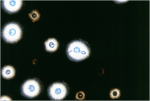 Figure: India ink preparation of CSF sample
India ink preparation is used as a rapid and inexpensive diagnostic tool for detecting cryptococcal infection in many institutions and resource-poor settings. Demonstration of heavily capsulated yeast cells (see the image) in CSF, exudates, and urine establishes the diagnosis.
India ink preparation when positive in CSF is diagnostic of cryptococcal meningitis but its sensitivity is low. Many diagnostic laboratories have replaced this test with a more sensitive cryptococcal latex agglutination test.
Culture
Colonies develop within a few days on most media (e.g., Sabouraud’s dextrose agar) at room temperature or 37°C. Cryptococcus neoformans are sensitive to cycloheximide so media containing cycloheximide should be avoided. Other culture media are Blood Agar, BHI Agar, Bird seed agar, etc. Colonies in SDA are creamy, white, and mucoid (because of capsule).
Cryptococcus neoformans produces melanin on birdseed (niger/Guizotia) agar via its phenoloxidase, forming brown to black colonies. This is a rapid presumptive identification and links directly to the phenoloxidase virulence factor above.
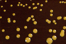 Image-2: Mucoid colonies of C. neoformans in Bird Seed Agar (source: The University of Adelaide)
Identification: Cryptococcus neoformans is identified by urease production and carbohydrate assimilation test, and confirmed by direct immunofluorescence using a fluorescein-labeled anti-neoformans antibody.
Cryptococcal Antigen (CrAg) Detection
Detecting the capsular antigen glucuronoxylomannan (GXM) in CSF or serum is now the cornerstone of cryptococcal diagnosis, and it has largely displaced India ink because it is far more sensitive and can be done on blood. Three formats exist:
Latex agglutination. Latex particles coated with anti-GXM antibody clump when the antigen is present. Sensitivity for cryptococcal meningitis is around 90%. It requires some technical skill and controls for rheumatoid factor.
Enzyme immunoassay (EIA). A plate-based antigen assay, useful for batch testing.
Lateral flow assay (LFA). A dipstick point-of-care test, cheap, rapid (about 10 minutes), needs no cold chain or specialized equipment, and works on serum, plasma, whole blood, or CSF. The LFA transformed cryptococcal diagnosis in resource-limited and high-HIV-burden settings, where it is now used to screen antigen in advanced HIV patients before symptoms develop. It is the format most likely to be used at the bench today.
Two points that matter at the bench:
A positive CrAg titer can also be used to gauge burden and follow response, since falling titers accompany successful treatment. Titers are reported as part of the result.
The prozone (postzone) effect is a genuine trap: at very high antigen concentrations, antibody can be overwhelmed and a strongly positive sample may read weakly positive or even negative. When the clinical suspicion is high but the CrAg is unexpectedly low or negative, the sample is diluted and retested. This is the same equivalence-zone phenomenon covered in antigen-antibody reactions.
Antibody detection has little diagnostic value (immunocompromised patients mount poor antibody responses), and molecular methods are not routine for diagnosis.
Key Facts
- Only pathogenic yeast known to have a polysaccharide capsule.
- The most common cause of fungal meningitis. Cryptococcal meningitis is a common cause of death among HIV/AIDS patients
- India ink preparation and cryptococcal antigen agglutination are commonly used diagnostic methods.
- Association with birds excreta (pigeon droppings) and rapid urease positive
How to Remember
Narrow-based budding = Cryptococcus; broad-based budding = Blastomyces. Both are budding yeasts in tissue, and this one feature separates them. Pair them in memory so the contrast sticks.
Neoformans needs a weak host; gattii doesn't. C. neoformans is the pigeon-dropping opportunist of HIV/AIDS patients. C. gattii is the tree-associated species that hits the immunocompetent.
The capsule does two jobs, and both are testable. It blocks phagocytosis (virulence), and its GXM antigen is what the CrAg test detects (diagnosis).
Black on birdseed = melanin = phenoloxidase. The same enzyme that makes the colonies black on birdseed agar is a virulence factor that protects the yeast from oxidative killing.
"India ink is out, CrAg is in." India ink is cheap and fast but insensitive; the CrAg lateral flow assay replaced it as the first-line test.
Key exam facts
| Feature | Detail |
|---|---|
| Morphology | Encapsulated yeast, narrow-based budding, 2 to 15 µm |
| Capsule polysaccharide | Glucuronoxylomannan (GXM); anti-phagocytic; the CrAg target |
| Two species | C. neoformans (birds, immunocompromised), C. gattii (trees, immunocompetent) |
| Ecology of neoformans | Pigeon and bird droppings |
| Key virulence factors | Capsule (GXM) and melanin (phenoloxidase) |
| Neurotropism | Preferentially disseminates to the CNS, causing meningitis |
| Rapid microscopy | India ink (clear halo in CSF); low sensitivity |
| First-line antigen test | CrAg lateral flow assay (LFA) |
| Birdseed (niger) agar | Brown-black colonies from melanin (phenoloxidase) |
| Urease | Positive |
| Sensitive to | Cycloheximide (avoid cycloheximide-containing media) |
| Species-differentiating medium | CGB agar (C. gattii turns it blue) |
References
- Rathore, S. S., Sathiyamoorthy, J., Lalitha, C., & Ramakrishnan, J. (2022). A holistic review on Cryptococcus neoformans. Microbial pathogenesis, 166, 105521. https://doi.org/10.1016/j.micpath.2022.105521
- Lin X. (2009). Cryptococcus neoformans: morphogenesis, infection, and evolution. Infection, genetics and evolution : journal of molecular epidemiology and evolutionary genetics in infectious diseases, 9(4), 401–416. https://doi.org/10.1016/j.meegid.2009.01.013
Frequently Asked Questions
How is Cryptococcus different from Candida on microscopy?
How is Cryptococcus different from Candida on microscopy?
Cryptococcus is an encapsulated yeast with narrow-based budding and no pseudohyphae, and it shows a clear halo on India ink. Candida has no significant capsule, buds without the narrow neck, and forms pseudohyphae (and true hyphae for some species). The capsule and the absence of pseudohyphae are the quickest distinguishing features.
Why does Cryptococcus cause meningitis specifically?
Why does Cryptococcus cause meningitis specifically?
What is the difference between C. neoformans and C. gattii?
What is the difference between C. neoformans and C. gattii?
C. neoformans is found in bird droppings and infects mainly immunocompromised people, especially those with advanced HIV. C. gattii is associated with trees, has a more tropical distribution (with a known outbreak in the Pacific Northwest), and characteristically infects immunocompetent people. They are separated in the lab on CGB agar, which C. gattii turns blue.
Why has the CrAg test replaced India ink?
Why does Cryptococcus grow black on birdseed agar?
Why does Cryptococcus grow black on birdseed agar?
Cryptococcus neoformans produces the enzyme phenoloxidase, which converts substrates in birdseed (niger seed) agar into melanin, turning the colonies brown to black. This is a rapid presumptive identification, and the same melanin production is one of the organism's virulence factors.

Tankeshwar Acharya, MSc (Medical Microbiology)
Tankeshwar Acharya is an Assistant Professor in the Department of Microbiology at Patan Academy of Health Sciences (PAHS), Nepal, where he has been teaching and practicing clinical microbiology for over 14 years. He is the founder of Microbe Online, one of the leading free microbiology education resources on the web, covering bacteriology, mycology, parasitology, immunology, and clinical laboratory diagnostics written from direct experience in both the classroom and the diagnostic laboratory.
Comments
No comments yet. Be the first to share your thoughts.
Leave a comment
All comments are reviewed before they appear.