
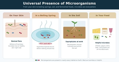
Universal Presence of Microorganisms: Why Microbes Are Everywhere
Microorganisms live on your skin, in boiling springs, and in every fermented food. Learn microbial ubiquity, normal flora, extremophiles, and why it matters.
Bacteria, viruses, fungi, and parasites explained the way the bench and the ward actually use them: how tests work, how to read results, and where diagnosis goes wrong. With interactive MCQs to test yourself.
Microorganisms live on your skin, in boiling springs, and in every fermented food. Learn microbial ubiquity, normal flora, extremophiles, and why it matters.
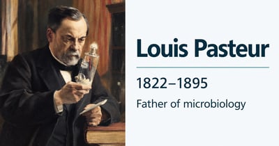
Who was Louis Pasteur and why is he called the father of microbiology? His work on germ theory, fermentation, pasteurization, and the first attenuated vaccines.
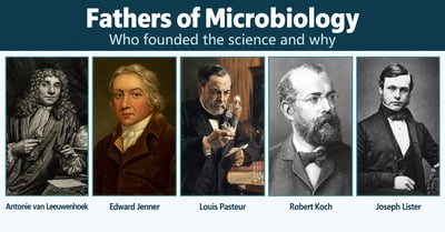
Who is the father of microbiology, bacteriology, and immunology? A clear guide to Pasteur, Koch, Leeuwenhoek, Lister, and Jenner, and what each is credited with.
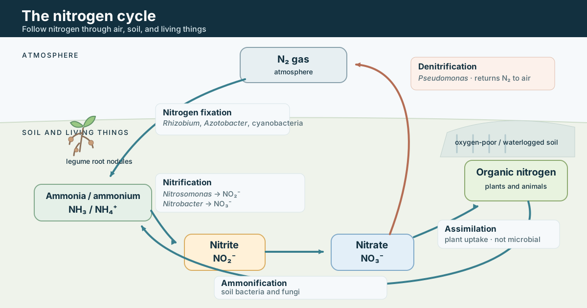
The nitrogen cycle explained step by step: fixation, nitrification, assimilation, ammonification, and denitrification, and the microbes that drive each stage.
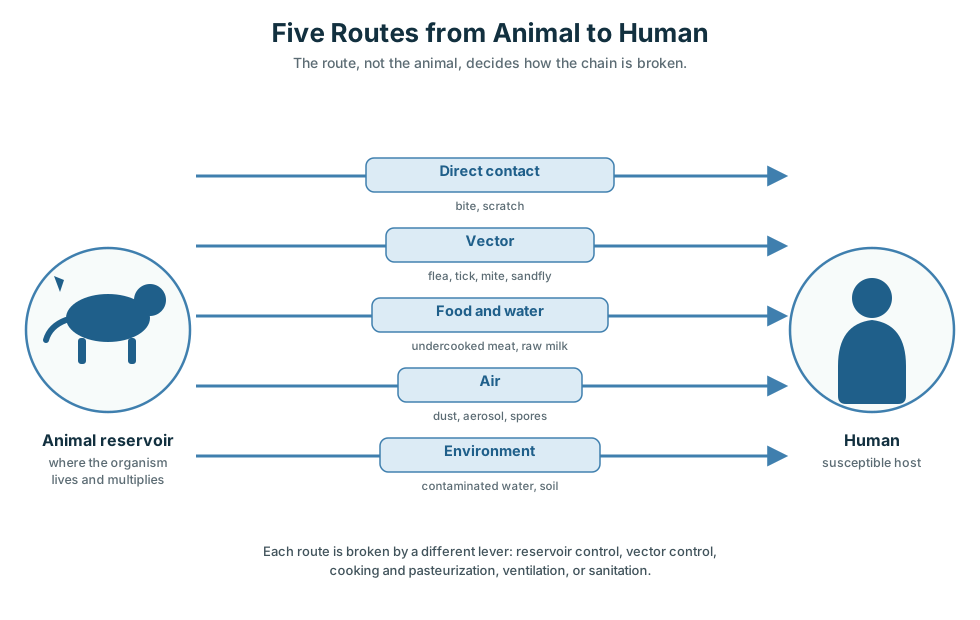
How zoonoses pass from animal reservoirs to humans, organized by the five transmission routes, why the reservoir decides control, with worked examples.
How automated specimen processors plant and streak cultures, how bead streaking compares with a manual loop, and where these instruments fit as the front end of lab automation.

How a thermocycler runs PCR: its parts, the Peltier block and heated lid, why ramp rate and gradient settings change your result, and common errors.

What a standard operating procedure is, why the microbiology laboratory runs on SOPs, and a step-by-step guide to writing a clear, usable SOP with the sections it must contain.
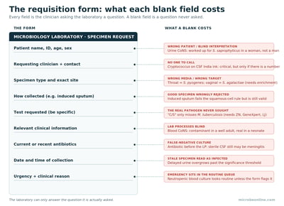
The requisition form is how a clinician talks to the microbiology laboratory. What each field means, what a blank field costs, and why filling it completely changes the diagnosis.
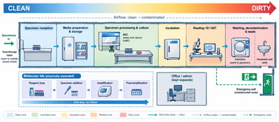
How a microbiology laboratory is laid out, the unidirectional clean-to-dirty workflow, zoning, biosafety cabinet placement, and the design principles that keep work safe and contam...
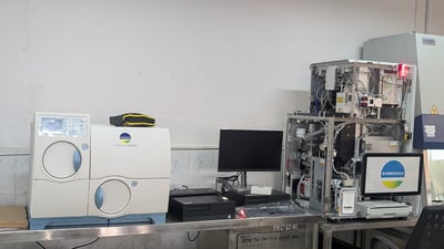
How each step of the microbiology workflow is automated, from media preparation to identification and susceptibility, and where standalone instruments end and full integration begi...

What it takes to run a microbiology laboratory, the facility, workflow from specimen to report, safety, equipment, and the quality system that governs it all, with a guide to each...

What laboratory accreditation is, how it differs from certification, what ISO 15189 requires, and how a microbiology laboratory achieves and maintains accreditation through bodies...
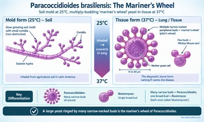
Paracoccidioides brasiliensis causes paracoccidioidomycosis (South American blastomycosis). Its tissue yeast forms the diagnostic "mariner's wheel" of multiple peripheral buds. Mor...