A patient on prolonged broad-spectrum antibiotics develops oral white plaques and a burning sensation. The Gram stain shows Gram-positive budding yeast with pseudohyphae. Candida albicans; an organism that normally lives harmlessly on mucosal surfaces has become a pathogen because the microbial competition was eliminated.
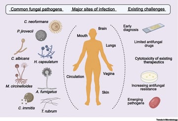
Fungi are eukaryotic organisms that cause infections ranging from superficial skin disease to life-threatening systemic illness. They are increasingly important in clinical practice because the patients most vulnerable to fungal infections (those on immunosuppressants, chemotherapy, or prolonged antibiotics, and those with HIV) are a growing population.
This section covers:
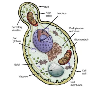
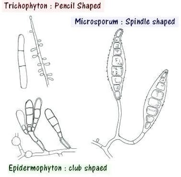
- Fungal structure and classification: yeasts, moulds, and dimorphic fungi; cell wall composition; hyphal morphology; and the clinical significance of these structural differences
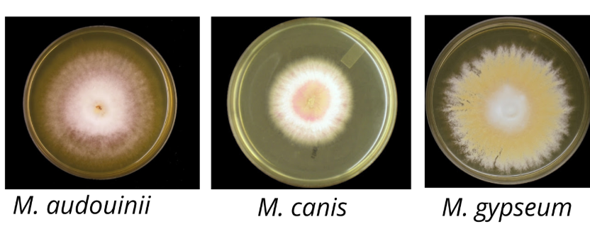
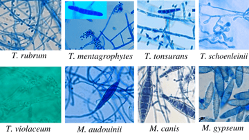
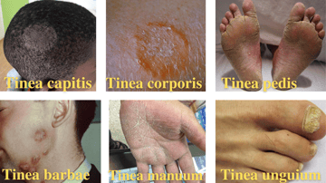
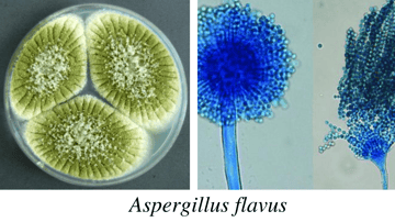
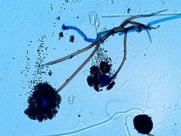
- Organism profiles: Candida, Aspergillus, Cryptococcus, Histoplasma, Coccidioides, Mucor, dermatophytes, and other clinically relevant genera
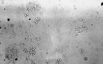
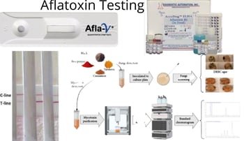
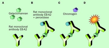
- Laboratory identification: direct microscopy (KOH preparation, India ink, Gram stain), culture on Sabouraud Dextrose Agar, germ tube test, biochemical identification, and antifungal susceptibility testing
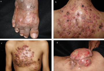
- Pathogenesis and clinical disease: the conditions that predispose to fungal infection, the mechanisms by which fungi cause tissue damage, and the major clinical syndromes
Mycology is often treated as a secondary topic in microbiology curricula, but its clinical importance in immunocompromised patients makes it exam-relevant and patient-care-relevant in equal measure.