

Bacteria grow on solid media as colonies. A colony is defined as a visible mass of microorganisms originating from a single mother cell. Key features of these bacterial colonies serve as important criteria for their identification.
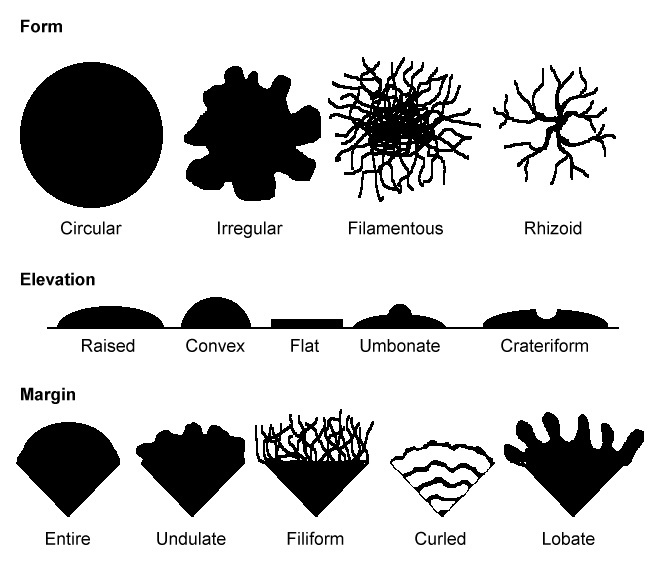 Figure: Characteristics of bacterial colonies
Colony morphology can sometimes be useful in bacterial identification. Colonies are described based on size, shape, texture, elevation, pigmentation, and effect on growth medium.
Why colony morphology matters in clinical microbiology
Colony morphology is the first observation a clinical microbiologist makes when examining an incubated culture plate — before any biochemical testing, before MALDI-TOF, before serology. Within seconds of opening an incubated plate, an experienced microbiologist can make immediate presumptive assessments that directly guide the next steps in identification.
The combination of:
- Medium (which agar the organism is growing on)
- Colony characteristics (size, color, hemolysis, texture, odor)
- Gram stain of a representative colony
...gives a preliminary identification in most cases before any further testing is performed.
This article covers the universal descriptors used to characterize any bacterial colony. For organism-specific colony appearances on selective and differential media, see the dedicated articles:
→ Blood Agar: Colony Morphology
→ MacConkey Agar: Colony Morphology
Find common criteria that are used to describe colony morphology;
Colony shape
It includes the form, elevation, and margin of the bacterial colony.
Form of the bacterial colony: The form refers to the shape of the colony. These four forms represent the most common colony shapes you are likely to encounter.
- circular,
- irregular,
- filamentous, and
- rhizoid
Elevation of the bacterial colony: It gives information about how much the colony rises above the agar. This describes the “side view” of a colony.
The six most common elevations of bacterial colonies are
- flat,
- raised,
- umbonate (having a knobby protuberance),
- crateriform,
- convex, and
- pulvinate (cushion-shaped).
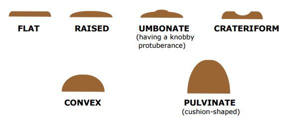 Figure: Elevation of Bacteria colony
The margin of the bacterial colony: The margin or edge of a colony may be a vital characteristic in identifying organisms. Examples are
- entire (smooth),
- irregular,
- undulate (wavy),
- lobate,
- curled, and
- filiform.
Colonies that are irregular in shape and/or have irregular margins are likely to be motile organisms. Highly motile organisms swarmed over the culture media, such as Proteus spp.
Size of the bacterial colony
The size of the colony can be a useful characteristic for identification. The diameter of a representative colony may be measured in millimeters or described in relative terms such as pinpoint, small, medium, and large.
 Figure: Punctiform and other types of bacterial colony
Tiny colonies are also referred to as punctiform (pin-point). Colonies larger than about 5 mm are likely to be motile organisms. Punctiform colonies are distinguished from circular colonies by their very small size.
Appearance of the colony surface
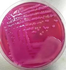 Figure: Mixed growth of mucoid Lactose fermenting colonies and NLF colonies in MacConkey Agar
Bacterial colonies are frequently shiny and smooth in appearance. Other surface descriptions might be: dull (opposite of glistening), veined, rough, wrinkled (or shriveled), or glistening. Bacillus species give dry, wrinkled colonies. Pseudomonas stutzeri also gives similar-appearing wrinkled colonies.
Consistency/Texture
Several terms that may be appropriate for describing the texture or consistency of bacterial growth are: dry, moist, viscid (sticks to loop, hard to get off), brittle/friable (dry, breaks apart), mucoid (sticky, mucus-like).
Color of the colonies (pigmentation)
Some bacteria produce pigment when they grow in the medium, e.g., green pigment produces by Pseudomonas aeruginosa, buff-colored colonies of Mycobacterium tuberculosis in L.J medium, and red-colored colonies of Serratia marcescens.
The opacity of the bacterial colony
The opacity of a bacterial colony can be described as transparent (clear), opaque (not transparent or clear), translucent (almost clear, but distorted vision–like looking through frosted glass), or iridescent (changing colors in reflected light). A pinpoint translucent β-hemolytic colonies on blood agar is most probably a Streptococcus species. Staphylococci give opaque, smooth, and circular colonies on the agar plate surface.
Some important terminologies
Draughtsman colonies
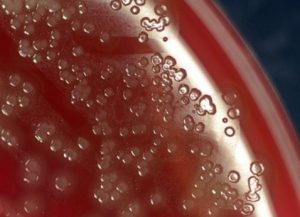 Figure: Draughtsman's colonies of S. pneumoniae
Young colonies of Streptococcus pneumoniae(pneumococci) have raised centers, but as the culture ages, they become flattened, with a depressed central part and raised edges giving them a ringed appearance also known as ‘draughtsman colonies’.
Colony Morphology of Clinically Important Bacteria
The following table summarizes the characteristic colony appearances of clinically important bacteria on blood agar (the most informative primary plating medium) and nutrient agar after 18–24 hours of aerobic incubation at 35–37°C, unless otherwise noted. Anaerobes (Clostridium, Bacteroides) and slow growers (Actinomyces) require anaerobic conditions and longer incubation, as indicated in their rows.
Gram-positive cocci
| Organism | Blood agar colony | Nutrient agar | Hemolysis | Key distinguishing features |
|---|---|---|---|---|
| Staphylococcus aureus | 2–3 mm, circular, convex, smooth, golden-yellow to cream, opaque | Similar; yellow pigment more prominent at room temperature | Beta (variable) | Golden pigment; coagulase positive; MRSA strains may lack pigment |
| Staphylococcus epidermidis | 1–2 mm, circular, convex, white to grey-white, smooth, opaque | White, non-pigmented | Gamma | White colonies; coagulase negative; common skin contaminant |
| Staphylococcus saprophyticus | 1–2 mm, circular, convex, white to off-white | White | Gamma | Similar to S. epidermidis; novobiocin resistant; UTI in young women |
| Streptococcus pyogenes (GAS) | 0.5–1 mm, grey-white, translucent, circular, dry | Tiny, grey-white | Beta — large, wide zone (2–4× colony diameter) | Large beta-hemolytic zone; tiny colonies; bacitracin sensitive |
| Streptococcus agalactiae (GBS) | 0.5–1 mm, grey-white, flat, translucent | Tiny, grey-white | Beta — narrow zone (barely exceeds colony) | Narrow beta zone; CAMP positive; hippurate positive |
| Streptococcus pneumoniae | 0.5–1.5 mm, grey, mucoid, umbilicated (depressed center at 48+ hours) | Tiny, mucoid | Alpha — green discoloration | Umbilicated colonies at 48 hours; bile soluble; optochin sensitive |
| Viridans streptococci | 0.3–0.5 mm, grey-white, non-mucoid | Tiny | Alpha — green discoloration | Bile insoluble; optochin resistant; multiple species |
| Enterococcus faecalis | 0.5–1 mm, grey-white, smooth | Small, grey-white | Gamma (rarely alpha or beta) | Growth in 6.5% NaCl; PYR positive; bile esculin positive |
| Micrococcus spp. | 1–2 mm, bright yellow to orange, circular, opaque, dry | Yellow to orange; pigment prominent | Gamma | Distinctive yellow/orange pigment; modified oxidase positive |
Gram-positive rods
Organism | Blood agar colony | Key features | Hemolysis | Notes |
|---|---|---|---|---|
Bacillus anthracis | 3–5 mm, grey-white, flat, irregular "Medusa head" or ground glass; non-hemolytic; tenacious — stands up when lifted with loop | Large, flat, irregular, rough | Gamma (non-hemolytic) | Non-hemolytic Bacillus — distinguishes from B. cereus |
Bacillus cereus | 3–5 mm, grey-white, spreading, irregular, waxy | Large, spreading, rough | Beta — wide zone | Beta-hemolytic; food poisoning organism |
Clostridium perfringens | 2–4 mm, grey-white, flat, irregular, ground glass texture | Anaerobic; large, flat | Double zone (inner beta, outer alpha) | Inner complete (theta toxin) and outer incomplete (alpha toxin/lecithinase) zone; characteristic, not strictly pathognomonic |
Clostridium tetani | Thin, swarming film across entire plate | Anaerobic; swarming, invisible | Beta (variable) | Swarming growth creates thin haze; terminal spore on gram stain |
Clostridium difficile |
| Anaerobic only; grows on CCFA | Gamma | Characteristic horse-barn odor; toxin detection required for diagnosis |
Listeria monocytogenes | 1–2 mm, grey-white, smooth, glistening | Small, glistening | Beta — narrow zone | Narrow beta zone; tumbling motility at 25°C |
Corynebacterium diphtheriae | 1–2 mm, grey-white, dry, granular | Small, grey, dry | Gamma (variable) | Best differentiated on tellurite medium (grey-black colonies); metachromatic granules on Albert stain |
Actinomyces spp. | Tiny, white-grey, rough, "molar tooth" appearance at 5–7 days | Anaerobic; slow growing | Gamma | Slow growth (5–7 days); rough "molar tooth" colonies; sulfur granules in pus |
Gram-negative cocci and coccobacilli
| Organism | Colony appearance | Key features |
|---|---|---|
| Neisseria gonorrhoeae | 0.5–1 mm, grey, translucent, convex, glistening; requires CO₂; grows on chocolate/Thayer-Martin agar, not blood agar reliably | Very small; fastidious; requires CO₂; does not grow on MacConkey |
| Neisseria meningitidis | Similar to gonococcus; 1–2 mm, grey, translucent, convex | Slightly larger than N. gonorrhoeae; mucoid capsulated strains |
| Moraxella catarrhalis | 1–2 mm, grey-white, opaque, convex; characteristic "hockey puck" — colony slides intact across plate when pushed with loop | "Hockey puck" sliding characteristic is distinctive |
| Haemophilus influenzae | Tiny (0.5–1 mm), grey, translucent, dewdrop-like; mousy or bleach-like odor; requires blood agar or chocolate agar | Characteristic mousy odor; satellitism around S. aureus on blood agar |
Gram-negative rods
| Organism | Colony appearance (blood agar) | Key features |
|---|---|---|
| Escherichia coli | 2–3 mm, grey, flat, smooth, sometimes mucoid; some strains hemolytic | Standard gram-negative rod appearance; hemolytic strains associated with UTI/diarrhea |
| Klebsiella pneumoniae | 3–5 mm, very mucoid, grey-white, dome-shaped; strings when touched with loop | Mucoid capsule dominant feature |
| Pseudomonas aeruginosa | 3–4 mm, flat, spreading, metallic sheen; blue-green pigmentation (pyocyanin + fluorescein); irregular edges | Blue-green pigment is virtually pathognomonic; sweet grape-like or corn tortilla odor |
| Proteus mirabilis | Swarming — concentric rings spread across entire plate on blood agar; reduced on MacConkey | Swarming is distinctive; characteristic putrid/fishy odor |
| Salmonella typhi | 1–2 mm, grey-white, smooth, non-hemolytic; non-swarming | Small, non-hemolytic; no distinctive color |
| Vibrio cholerae | 2–3 mm, grey, smooth, moist, translucent; El Tor biotype beta-hemolytic, classical biotype non-hemolytic | Prefers alkaline pH (selective on TCBS, enriched in alkaline peptone water |
| Acinetobacter baumannii | 1.5–2.5 mm, pale to white, opaque, convex, smooth | Non-pigmented; important MDR nosocomial pathogen |
| Bacteroides fragilis | 1–3 mm, grey, non-hemolytic, circular, convex; anaerobic; relatively rapid growth for an anaerobe | Non-hemolytic; bile-resistant; grows on BBE agar (esculin hydrolysis → black colonies) |
How Colony Morphology Guides Clinical Identification
When a clinical microbiologist opens an incubated culture plate, they follow a systematic approach:
Step 1: Assess overall plate appearance
- Is there one type of colony or a mixture? (pure culture vs mixed growth)
- What media are present and what are the selective agents?
- Any unusual odors? (Pseudomonas grape-like, Proteus putrid, C. difficile horse-barn)
Step 2: Examine individual colonies
- On blood agar: is there hemolysis? What type and zone size?
- On MacConkey: lactose fermenter (pink) or non-fermenter (colorless)?
- Size: pinpoint (<0.5mm), small (0.5–1mm), medium (1–3mm), large (>3mm)?
- Surface: smooth/glistening (typical), rough/dry (Bacillus, Nocardia), mucoid (encapsulated organisms)?
- Pigmentation: yellow (S. aureus, Micrococcus), blue-green (Pseudomonas), red (Serratia at 25°C)?
- Swarming: Proteus on blood agar?
Step 3: Gram stain a representative colony The combination of colony morphology + gram stain reaction + morphology gives a preliminary identification:
| Blood agar + MacConkey + Gram stain result | Presumptive identification |
|---|---|
| Beta-hemolytic on blood + tiny colonies + GPCi chains | Streptococcus pyogenes or S. agalactiae |
| Alpha-hemolytic + tiny mucoid + GPCi pairs (lancet-shaped) | Streptococcus pneumoniae |
| Beta-hemolytic + medium yellow colonies + GPCi clusters | Staphylococcus aureus |
| Pink on MacConkey + grey on blood + GNR | Lactose-fermenting Enterobacteriaceae (E. coli, Klebsiella) |
| Colorless on MacConkey + grey on blood + GNR | Non-lactose fermenter (Salmonella, Shigella, Proteus, Pseudomonas) |
| Blue-green pigment + spreading + GNR + grape odor | Pseudomonas aeruginosa |
| Swarming on blood + GNR + putrid odor | Proteus mirabilis |
| No growth on MacConkey + tiny grey + GNC (diplococci) | Neisseria spp. |
Step 4: Select appropriate biochemical tests Based on the preliminary identification, choose a targeted battery of biochemical tests (catalase, coagulase, oxidase, TSI, urease, etc.) or send for MALDI-TOF identification.
Chromogenic Media: Colony Color as Direct Identification
Modern chromogenic media incorporate enzyme-specific substrates that produce colored products when cleaved by enzymes produced by specific organisms — allowing presumptive species-level identification directly from primary culture based on colony color alone:
| Medium | Organism | Colony color |
|---|---|---|
| CHROMagar Candida | Candida albicans | Green/teal |
| CHROMagar Candida | Candida tropicalis | Metallic blue-grey |
| CHROMagar Candida | Candida krusei | Pink, flat, spreading |
| CHROMagar MRSA | MRSA | Mauve/pink |
| CHROMagar MRSA | MSSA and other organisms | Blue, white, or inhibited |
| CHROMagar Orientation (urine) | E. coli | Pink-red |
| CHROMagar Orientation (urine) | Klebsiella/Enterobacter | Metallic blue |
| CHROMagar Orientation (urine) | Proteus spp. | Brown halo |
| CHROMagar Orientation (urine) | Enterococcus spp. | Turquoise/teal |
| CPS ID3 | E. coli | Pink-red |
| CPS ID3 | Enterococcus spp. | Teal |
Chromogenic media significantly reduce the time to organism identification in high-volume settings such as urine culture laboratories from 2–3 days with conventional methods to 18–24 hours. They also allow detection of mixed infections (two different organisms) by producing different colored colonies on the same plate.
→ CHROMagar Candida for Candida Species Identification
References
- Procop GW, Koneman EW. Koneman's Color Atlas and Textbook of Diagnostic Microbiology. 7th ed. Wolters Kluwer; 2017.
- Tille PM. Bailey & Scott's Diagnostic Microbiology. 15th ed. Elsevier; 2022.
- Leber AL, editor. Clinical Microbiology Procedures Handbook. 4th ed. Washington, DC: ASM Press; 2016.
- Madigan MT, Bender KS, Buckley DH, Sattley WM, Stahl DA. Brock Biology of Microorganisms. 15th ed. Pearson; 2018.

Comments
No comments yet. Be the first to share your thoughts.
Leave a comment
All comments are reviewed before they appear.