
Teichoic Acid: Structure, Types, and Functions
Teichoic acid: wall teichoic acid (WTA) and lipoteichoic acid (LTA), structure, functions, and why they matter clinically: gram-positive sepsis, S. aureus nasal colonization, antibiotic resistance, and new drug targets.

On this page
Many gram-positive bacteria have acidic components called teichoic acids embedded in their cell wall. Teichoic acids were discovered in 1958 by Armstrong and co-authors.
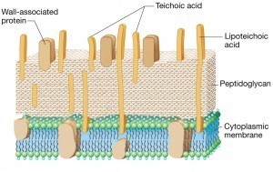 Figure: Gram Positive cell wall with Teichoic acid
The term teichoic acid encompasses a diverse family of cell surface glycopolymers containing phosphodiester-linked polyol repeat units. Teichoic acids are fibers of glycerol phosphate (glycerol teichoic acid) or ribitol phosphate (ribitol teichoic acid).
Teichoic acids are located in the outer layer of certain Gram-positive bacteria (such as Staphylococci, Streptococci, Lactobacilli, and Bacillus spp.). Teichoic acids are absent from gram-negative bacteria.
Why teichoic acids matter?
Teichoic acid is one of those topics that appears in every microbiology textbook, gets tested in every exam, and yet leaves students wondering: "Why does this obscure cell wall polymer deserve a whole lecture?"
Three reasons teichoic acid is genuinely important for a medical professional to understand:
1. Gram-positive sepsis kills patients, and teichoic acid is the trigger.
When we think of bacterial sepsis, we often think of gram-negative endotoxin (LPS). But gram-positive bacteria such as Staphylococcus aureus, Streptococcus pneumoniae, and Enterococcus are equally common causes of life-threatening sepsis.
The inflammatory mediator responsible for gram-positive septic shock is primarily lipoteichoic acid (LTA), which triggers TLR2 on macrophages and monocytes in the same way LPS triggers TLR4.
A student who understands this can immediately explain why a patient with gram-positive bacteremia develops the same clinical picture of fever, hypotension, and cytokine storm as a patient with gram-negative sepsis, despite having no LPS.
2. Teichoic acid determines where S. aureus lives, and therefore who gets infected.
S. aureus colonizes the anterior nares (nostrils) of approximately 30% of healthy adults. The reason it prefers the nose over other body surfaces is largely due to wall teichoic acid. The ribitol-phosphate WTAs of S. aureus are required for binding to receptors on nasal epithelial cells (the host scavenger receptor SREC-I has been implicated), and WTA-deficient mutants show markedly reduced nasal colonization in animal models.
This adhesion is the first step in nasal colonization, and nasal colonization is the primary risk factor for subsequent S. aureus infection (surgical site infections, MRSA bacteremia). Blocking WTA-mediated adhesion is therefore a genuine infection prevention strategy.
3. Teichoic acids are emerging antibiotic drug targets.
The enzymes that synthesize wall teichoic acids (particularly the enzyme TarO, which catalyzes the first committed step in WTA biosynthesis in S. aureus) are essential for bacterial survival in the host and importantly, they have no human homologues. This makes them highly selective targets for novel antibiotics. Compounds that inhibit WTA biosynthesis are in active development as treatments for MRSA, where conventional antibiotic options are limited.
Types of Teichoic Acids
- Lipoteichoic acids (LTAs): Teichoic acids that are covalently linked to the lipid in the cytoplasmic membrane.
- Wall teichoic acids (WTAs): Teichoic acids that are covalently attached to muramic acid in the wall peptidoglycan.
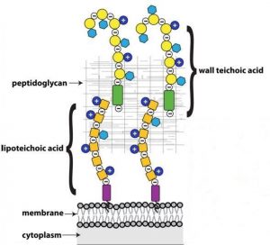 Figure: Wall Teichoic acid and Lipoteichoic acid
Wall teichoic acids are intimately involved in many aspects of cell division and are essential for maintaining cell shape in rod-shaped organisms. WTAs are required for beta-lactam resistance in methicillin-resistant S. aureus (MRSA), and they modulate susceptibility to cationic antibiotics in several organisms.
The analogy that makes WTA vs LTA location unforgettable
Think of the gram-positive cell wall as a brick wall with electrical wiring running through it.
- The peptidoglycan is the brick structure, the rigid framework
- The wall teichoic acids (WTAs) are the wiring embedded within the bricks. They are covalently attached to the peptidoglycan itself, running through the wall, with their ends protruding from the outer surface
- The lipoteichoic acids (LTAs) are the wiring anchored at the electrical panel (the plasma membrane). These are rooted in the membrane lipid bilayer and extend outward through the peptidoglycan to the surface.
Both types of "wiring" carry the same negative charge (their electrical current, so to speak), but they are anchored at different points. This physical arrangement explains why they have somewhat different functions: WTAs interact more with the external environment (phage receptors, adhesion), while LTAs interact more with the plasma membrane and regulate internal processes (autolysis, ion transport).
Clinical Significance of Teichoic Acids
1. Gram-positive septic shock: LTA as the trigger
When gram-positive bacteria are killed in the bloodstream (whether by antibiotics or host immune cells) they release their cell wall components, including large quantities of lipoteichoic acid. LTA binds to TLR2 (Toll-like receptor 2) on the surface of macrophages and monocytes, triggering the same cytokine cascade (TNF-α, IL-1, IL-6) that LPS triggers through TLR4.
This means the clinical picture of gram-positive septic shock (high fever, hypotension, tachycardia, risk of multi-organ failure) is mediated by LTA in the same way gram-negative septic shock is mediated by LPS.
The important nuance: LTA is generally considered less potent than LPS as an inflammatory stimulus. It requires approximately 1,000 times higher concentrations to produce equivalent cytokine release. This partly explains why gram-positive sepsis, while serious, tends to have a somewhat lower case fatality rate than gram-negative sepsis. But at the concentrations released during a bloodstream infection, LTA is more than sufficient to produce life-threatening systemic inflammation.
2. S. aureus nasal colonization and MRSA infection risk
The anterior nares are the ecological niche of Staphylococcus aureus. Approximately:
- 30% of healthy adults are persistent nasal carriers
- 30% are intermittent carriers
- 40% are non-carriers
This colonization is not random. It depends on ribitol-phosphate wall teichoic acids on the S. aureus surface binding to specific receptors on nasal epithelial cells. Non-carrier individuals have nasal microbiota that competitively exclude S. aureus partly by competing for the same adhesion sites.
Why this matters clinically: Nasal carriage of S. aureus (especially MRSA) is the single most important risk factor for subsequent S. aureus infection. MRSA surgical site infection rates are substantially higher in patients colonized with nasal MRSA at the time of surgery. This is why preoperative MRSA nasal decolonization with mupirocin nasal ointment (which inhibits bacterial protein synthesis and reduces nasal carriage) is recommended before high-risk procedures like cardiac surgery and joint replacement. Understanding that WTA drives this colonization is understanding why this prevention strategy targets the nose specifically.
3. D-alanine modification and antibiotic resistance
The teichoic acid backbone can be modified by the addition of D-alanine residues by a set of enzymes encoded by the dlt operon (DltA, DltB, DltC, DltD). These D-alanine residues reduce the overall negative charge of the teichoic acid and the cell surface.
Why this matters: Many host antimicrobial peptides (defensins, cathelicidins) work by being attracted to the negatively charged bacterial surface — they are cationic (positively charged) and bind electrostatically to the anionic bacteria, then insert into and disrupt the cell membrane. By adding D-alanine to teichoic acids, bacteria reduce the negative charge of their surface, reducing electrostatic attraction and making themselves significantly more resistant to these host antimicrobial peptides.
Bacteria with mutations in the dlt operon (no D-alanine incorporation) are:
- More susceptible to cationic antimicrobial peptides (defensins)
- More susceptible to vancomycin (slightly)
- Less virulent in animal infection models
This resistance mechanism is one reason gram-positive pathogens can survive in host environments that should be lethal, tissues and body fluids are rich in antimicrobial peptides.
4. Bacteriophage receptor
Wall teichoic acids serve as the primary receptor for many bacteriophages that infect gram-positive bacteria. The phage tail fibers recognize and bind to the specific WTA structure of their host bacterium. This specificity is so precise that the same bacterial species with different WTA structures (due to different glycosylation patterns) can be completely resistant to a phage that infects the parent strain.
Why this matters:
- Phage typing (historically used for S. aureus epidemiological typing) was entirely based on the specificity of phage-WTA binding
- Phage therapy (using bacteriophages to treat antibiotic-resistant infections) depends on matching the correct phage to the WTA structure of the target bacterium
- Anti-WTA strategies that modify or block WTA synthesis can sensitize normally phage-resistant bacteria to phage killing; a potential synergistic therapeutic approach for MRSA
5. Cation regulation and cell physiology
The high negative charge of teichoic acids serves as a cation reservoir at the cell surface. They bind and concentrate divalent cations (Ca²⁺, Mg²⁺) for regulated transport into the cell. These cations are essential cofactors for many membrane enzymes, including the autolysins that remodel peptidoglycan during cell division.
Teichoic acids also regulate autolysis; the controlled breakdown of peptidoglycan that is essential for cell wall remodeling during growth and division. If teichoic acid synthesis is inhibited, autolytic activity becomes dysregulated, leading to aberrant cell wall structure and eventually cell death. This is one reason why WTA biosynthesis inhibitors are being explored as antibiotics; inhibiting WTA indirectly disrupts autolysis and kills the bacterium through a mechanism entirely different from beta-lactams or glycopeptides.
Structure of Teichoic Acid
TAs are glycopolymers rich in phosphates present in the peptidoglycan layers of gram-positive bacteria (pathogenic and nonpathogenic). The structure of teichoic acid varies based on its type. Below is a brief description of the wall- and lipo-teichoic acid structure.
Structure of Wall Teichoic Acid (WTA)
In WTAs, the anionic glycopolymers attach covalently to the N-acetylmuramic acid of peptidoglycan through a phosphodiester linkage. Teichoic acids can account for a substantial fraction of the cell wall dry weight, up to roughly half in some gram-positive species, though the exact proportion varies widely by organism and growth conditions.
 Figure: Structure of Teichoic acid from Micrococcaceae
The structure of WTA varies between organisms, but the core is a long chain of repeating glycerol-phosphate or ribitol-phosphate units, joined to the peptidoglycan through a short linkage unit. Staphylococcus aureus WTA is mainly ribitol phosphate, while Bacillus subtilis may use glycerol phosphate, ribitol phosphate, or both depending on the strain.
These repeating units are then decorated with D-alanine and sugars such as glucose or N-acetylglucosamine. These modifications are not cosmetic: they change the cell's surface charge and strongly affect how the organism interacts with the host and how susceptible it is to antibiotics. The D-alanine modification in particular is the one covered in the antimicrobial-resistance section above.
Structure of Lipoteichoic Acid (LTA)
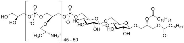 Figure: Lipoteichoic acid
LTAs are amphiphilic molecules with an alditol-phosphate hydrophilic chain. The classical and by far most common LTA is poly(glycerol phosphate), often called type I LTA. Its structure is more conserved across species than that of WTA. A glycolipid (in most cases) forms the hydrophobic moiety of the LTA, the hydrophilic part links to the glycolipid by a phosphodiester bridge. The glycolipid anchors all the molecules in the cytoplasmic membrane.
How is teichoic acid related to the Gram stain?
Teichoic acids are a defining component of the thick gram-positive cell wall and contribute much of its negative surface charge. They are absent from gram-negative bacteria.
While the crystal violet retention that makes a cell "gram-positive" is due mainly to the thick peptidoglycan layer, teichoic acids are part of what makes that wall distinctively gram-positive, and their presence is one of the structural features that separates the two groups.
WTA vs LTA
| Feature | Wall Teichoic Acid (WTA) | Lipoteichoic Acid (LTA) |
|---|---|---|
| Anchored to | Peptidoglycan (muramic acid) | Plasma membrane (via lipid anchor) |
| Location | Embedded in and protruding from cell wall | Spans from membrane through peptidoglycan |
| Chemical backbone | Ribitol phosphate (S. aureus) or glycerol phosphate | Glycerol phosphate (most species) |
| Primary functions | Bacteriophage receptor; S. aureus nasal adhesion; surface charge regulation; antibiotic resistance target | Immune stimulation (TLR2); cell division regulation; autolysin regulation; cation transport |
| Role in sepsis | Less direct | Primary gram-positive inflammatory mediator (TLR2 agonist) |
| Antibiotic target potential | High. TarO inhibitors being developed; no human homologue | Moderate |
| Found in | Gram-positive bacteria only | Gram-positive bacteria only |
| Present in gram-negative bacteria? | No | No (LPS serves analogous roles instead) |
How to Learn and Remember Teichoic Acids
One sentence that captures the entire clinical relevance
"Teichoic acid is to gram-positive bacteria what LPS is to gram-negative: it mediates colonization, drives inflammation in sepsis, and its synthesis enzymes are the next frontier of MRSA drug targets."
Clinical stories that make teichoic acid memorable
Story 1: The pre-op MRSA swab
A patient is admitted for elective hip replacement. Routine pre-operative MRSA screening swabs the anterior nares. The swab comes back positive, MRSA nasal carriage. Surgery is postponed for 5 days while the patient applies mupirocin nasal ointment twice daily to decolonize the nose. Why the nose?
Because S. aureus WTA binds specifically to nasal epithelial cell receptors. Why mupirocin? Because it kills the colonizing bacteria, removing the source. Every MRSA decolonization protocol in every hospital in the world exists because of the teichoic acid-mediated nasal adhesion of S. aureus.
Story 2: The septic patient who "should" have LPS
A junior doctor sees a patient in septic shock with high fever, hypotension, and rising inflammatory markers. Blood cultures grow gram-positive cocci in clusters: S. aureus bacteremia. The junior asks: "But I thought septic shock was caused by endotoxin, shouldn't this be gram-negative?" The answer is lipoteichoic acid. TLR2, not TLR4. Same cytokine storm, different trigger, similar clinical picture. Understanding LTA is understanding why gram-positive sepsis is just as dangerous as gram-negative.
Key exam facts in one table
| Question | Answer |
|---|---|
| Where is WTA anchored? | Peptidoglycan (muramic acid) |
| Where is LTA anchored? | Plasma membrane (lipid anchor) |
| Which TLR does LTA activate? | TLR2 (not TLR4, which LPS uses) |
| Which TLR does LPS activate? | TLR4 |
| What cytokines does LTA stimulate? | TNF-α, IL-1, IL-6 (same as LPS) |
| What is the gram-positive equivalent of endotoxin? | Lipoteichoic acid (LTA) |
| What do D-alanine modifications on teichoic acid do? | Reduce surface negative charge → resistance to cationic antimicrobial peptides |
| What is the role of WTA in S. aureus pathogenesis? | Nasal colonization adhesion (binds nasal epithelial cell receptors) |
| Are teichoic acids found in gram-negative bacteria? | No. Absent from gram-negative bacteria |
| Which biosynthesis enzyme is a new antibiotic drug target? | TarO (first committed step in WTA biosynthesis in S. aureus) |
References
- Madigan, M. T., Bender, K. S., Buckley, D. H., Sattley, W. M., & Stahl, D. A. (2021). Brock Biology of Microorganisms (16th ed.). Pearson.
- Tille, P. M. (2022). Bailey & Scott's Diagnostic Microbiology (15th ed.). Elsevier.
- Brown, S., Santa Maria, J. P., & Walker, S. (2013). Wall teichoic acids of gram-positive bacteria. Annual Review of Microbiology, 67, 313–336. https://doi.org/10.1146/annurev-micro-092412-155620
- Weidenmaier, C., & Peschel, A. (2008). Teichoic acids and related cell-wall glycopolymers in Gram-positive physiology and host interactions. Nature Reviews Microbiology, 6(4), 276–287. https://doi.org/10.1038/nrmicro1861
Frequently Asked Questions
What is the difference between wall teichoic acid and lipoteichoic acid?
Why is lipoteichoic acid called the gram-positive equivalent of endotoxin?
LPS activates TLR4; LTA activates TLR2, both trigger the same cytokine cascade (TNF-α, IL-1β, IL-6) causing septic shock. LTA is ~1000x less potent per molecule than LPS, but sufficient quantities released during bacteremia still cause life-threatening inflammation.
How does teichoic acid contribute to S. aureus nasal colonisation?
Ribitol-phosphate WTAs on S. aureus bind specific receptors on nasal epithelial cells, explaining preferential nasal colonization (~30% persistent carriers). Nasal carriage is the most important risk factor for subsequent infection. This is why mupirocin nasal decolonization is used before high-risk surgery.
How do teichoic acids help bacteria resist antimicrobial peptides?
Why are teichoic acid synthesis enzymes being developed as antibiotic targets?
TarO (first step in WTA biosynthesis) is essential for S. aureus virulence, has no human homologue, and inhibiting it sensitizes MRSA to beta-lactam antibiotics. This makes anti-WTA compounds potential beta-lactam sensitizers, not just standalone antibiotics.
Do gram-negative bacteria have teichoic acids?
No, teichoic acids are exclusive to gram-positive bacteria. Gram-negative bacteria have LPS in their outer membrane serving analogous roles (surface charge, immune stimulation, phage receptor) but with completely different chemistry.
What is the role of teichoic acid in bacteriophage infection?
WTA serves as the primary phage receptor for gram-positive bacteria. Phage tail fibers recognize specific WTA glycosylation patterns. This strain-specific variation explains why a phage effective against one S. aureus strain may completely fail against another with different WTA structure.
What regulates autolysis and why do teichoic acids matter?
Teichoic acids (particularly LTA) regulate autolysin enzyme activity, controlling where and when self-digestion of peptidoglycan occurs during growth/division. Inhibiting teichoic acid synthesis dysregulates autolysis, causing aberrant morphology and death: a mechanism independent of beta-lactams, explaining anti-WTA activity against resistant MRSA.

Tankeshwar Acharya, MSc (Medical Microbiology)
Tankeshwar Acharya is an Assistant Professor in the Department of Microbiology at Patan Academy of Health Sciences (PAHS), Nepal, where he has been teaching and practicing clinical microbiology for over 14 years. He is the founder of Microbe Online, one of the leading free microbiology education resources on the web, covering bacteriology, mycology, parasitology, immunology, and clinical laboratory diagnostics written from direct experience in both the classroom and the diagnostic laboratory.
Comments
No comments yet. Be the first to share your thoughts.
Leave a comment
All comments are reviewed before they appear.