
Candida albicans: Gram Stain, Morphology, Pathogenesis, and Lab Diagnosis
Candida albicans stains Gram-positive and appears as budding yeast cells with pseudohyphae. Learn its microscopy, why it stains as it does, virulence and pathogenesis, the diseases it causes, and how it is identified in the lab.

On this page
A high vaginal swab grows creamy white colonies on blood agar, and at a glance they look just like Staphylococcus. A Gram stain settles it in seconds: instead of clusters of cocci, the slide shows large Gram-positive budding cells with elongated chains pinched at regular points. Those are Candida albicans yeast cells and pseudohyphae. Learning to recognize that picture, and knowing why a yeast stains Gram-positive at all, is the most useful skill.
Candida albicans is part of the normal human flora, carried by roughly 40 to 80% of healthy people in the gastrointestinal tract, the oropharynx, and the female genital tract. It is the most common cause of candidiasis (also called moniliasis). Other clinically important species include Candida tropicalis, Candida parapsilosis, Candida glabrata, Candida krusei, and Candida auris. Candida auris deserves special mention because it is often multidrug-resistant, spreads in hospitals, and is difficult to identify by routine methods. The World Health Organization lists it as a critical-priority fungal pathogen.
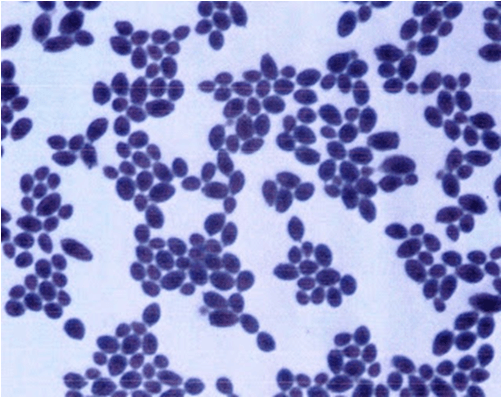 Figure: Candida albicans in Gram Staining
Candida on Gram stain and under the microscope
Candida albicans stains Gram-positive. On a Gram-stained smear it appears deep purple. However, there is an important nuance behind that simple answer. Yeasts are not truly classified by the Gram stain the way bacteria are, because the Gram reaction depends on a peptidoglycan cell wall, which fungi do not have.
Instead, the thick fungal cell wall (made of glucans, mannans, and chitin) retains the crystal violet, so Candida stains Gram-positive. Because the stain uptake can be uneven, Candida is sometimes described as Gram-variable, staining more strongly in some cells than others.
What Candida albicans looks like under the microscope
On a Gram stain or a wet mount of a clinical sample, look for three features:
- Budding yeast cells (blastoconidia): oval cells, larger than bacteria (about 3 to 6 micrometers), each with a single bud attached by a narrow neck.
- Pseudohyphae: chains of elongated yeast cells that stay joined end to end, with visible constrictions (pinch points) at each junction. The regular constrictions are the key that tells pseudohyphae apart from true hyphae, which have parallel walls and no constrictions.
- True hyphae: Candida albicans can also form true hyphae when it invades tissue, so a mix of yeast cells, pseudohyphae, and true hyphae in tissue points toward invasive disease rather than simple colonization.
How to tell it apart from what it mimics
- From bacteria: Candida cells are much larger than cocci or bacilli and show budding. A colony that looks like Staphylococcus on blood agar, especially from a high vaginal swab, is quickly resolved by a Gram stain or wet mount, where large budding yeast cells replace the small cocci you would expect from Staphylococcus aureus. This look-alike trap is a common cause of misreading vaginal swab cultures.
- From other yeasts: the presence of pseudohyphae with regular constrictions favors Candida over Cryptococcus, which shows narrow-based budding with a wide capsule and no pseudohyphae. For a full feature-by-feature key to reading fungi under the microscope, see the guide to microscopic identification of fungi.
Virulence factors
Candida albicans is a commensal that turns pathogenic when host defenses or normal flora are disturbed. Its ability to cause disease rests on several factors, each tied to what it does for the organism.
- Adhesins and the fibronectin receptor. What: surface proteins, including a fibronectin-binding receptor, and hydrophobic surface molecules. Why: they let Candida stick to host epithelium and to fibronectin in the extracellular matrix, the essential first step before invasion. Without adherence, the yeast is shed before it can establish infection.
- Yeast-to-hypha switching (morphogenesis). What: the ability to switch between the budding yeast form and a filamentous hyphal form. Why: yeast cells spread and disseminate, while hyphae penetrate epithelial surfaces and resist being engulfed by phagocytes. This reversible switch is central to invasion, which is why finding hyphae in tissue signals active disease.
- Secreted aspartyl proteases (SAPs). What: enzymes that digest proteins. Why: they break down host tissue barriers and immune proteins, aiding invasion and evading defenses. SAPs increase the organism's ability to cause disease in experimental models.
- Phospholipases. What: enzymes that damage host cell membranes. Why: they help the hyphal tip penetrate epithelial cells, promoting tissue invasion.
- Phenotypic switching. What: the ability to change colony and cell characteristics rapidly and reversibly. Why: it lets the organism adapt to different host sites and evade the immune response.
- Biofilm formation. What: communities of yeast and hyphae encased in a matrix, especially on catheters and prosthetic devices. Why: biofilms resist antifungals and host defenses, which is why device-related candidemia is hard to clear without removing the device.
Pathogenesis: putting it together
The sequence follows the same logic as any invasive infection. First, Candida attaches to the epithelium using its adhesins and fibronectin receptor. Next, when host defenses are weakened, it switches from yeast to hyphae and uses secreted proteases and phospholipases to breach the epithelial barrier and resist phagocytosis. It then damages and invades the tissue, and on catheters or damaged surfaces it forms protective biofilms. Finally, if it reaches the bloodstream, it spreads to seed deep organs, causing disseminated candidiasis.
Intact skin and mucous membranes are the first barrier against this sequence. Cell-mediated immunity is the key defense: Th1 cells produce gamma-interferon that activates macrophages to kill the organism. This is why most serious Candida infection is opportunistic, appearing when that immunity or the normal flora is disturbed.
Who is at risk
The more the host is weakened, the more invasive the disease. Major risk factors include neutropenia (high risk of disseminated infection), prolonged broad-spectrum antibiotics or steroids (which disturb normal flora), diabetes, invasive procedures such as surgery and indwelling catheters, and HIV/AIDS. Candidiasis is the most common fungal infection in people living with HIV.
Diseases
C. albicans is responsible for several infections in healthy and immunocompromised patients.
Overgrowth of C. albicans produces white ‘cottage cheese-like film’ called thrush. This form of candidiasis is known as pseudomembranous candidiasis and is the classic sign of acute infection.
Main diseases include;
- Oropharyngeal candidiasis (oral thrush): Common in those with HIV/AIDS.
- Vulvovaginal candidiasis (vaginal thrush): Overgrowth of C. albicans can cause vulvovaginal candidiasis. It is a common infection during pregnancy and in diabetic patients. Suppression of growth of vaginal lactobacilli by antimicrobial therapy also leads to overgrowth of C. albicans causing Candida vaginitis.
Other diseases caused by Candida are paronychia, onychomycosis, endocarditis, eye infection, intertriginous candidiasis, etc. Disseminated infection of Candida and meningitis is seen mostly in immunocompromised and/or seriously ill patients.
Laboratory Diagnosis
Specimen depends on disease presentation. The common submitted sample includes; urine (in case of UTI), vaginal discharge (suspected cases of vaginal thrush) or CSF (when meningitis is suspected), sputum (when pneumonia is suspected), and blood, or other exudates from the mucosal surface.
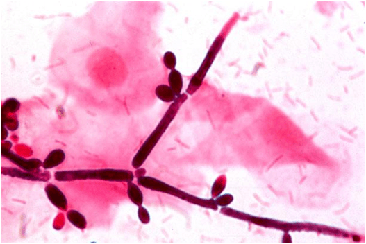 Figure: Pseudohyphae of Candida albicans
Note: Candida is rarely a true cause of pneumonia. Growth of Candida in lower respiratory specimens usually reflects contamination from the mouth, where yeasts are normal inhabitants, rather than genuine lung infection. Even in severely immunocompromised patients, a positive respiratory culture for Candida does not by itself confirm Candida pneumonia.
Culture
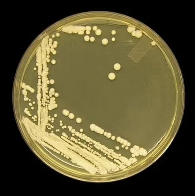 Figure: Candida albicans on Sabouraud-Dextrose Agar at 48 hours at 30°C
Candida albicans grows on Sabouraud dextrose agar and on routine bacterial media, producing cream-colored, pasty colonies with a yeast smell after 24 to 48 hours at 25 to 37°C. On blood-containing media it can form colonies with hyphal projections ("feet," sometimes star-shaped) within 48 hours.
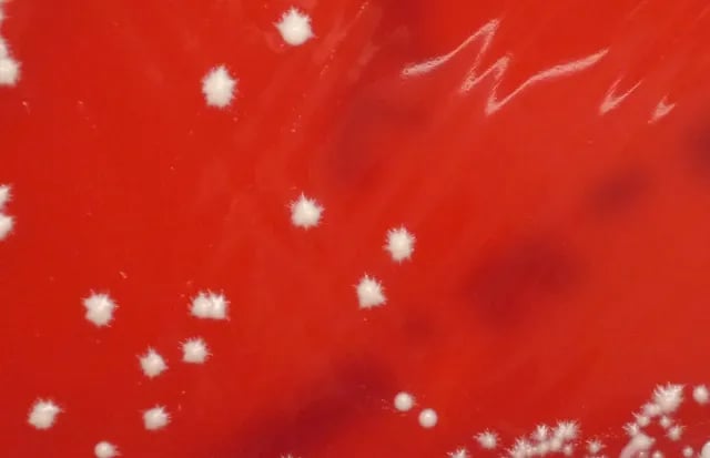 Figure: Star-shaped colonies of Candida albicans in blood agar.
On blood agar the creamy white colonies can be mistaken for Staphylococcus, so any suspicious colony, especially from a high vaginal swab, should be checked by wet mount or Gram stain.
Presumptive identification
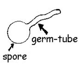 Figure: Germ tube of Candida albicans
- Germ tube test: a positive germ tube (a short hyphal outgrowth with no constriction at its base, formed when the yeast is incubated in serum) gives a presumptive identification of Candida albicans. This, together with "feet" on blood agar, is enough for a presumptive call. For the method and how to read it, see the germ tube test article.
- Chlamydospore formation: Candida albicans forms chlamydospores on cornmeal agar. Note that Candida dubliniensis also forms both germ tubes and chlamydospores, so these two features do not separate them. The standard way to tell them apart is growth at 45°C, at which C. albicans grows and C. dubliniensis does not (some sources cite 42°C), along with CHROMagar color and molecular methods.
- Carbohydrate assimilation and fermentation: these help differentiate C. albicans from other species such as C. tropicalis, C. parapsilosis, C. krusei, and C. glabrata.
Definitive and rapid identification
Automated systems, MALDI-TOF mass spectrometry, and molecular methods give species-level identification. Directly from positive blood cultures, a PNA FISH assay can rapidly distinguish the common Candida species causing candidemia, which speeds up appropriate treatment.
Culture
Routine bacterial culture is sufficient for detecting Candida species whether aerobic blood culture bottle or agar media are used.
Candida albicans grows well on Sabouraud dextrose agar and in the most routinely used bacteriological media. Cream-colored pasty colonies usually appear after 24-48 hours of incubation at 25-37°C. The colonies have a distinctive yeast smell and the budding cells can be easily seen by direct microscopy in stained or unstained preparations.
Candida albicans can be recognized by the formation of hyphal elements radiating from colonies on blood-containing media within 48 h of initial incubation. These colonies with “feet,” which can also resemble stars.
In Blood Agar, Candida albicans gives white, creamy-colored colonies, which can be mistaken for Staphylococcus spp. Whenever you are analyzing the culture report of ‘high vaginal swab,’ take extra care as the colony you are observing can be of Candida albicans instead of Staphylococcus aureus or vice versa (a quick solution for this is to perform wet mount or gram staining and observing under a microscope).
The carbohydrate fermentation test can differentiate C. albicans from other pathogenic species of Candida, such as Candida tropicalis, Candida parapsilosis, Candida krusei, and C. glabrata.
Treatment
Treatment depends on the site and severity of infection, and this article gives drug-of-choice and class-level guidance only.
Mucocutaneous disease (oral or vaginal thrush, skin): topical azoles such as clotrimazole and miconazole, or oral fluconazole for more extensive or recurrent disease.
Invasive candidiasis and candidemia: an echinocandin (for example caspofungin, micafungin, or anidulafungin) is now the first-line treatment. Echinocandins work by blocking synthesis of beta-1,3-glucan in the fungal cell wall. Fluconazole is used as step-down therapy or for stable patients with a susceptible isolate. Removing infected catheters and other devices is an important part of treatment.
Resistance matters. Candida krusei is intrinsically resistant to fluconazole, Candida glabrata often has reduced azole susceptibility, and Candida auris is frequently multidrug-resistant. This is why species identification and, when needed, susceptibility testing guide therapy.
How to Remember
Gram-positive, but a yeast. Candida stains Gram-positive (purple), but it is a yeast staining that way, not a Gram-positive bacterium. It has no peptidoglycan.
Pinch points mean pseudohyphae. Regular constrictions along a chain of cells are pseudohyphae. True hyphae have parallel walls and no pinch points.
Germ tube, think albicans. A positive germ tube is the quick presumptive clue for Candida albicans.
Yeast spreads, hyphae invade. The yeast form disseminates; the hyphal form penetrates tissue. Hyphae in tissue mean invasion.
Echinocandins for the bloodstream, azoles for the surface. Invasive disease and candidemia get an echinocandin first. Mucocutaneous disease gets topical or oral azoles.
Key exam facts
| Item | Fact |
|---|---|
| Organism | Candida albicans, a yeast |
| Gram reaction | Gram-positive (stains purple; Gram-variable; no peptidoglycan) |
| Microscopy | Budding yeast cells (blastoconidia), pseudohyphae with constrictions, true hyphae in tissue |
| Normal habitat | GI tract, oropharynx, female genital tract (commensal) |
| Presumptive ID | Germ tube positive; "feet" on blood agar; chlamydospores on cornmeal |
| Distinguishes from C. dubliniensis | C. albicans grows at 45°C, C. dubliniensis does not; both are germ-tube and chlamydospore positive |
| Key virulence factors | Adhesins, yeast-hypha switching, secreted aspartyl proteases, phospholipases, biofilm |
| Main defense | Cell-mediated immunity (Th1, gamma-interferon, macrophages) |
| Classic lesion | Pseudomembranous "cottage cheese" thrush |
| Invasive treatment | Echinocandin first-line |
| Mucocutaneous treatment | Topical or oral azoles |
| Notable resistant species | C. krusei (fluconazole-resistant), C. auris (multidrug-resistant) |
Where Students Get Confused
"Candida is a Gram-positive bacterium." No. Candida is a yeast (a fungus). It stains Gram-positive, but it has no peptidoglycan cell wall, so it is not a Gram-positive bacterium.
"Pseudohyphae and true hyphae are the same." No. Pseudohyphae are chains of elongated yeast cells with constrictions at the junctions. True hyphae have parallel walls and no constrictions. The pinch points are the distinguishing feature.
"Candida growing in sputum means Candida pneumonia." Rarely. Yeasts are normal in the mouth, so respiratory cultures usually reflect contamination. Candida pneumonia is uncommon even in immunocompromised patients.
"A positive germ tube confirms Candida albicans." It is presumptive, not definitive. Candida dubliniensis is also germ-tube positive. Growth at 45°C and other tests separate the two.
"Fluconazole is the first-line drug for all Candida infections." No. Echinocandins are first-line for invasive candidiasis and candidemia. Azoles are used for mucocutaneous disease and as step-down therapy, and some species are azole-resistant.
Resumen en español
Candida albicans es una levadura que forma parte de la flora normal del cuerpo humano y es la causa más común de candidiasis. En la tinción de Gram se tiñe como Gram positiva (color morado), aunque es una levadura y no una bacteria Gram positiva, ya que no tiene peptidoglicano en su pared celular. Al microscopio se observa como células de levadura en gemación (blastoconidios) junto con pseudohifas que presentan constricciones en las uniones.
Sus principales factores de virulencia incluyen la adhesión al epitelio, el cambio de forma de levadura a hifa, las proteasas y fosfolipasas, y la formación de biopelículas. Causa candidiasis oral y vaginal (algodoncillo), y en pacientes inmunocomprometidos puede causar candidemia e infección diseminada. La prueba del tubo germinal positiva sugiere de forma presuntiva Candida albicans. El tratamiento de la enfermedad invasiva es con equinocandinas como primera línea, y con azoles para la enfermedad mucocutánea.
References and further reading
- Tille, P. M. (2022). Bailey and Scott's Diagnostic Microbiology (15th ed.). Elsevier.
- Procop, G. W., et al. (2017). Koneman's Color Atlas and Textbook of Diagnostic Microbiology (7th ed.). Wolters Kluwer.
- Leber, A. L. (Ed.). (2016). Clinical Microbiology Procedures Handbook (4th ed.). ASM Press. https://doi.org/10.1128/9781683670438.CMPH
Frequently Asked Questions
Is Candida albicans Gram-positive or Gram-negative?
Is Candida albicans Gram-positive or Gram-negative?
It stains Gram-positive (purple). But Candida is a yeast, not a bacterium, and has no peptidoglycan cell wall, so it is best understood as a yeast that stains Gram-positive rather than a true Gram-positive bacterium. Its staining can be uneven (Gram-variable).
What does Candida albicans look like under the microscope?
What does Candida albicans look like under the microscope?
Oval budding yeast cells (blastoconidia) about 3 to 6 micrometers, often with pseudohyphae, which are chains of elongated cells with constrictions at the junctions. In tissue it can also form true hyphae.
What is the difference between pseudohyphae and true hyphae?
What is the difference between pseudohyphae and true hyphae?
Pseudohyphae are chains of elongated yeast cells with constrictions where the cells join. True hyphae have parallel walls and no constrictions.
How is Candida albicans identified in the lab?
How is Candida albicans identified in the lab?
Presumptively by a positive germ tube test and "feet" on blood agar, and by chlamydospore formation on cornmeal agar. Definitive identification uses automated systems, MALDI-TOF, or molecular methods.
What is the germ tube test?
What is the germ tube test?
A rapid presumptive test where Candida albicans forms a short hyphal outgrowth in serum. A positive result suggests C. albicans, though C. dubliniensis can also be positive.
What diseases does Candida albicans cause?
What diseases does Candida albicans cause?
Oral thrush, vaginal thrush, skin and nail infections, and, in immunocompromised or seriously ill patients, candidemia and disseminated infection affecting deep organs.
How is Candida infection treated?
How is Candida infection treated?
Mucocutaneous disease is treated with topical or oral azoles. Invasive candidiasis and candidemia are treated first-line with an echinocandin. Some species, such as C. krusei and C. auris, are drug-resistant, so identification guides treatment.
Why can Candida be mistaken for Staphylococcus on culture?
Why can Candida be mistaken for Staphylococcus on culture?
On blood agar both can form creamy white colonies. A Gram stain or wet mount quickly distinguishes them, since Candida shows large budding yeast cells rather than clusters of cocci. This matters most with high vaginal swabs.

Tankeshwar Acharya, MSc (Medical Microbiology)
Tankeshwar Acharya is an Assistant Professor in the Department of Microbiology at Patan Academy of Health Sciences (PAHS), Nepal, where he has been teaching and practicing clinical microbiology for over 14 years. He is the founder of Microbe Online, one of the leading free microbiology education resources on the web, covering bacteriology, mycology, parasitology, immunology, and clinical laboratory diagnostics written from direct experience in both the classroom and the diagnostic laboratory.
Comments
No comments yet. Be the first to share your thoughts.
Leave a comment
All comments are reviewed before they appear.