Germ Tube Test: Principle, Procedure, Results
Germ Tube Test: Principle, Procedure, Results
Germ tube test is a screening test for the presumptive identification of Candida albicans. Candida dubliniensis can also form germ tubes and Candida tropicalis can produce pseudohyphae that closely resemble germ tubes.
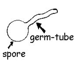 Figure: Germ Tube
Germ tube formation was first reported by Reynolds and Braude and hence the germ tube test is also known as Reynolds-Braude phenomenon.
Buds and pseudo-hyphae can be distinguished from germ tubes by the constricted attachment at the point of origin. Germ tubes don’t show constriction at the point of origin.
Principle of Germ Tube Test
Germ tubes are short outgrowths, non-septate germinating hyphae. They are ½ the width and 3 – 4 times the length of the cell from which they arise. When cells of Candida are incubated in serum at 37°C for 2-4 hours, Candida albicans and Candida dubliniens is produce short, slender, tube-like structures called germ tubes.
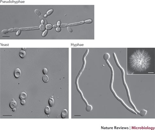 Figure: Characteristics morphology of Candida albicans
A germ tube appears as a short lateral extension from the yeast cell and does not have a constriction (septum) where it meets the yeast cell or any constriction at the septum along the tube.
The formation of germ tubes is associated with increased synthesis of protein and ribonucleic acid. Various media like fetal bovine serum may be used as a substitute for human pooled serum.
Requirements
- Culture: Suspected Candida colonies from sabouraud dextrose agar (SDA).SDA is the best medium to isolate yeasts for germ tube production; sheep blood agar is an acceptable substitute
- Reagent: Serum (human, sheep, fetal bovine) or other commercially produced media for germ tube testing
- Others: Test tubes, loop or wooden applicator, Pasteur pipettes, slides, cover slips.
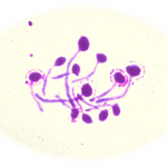 Figure: Candida albicans: Germ Tube Positive
Quality Control
The following yeasts should be included each time the test is performed.
- Germ tube positive, C. albicans ATCC 14053
- Germ tube negative, Candida tropicalis ATCC 66029
Procedure
- Lightly touch a yeast colony with a wooden applicator stick.
- Suspend the yeast cells in an appropriately labeled tube of fetal bovine serum (make a light suspension).
- Incubate the tube for 2-3 hours in a 35 – 37°C incubator.
- Place a drop of the suspension on a slide using a Pasteur pipette.
- Place a coverslip over the suspension.
- Examine the wet mount microscopically (at 40X) for the presence or absence of germ tubes.
Result and interpretation
Positive germ tube test: A short hyphal (filamentous) extension arising laterally from a yeast cell with no constriction at the point of origin is identified as C.albicans or C. dubliniensis. Some C. tropicalis isolates produce pseudohyphae that require careful observation to discriminate from those of C. albicans.
A minimum of five germ tubes should be observed before calling an organism positive, because rare tubes may be produced by other species. Growth at 45°C separates C. albicans from Candida dubliniensis.
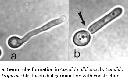 Figure: Difference between pseudohyphae and germ tube
Negative germ tube test: No hyphal extension arising from a yeast cell or a short hyphal extension with constriction at the point of origin. A constriction where the lateral extension meets the yeast cell is produced by pseudohyphae or budding cells.
Limitations
- Candida tropicalis may produce pseudo-germ tubes after 3 hrs of incubation but they show constriction at the point of origin.
- Too heavy inoculum will inhibit germ tube formation.
References and further readings
- Sudbery, P. E. (2011). Growth of Candida albicans hyphae. Nature Reviews Microbiology, 9(10), 737–748. https://doi.org/10.1038/nrmicro2636
- Sudbery, P. E. (2001). The germ tubes of Candida albicans hyphae and pseudohyphae show different patterns of septin ring localization. Molecular Microbiology, 41(1), 19–31. https://doi.org/10.1046/j.1365-2958.2001.02459.x
- Clinical Microbiology Procedures Handbook, Fourth Edition. (2016). American Society of Microbiology.

Tankeshwar Acharya, MSc (Medical Microbiology)
Tankeshwar Acharya is an Assistant Professor in the Department of Microbiology at Patan Academy of Health Sciences (PAHS), Nepal, where he has been teaching and practicing clinical microbiology for over 14 years. He is the founder of Microbe Online, one of the leading free microbiology education resources on the web, covering bacteriology, mycology, parasitology, immunology, and clinical laboratory diagnostics written from direct experience in both the classroom and the diagnostic laboratory.