
Dimorphic Fungi: Examples, Thermal Dimorphism, and Mnemonic
Dimorphic fungi grow as mold in the environment and yeast in the body. See the six classic examples compared, why the 37°C switch is a virulence factor, their tissue forms, geography, and the mnemonic to remember them.

On this page
Fungi can be broadly classified into two groups; yeasts and molds. Yeasts are single-celled organisms that reproduce by budding, but molds grow as filament-like structures and reproduce primarily using asexual reproductive spores such as conidiospores, sporangiospores, and arthrospores.
Properties
Dimorphic fungi exist as molds in the environment at ambient temperature (25°C– 30°C) and as yeasts (or other structures, e.g., spherules in case of Coccidioides immitis)* in human tissues at body temperature (35°C -37°C). Many medically important fungi show this unique behavior based on the temperature of growth and habitat.
*Note: Coccidioides is now recognized as two species, C. immitis in California and C. posadasii elsewhere in the endemic range; they are clinically identical and handled the same way.
The quick mnemonic
Mold in the Cold, Yeast in the Heat. This one line captures the whole concept: in the cold environment these fungi grow as mold; at warm body temperature they switch to yeast. ("Beast" is sometimes added to the end to complete the rhyme: "yeast in the heat, in the beast," the beast being the warm-blooded host.)
To remember which fungi are dimorphic, use: Body Heat Probably Changes ShaPe, for B*lastomyces, Histoplasma, Paracoccidioides, Coccidioides, Sporothrix,* and Penicillium (now Talaromyces) marneffei.
Why thermal dimorphism matters
Why would a fungus bother changing its entire morphology based on temperature? The answer reveals the core survival strategy of these organisms, and understanding it makes the whole topic click into place:
In the environment (25–30°C): these fungi grow as molds, producing abundant spores (conidia) that are lightweight and easily aerosolized, ideal for environmental dispersal and finding new soil/organic matter to colonize.
Inside the human body (35–37°C): the same fungus converts to yeast form. Yeast cells are smaller, can survive and even replicate inside macrophages, and are far better suited to surviving the host immune response than the mold form would be. The conversion is therefore not incidental; it is a deliberate virulence switch. A fungus that failed to convert to yeast form inside the host would be cleared far more easily by phagocytes.
This explains a critical laboratory safety point: the mold form is the infectious form for laboratory personnel. Inhaling aerosolized conidia from a culture plate is how laboratory-acquired infections occur. This is precisely why all suspected dimorphic fungal cultures must be handled in a biological safety cabinet, and why many reference laboratories restrict culture of these organisms to specialized BSL-3 facilities once dimorphic fungal infection is suspected.
Most dimorphic fungi cause systemic mycoses and Sporothrix (one of the dimorphic fungi) causes subcutaneous mycoses.
Note: All molds should be handled within appropriate biological safety cabinets to avoid a laboratory-acquired infection.
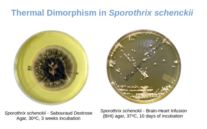 Figure: Sporothrix schenckii gives mold colonies at environmental temperature and yeast-like colonies are 37°C
The six dimorphic fungi compared
| Organism | Tissue (yeast) form & diagnostic clue | Disease | Endemic region | Environmental source |
|---|---|---|---|---|
| Histoplasma capsulatum | Small yeast (2-4 μm) inside macrophages; no capsule despite the name | Histoplasmosis | Ohio/Mississippi River valleys; Latin America, Africa, Asia | Bird and bat droppings (caves, chicken coops) |
| Blastomyces dermatitidis | Yeast with a single broad-based bud; thick, doubly refractile wall | Blastomycosis | Ohio/Mississippi valleys, Great Lakes, Canada | Moist soil with decaying wood |
| Coccidioides immitis / posadasii | Spherule filled with endospores (not a yeast); 20-200 μm | Coccidioidomycosis ("Valley fever") | Southwestern USA, Mexico, Central/South America | Arid desert soil |
| Paracoccidioides brasiliensis | Yeast with multiple peripheral buds ("mariner's wheel") | Paracoccidioidomycosis | Rural Latin America (esp. Brazil) | Soil; farming/agriculture |
| Sporothrix schenckii | Cigar-shaped yeast (oval, elongated) | Sporotrichosis ("rose gardener's disease") | Worldwide; classically gardeners/farmers | Vegetation, rose thorns, sphagnum moss |
| Talaromyces marneffei | Yeast dividing by fission (central septum), not budding | Talaromycosis | Southeast Asia (Thailand, Vietnam, S. China) | Bamboo rats; soil |
Two of the six break the "becomes a yeast" rule, and they are the favorite exam traps: Coccidioides forms a spherule, not a yeast, and Talaromyces divides by fission, not budding. If you remember those two exceptions, the rest follow the standard mold-to-yeast pattern.
The six fungi at a glance
Histoplasma capsulatum. Grows as mold in soil (bird/bat droppings) and as a small intracellular yeast in tissue; causes histoplasmosis. Despite the name, it has no capsule. For depth, see our article on Histoplasma / histoplasmosis.
Blastomyces dermatitidis. Soil mold; tissue yeast shows a single broad-based bud and a thick, doubly refractile wall. Causes blastomycosis, often with skin and lung involvement. For depth, see our article on Blastomyces / blastomycosis.
Coccidioides immitis / posadasii. Soil mold that, uniquely, forms a spherule filled with endospores in tissue rather than a yeast. Causes coccidioidomycosis ("Valley fever"). For depth, see our article on Coccidioides / coccidioidomycosis.
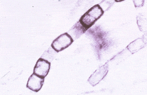 Figure: Coccidioides immitis– arthroconidia Dr Arthur DiSalvo
Paracoccidioides brasiliensis. Soil mold; tissue yeast shows multiple peripheral buds (the "mariner's wheel"). Causes paracoccidioidomycosis, formerly "South American blastomycosis." For depth, see our article on Paracoccidioides.
Sporothrix schenckii. The subcutaneous exception: usually acquired by traumatic inoculation (rose thorns, plant matter), not inhalation, causing sporotrichosis. Tissue form is a cigar-shaped yeast. For depth, see our article on Sporothrix / sporotrichosis.
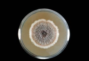 Figure: Sabouraud’s dextrose agar plate culture is growing the fungus Sporothrix schenckii CDC/Dr. Lucille K. Georg
Talaromyces marneffei. Formerly Penicillium marneffei; a mold in the environment and a fission yeast in tissue. An important opportunistic pathogen in AIDS patients in Southeast Asia, causing talaromycosis. For depth, see our article on Talaromyces marneffei.
Colony Morphology of Dimorphic Fungi
- If the organism is dimorphic fungi, you may observe mixtures of yeast and mold forms. In slow-growing dimorphic fungi, mold appears hair-like or cobweb-like consistency. The mold form can be converted to yeast by incubating subcultures on enriched media at 37°C.
- During a mold-to-yeast conversion of Blastomyces dermatitidis, an intermediate form known as the “prickly stage” can be seen. A microscopic preparation of such stage may show a mixture of hyphae, budding yeasts, and forms intermediate between the two.
- Yeast forms of dimorphic fungi are similar to other true yeasts. In contrast to yeasts, they are generally slow-growing, and the colonies remain quite small.
How to Remember Dimorphic Fungi
Mnemonic 1
Mold in the Cold, Yeast in the Heat (Beast)
Captures the universal behavioral rule; environment = mold, body = yeast.
Mnemonic 2
Body Heat Probably Changes ShaPe of the dimorphic fungi
B*lastomyces, Histoplasma, Paracoccidioides, Coccidioides, Sporothrix, Penicillium (Talaromyces) marneffei*
A common confusion: Candida and Cryptococcus are not thermally dimorphic fungi. Cryptococcus neoformans is a true, encapsulated yeast (it stays a yeast at all temperatures), causing pneumonia and cryptococcal meningitis in immunocompromised patients. Candida is often called "dimorphic" in a different sense: it switches between a yeast form and hyphae/pseudohyphae, but this is not the temperature-driven mold-to-yeast switch that defines the classic dimorphic fungi in this article. Neither undergoes the 25°C-mold / 37°C-yeast conversion, so neither belongs on the list of six above.
Memory trick for the two exceptions: Coccidioides and Talaromyces both break the "all dimorphic fungi become yeast" rule: Coccidioides becomes a spherule, not a yeast, and Talaromyces divides by fission rather than budding.
Two clinical stories that make this unforgettable
Story 1: The Arizona construction worker A construction worker in Phoenix, Arizona develops fever, cough, and chest pain after a dust storm during excavation work. Chest X-ray shows patchy infiltrates. A clinician unfamiliar with regional mycoses might treat this as bacterial pneumonia and see no improvement.
The diagnosis is coccidioidomycosis ("Valley Fever") which is extremely common in this specific geographic area following soil disturbance, and the patient's occupational exposure (excavation, disturbing desert soil) is the key clue that should have been asked about from the start.
Story 2: The laboratory technician's near-miss A clinical laboratory receives a sputum sample from a patient with suspected fungal pneumonia. A junior technician opens the culture plate on the open bench to examine the mold growth more closely under a dissecting microscope; not realizing the colony's slow growth rate and cottony morphology are consistent with a dimorphic fungus.
Senior staff immediately intervene, transfer the plate to a biological safety cabinet, and the culture is later confirmed as Blastomyces dermatitidis. Inhaling even a small number of aerosolized conidia from an open culture plate can cause laboratory-acquired blastomycosis or histoplasmosis. This is precisely why suspected dimorphic mold cultures must never be examined outside a biosafety cabinet.
Key exam facts
| Question | Answer |
|---|---|
| What triggers the mold-to-yeast conversion? | Temperature increase to 37°C (body temperature) |
| Why is this conversion considered a virulence factor? | Yeast form survives intracellularly and evades immune clearance better than mold form |
| Which dimorphic fungus does NOT form a true yeast in tissue? | Coccidioides immitis forms a spherule instead |
| Which dimorphic fungus divides by fission, not budding? | Talaromyces marneffei |
| What is the "prickly stage"? | Intermediate form seen during Blastomyces mold-to-yeast conversion, which is a mixture of hyphae and budding yeast |
| Are Candida and Cryptococcus dimorphic fungi? | No. Candida is yeast-like (yeast + pseudohyphae); Cryptococcus is a true yeast; neither undergoes the classic mold-yeast thermal switch |
| Why must dimorphic fungal cultures be handled in a biosafety cabinet? | The mold form's airborne conidia are the infectious form, which has inhalation risk to lab personnel |
Where Students Get Confused
"Which form is infectious, the mold or the yeast?" The mold form is infectious to people: its airborne conidia are inhaled from the environment (or from a culture plate, hence the biosafety rule). Once inside the warm body, the fungus converts to the yeast form, which is the invasive, tissue form. So the mold form transmits, the yeast form invades.
"Is Candida a dimorphic fungus?" Not in the sense meant here. The six classic dimorphic fungi switch between mold (25°C) and yeast (37°C) based on temperature. Candida switches between yeast and hyphae/pseudohyphae, but not by that temperature-driven mold-to-yeast mechanism, so it is not grouped with the classic thermally dimorphic fungi.
"Why does Coccidioides not fit the mold-to-yeast rule?" Because in tissue it forms a spherule (a large structure packed with endospores), not a yeast. It is still dimorphic (mold in the environment, spherule in tissue), but the tissue form is a spherule, not a yeast. This is one of the two classic exceptions.
"How does temperature actually cause the switch?" The shift to 37°C (plus host signals like CO₂ levels) triggers a change in the fungus's gene expression that remodels its shape and metabolism from the filamentous mold to the single-celled yeast (or spherule). The conversion is an active, regulated virulence program, not a passive response, which is why a fungus unable to convert is far less pathogenic.
"Why is a travel history so important?" Because dimorphic fungi are geographically restricted. The same lung findings point to different fungi depending on where the patient has been: Arizona/desert points to Coccidioides; Ohio River valley points to Histoplasma or Blastomyces; Southeast Asia points to Talaromyces. Geography narrows the differential more than almost any other single clue.
References
- Klein BS, Tebbets B. Dimorphism and virulence in fungi. Curr Opin Microbiol. 2007;10(4):314-319. https://doi.org/10.1016/j.mib.2007.04.002
- Rappleye CA, Goldman WE. Defining virulence genes in the dimorphic fungi. Annu Rev Microbiol. 2006;60:281-303. https://doi.org/10.1146/annurev.micro.59.030804.121055
- Sil A, Andrianopoulos A. Thermally dimorphic human fungal pathogens: polyphenetic pathways of dimorphism and virulence. Cold Spring Harb Perspect Med. 2015;5(8):a019794. https://doi.org/10.1101/cshperspect.a019794
- Brock Biology of Microorganisms. 16th ed. Pearson; 2021.
Frequently Asked Questions
Why do dimorphic fungi change shape based on temperature?
Thermal dimorphism is a virulence adaptation. Mold form (25-30°C) optimises environmental spore dispersal. Yeast form (35-37°C, body temp) is smaller, survives within macrophages, and resists immune clearance better, a deliberate survival strategy.
Why must dimorphic fungal cultures be handled in a biosafety cabinet?
The mold form's airborne conidia are the infectious form for humans. Examining cultures on an open bench risks aerosolizing conidia and causing laboratory-acquired infection.
Which dimorphic fungi do NOT follow the typical mold-to-yeast pattern?
Coccidioides immitis forms a spherule (20-200 μm, filled with endospores) instead of a true yeast. Talaromyces marneffei divides by fission (central septum) rather than budding. Both are common exam traps precisely because they break the general pattern.
Why is geographic/travel history important when dimorphic fungal infection is suspected?
Each organism has highly specific environmental niches: Histoplasma (Ohio/Mississippi valleys, bird/bat droppings), Coccidioides (SW USA arid soil), Blastomyces (Great Lakes/Ohio region), Paracoccidioides (rural Latin America), Talaromyces (Southeast Asia). Travel/occupational history dramatically narrows the differential before any test.
Is Sporothrix schenckii a systemic or subcutaneous pathogen?
Is Sporothrix schenckii a systemic or subcutaneous pathogen?
Primarily subcutaneous (sporotrichosis). Acquired by traumatic inoculation (rose thorns, splinters, so called 'rose gardener's disease'), typically remains localized with lymphocutaneous spread. Systemic dissemination is rare, occurring almost exclusively in severe immunocompromise.

Tankeshwar Acharya, MSc (Medical Microbiology)
Tankeshwar Acharya is an Assistant Professor in the Department of Microbiology at Patan Academy of Health Sciences (PAHS), Nepal, where he has been teaching and practicing clinical microbiology for over 14 years. He is the founder of Microbe Online, one of the leading free microbiology education resources on the web, covering bacteriology, mycology, parasitology, immunology, and clinical laboratory diagnostics written from direct experience in both the classroom and the diagnostic laboratory.
Comments
No comments yet. Be the first to share your thoughts.
Leave a comment
All comments are reviewed before they appear.