
Candida auris: Transmission, Risk, Diagnosis, Treatment
Candida auris is an emerging, often multidrug-resistant yeast that spreads in hospitals and is easily misidentified. Learn how it spreads, who is at risk, how it differs from Candida albicans, and how it is diagnosed and treated.

On this page
A patient who has spent weeks in intensive care, with a central line and several courses of antibiotics behind them, develops a bloodstream infection. The yeast that grows is resistant to the usual antifungal, and the same organism is soon found on the skin of two other patients on the ward. This is Candida auris, a yeast that colonizes skin, survives on surfaces, and moves between patients in a way ordinary Candida does not. Recognizing it quickly is as much an infection-control emergency as a treatment decision.
Candida auris (C. auris) causes a serious and sometimes fatal fungal infection that is emerging worldwide. First identified in 2009 in Japan, this drug-resistant yeast has now been reported in more than 40 countries across six continents. It is a particular threat to vulnerable patients, especially those who are hospitalized or critically ill. The World Health Organization lists C. auris as a critical-priority fungal pathogen, its highest category of concern.
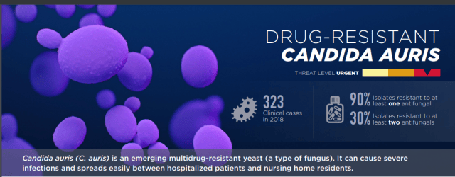 The reported numbers may understate the true spread, because identifying C. auris requires specialized laboratory methods that many labs do not have. Whole-genome sequencing shows that C. auris emerged as several distinct genetic groups (clades) in different regions of the world at roughly the same time, which is unusual and still not fully explained.
Why Candida auris matters
Three features make C. auris a serious public-health concern, and together they explain why it is treated as an infection-control emergency.
- It is often drug-resistant. Most isolates are resistant to the azoles (such as fluconazole), a substantial share are resistant to amphotericin B, and a smaller but rising number are resistant to the echinocandins, the first-line drugs. Some isolates are resistant to all three classes (pan-resistant), which leaves very few treatment options.
- It spreads in healthcare settings. Unlike most Candida, C. auris colonizes the skin and survives for long periods on surfaces and equipment. It moves from patient to patient in hospitals and long-term care facilities, and some common disinfectants do not reliably kill it. This is why a single case triggers a containment response.
- It is easily misidentified. Standard laboratory methods often mistake C. auris for other yeasts, which delays both correct treatment and infection-control measures. Reliable identification usually requires MALDI-TOF mass spectrometry or molecular methods.
Transmission
C. auris spreads mainly in healthcare settings, in three linked ways. First, it colonizes the skin of patients, often without causing any symptoms. A colonized patient carries the organism on their skin and can pass it on without being sick themselves. Second, it contaminates the environment. C. auris survives for weeks on bed rails, equipment, and other surfaces, and resists some routine disinfectants, so it persists in the patient's surroundings. Third, it transfers by contact, from patient to patient on the hands of healthcare workers, on shared equipment, and through the contaminated environment.
Colonization is the key to understanding its spread. Skin colonization is silent, so the organism moves through a ward before anyone is obviously infected. This is why control depends on screening at-risk patients on admission, isolating those who are colonized or infected, strict hand hygiene, and thorough cleaning with disinfectants proven to work against C. auris.
It is not spread through casual community contact in the way respiratory viruses are. The risk is concentrated in hospitals and long-term care facilities, among patients with serious underlying illness.
People at risk
Patients who have been hospitalized in a healthcare facility a long time, have a central venous catheter, or other lines or tubes entering their body, or have previously received antibiotics or antifungal medications, appear to be at the highest risk of infection with this yeast.
Candida auris vs Candida albicans
You may be confused C. auris with the familiar Candida albicans. They are both Candida yeasts, but they behave very differently, and the differences are exactly what make C. auris dangerous.
| Feature | Candida albicans | Candida auris |
|---|---|---|
| Familiarity | Common, well known, taught in detail | Emerging, recognized since 2009 |
| Normal habitat | Human commensal (gut, mouth, genital tract) | Colonizes skin; survives on surfaces |
| Drug resistance | Usually susceptible to first-line drugs | Often multidrug-resistant; some pan-resistant |
| Hospital spread | Rarely spreads between patients | Spreads readily in healthcare settings |
| Identification | Straightforward (germ tube, routine methods) | Easily misidentified; needs MALDI-TOF or molecular |
| Germ tube test | Positive | Negative |
| Main concern | Individual infection | Outbreaks and infection control |
The practical message: C. albicans is mainly a problem for the individual patient, while C. auris is also a problem for the whole ward. For the general biology of Candida and how C. albicans is identified, see the main article on Candida albicans.
Infections caused by C. auris
- Blood stream infection (in some patients it can spread throughout the body causing serious invasive infections).
- Wound infection
- Ear infection
C. auris got its name because it was isolated from a patient with an ear infection in Japan. Auris is the Latin word for ear.
More than 1 in 3 patients with invasive C. auris infection (for example, an infection that affects the blood, heart, or brain) die. It also has been isolated from respiratory and urine specimens, but it is unclear if it causes infections in the lung or bladder.
Laboratory Diagnosis
Sample: Blood or other body fluids
Candida auris under the microscope
C. auris looks like a fairly ordinary yeast. It appears as oval budding yeast cells and, like other Candida, stains Gram-positive on a Gram stain. It usually does not form true hyphae, and unlike Candida albicans it is germ-tube negative.
The important point is that microscopy alone cannot identify C. auris or distinguish it from other Candida species. Its appearance is not distinctive enough, which is exactly why it is so often misidentified and why species-level identification depends on MALDI-TOF mass spectrometry or molecular methods.
Culture
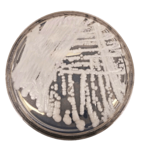 Figure: Candida auris colonies
Culture grows the organism, but telling C. auris apart from other yeasts, particularly Candida haemulonii, is difficult. C. auris is frequently misidentified as other Candida species and related yeasts by traditional phenotypic identification systems such as VITEK 2 YST, API 20C, BD Phoenix, and MicroScan. Because of this, any yeast that cannot be confidently identified, or that is reported as Candida haemulonii or another uncommon species from an at-risk patient, should raise suspicion for C. auris and be sent for confirmatory testing.
MALDI-TOF
Special laboratory tests are needed to identify C. auris. Diagnostic devices based on matrix-assisted laser desorption/ionization time-of-flight (MALDI-TOF) can differentiate C. auris from other Candida species.
Treatment
For invasive C. auris infection, an echinocandin is the recommended first-line treatment, started before susceptibility results are available. Treatment is guided by susceptibility testing once it is available, because resistance is common. Some isolates are resistant to all three main antifungal classes (azoles, echinocandins, and polyenes), and these pan-resistant infections are very difficult to treat and may require combinations of antifungals under specialist guidance.
An important distinction: antifungal treatment is for patients with actual C. auris infection, not for patients who are only colonized. Colonized patients are managed with infection-control measures, not antifungal drugs. This article gives class-level guidance only; regimens are decided by the treating clinician.
How to Remember
Auris means ear. C. auris was first isolated from an ear infection in Japan; "auris" is Latin for ear.
A ward problem, not just a patient problem. Unlike ordinary Candida, C. auris colonizes skin, survives on surfaces, and spreads between patients, so it is an infection-control emergency.
Silent skin colonization drives spread. Patients carry it on their skin without symptoms, which is why it moves through a ward before anyone looks sick.
Echinocandin first, but check susceptibility. Echinocandins are first-line for infection, but resistance is common, so susceptibility testing guides therapy. Treat infection, not colonization.
Easily missed. Routine lab methods misidentify it. MALDI-TOF or molecular methods are needed.
Key exam facts
| Item | Fact |
|---|---|
| Organism | Candida auris, an emerging yeast |
| First identified | 2009, Japan (named for an ear isolate) |
| WHO status | Critical-priority fungal pathogen |
| Key trait | Multidrug-resistant; often azole-resistant, some pan-resistant |
| Spread | Skin colonization, surface survival, person-to-person in healthcare |
| Germ tube | Negative (unlike C. albicans) |
| Gram stain | Gram-positive budding yeast (not distinctive) |
| Identification | MALDI-TOF or molecular methods (routine methods misidentify it) |
| Commonly confused with | Candida haemulonii and other Candida species |
| First-line treatment | Echinocandin (for infection, not colonization) |
| Mortality (invasive) | Roughly 30 to 60% |
| Main sites | Bloodstream, wounds, ears |
Where Students Get Confused
"Candida auris is just another Candida albicans." No. Both are Candida yeasts, but C. auris is often multidrug-resistant, colonizes skin, survives on surfaces, and spreads between patients. C. albicans rarely does these things.
"You treat colonized patients with antifungals." No. Antifungals are for actual infection. Colonized patients (carrying the organism without infection) are managed with infection-control measures.
"A negative germ tube rules out Candida." No. It helps rule out C. albicans, which is germ-tube positive. C. auris is germ-tube negative, so a negative result in the right setting should prompt further identification.
"Microscopy can identify Candida auris." No. It looks like an ordinary budding yeast and stains Gram-positive. It cannot be identified by appearance, which is why MALDI-TOF or molecular methods are needed.
"Candida auris is spread in the community like a cold." No. It spreads mainly in hospitals and long-term care facilities, among patients with serious underlying illness, through skin colonization, contaminated surfaces, and contact.
References and further readings
- Centers for Disease Control and Prevention. Candida auris information for healthcare professionals. https://www.cdc.gov/candida-auris/
- World Health Organization. (2022). WHO fungal priority pathogens list. https://www.who.int/publications/i/item/9789240060241
- Sanyaolu, A., et al. (2025). Candida auris: a continuing threat. (Review; see PubMed Central.)
Frequently Asked Questions
What is Candida auris?
What is Candida auris?
Candida auris is an emerging yeast that often resists antifungal drugs and spreads in healthcare settings. It was first identified in 2009 and is now found in more than 40 countries. The World Health Organization lists it as a critical-priority fungal pathogen.
How does Candida auris spread?
How does Candida auris spread?
Mainly in hospitals and long-term care facilities. It colonizes the skin of patients (often without symptoms), survives on surfaces and equipment for long periods, and passes from patient to patient by contact, including on the hands of healthcare workers.
Is Candida auris contagious in the community?
Is Candida auris contagious in the community?
The risk is concentrated in healthcare settings among seriously ill patients. It does not spread through casual community contact the way respiratory viruses do.
How is Candida auris different from Candida albicans?
How is Candida auris different from Candida albicans?
C. albicans is a common commensal that is usually drug-susceptible and rarely spreads between patients. C. auris is often multidrug-resistant, colonizes skin, survives on surfaces, spreads in hospitals, and is germ-tube negative. C. auris is an infection-control concern, not just an individual infection.
Why is Candida auris hard to identify?
Why is Candida auris hard to identify?
It looks like an ordinary yeast under the microscope and is easily misidentified by routine laboratory systems, often as Candida haemulonii. Reliable identification needs MALDI-TOF mass spectrometry or molecular methods.
How is Candida auris treated?
How is Candida auris treated?
Invasive infection is treated first-line with an echinocandin, guided by susceptibility testing because resistance is common. Some isolates resist all antifungal classes. Colonized patients without infection are managed with infection-control measures, not antifungals.

Tankeshwar Acharya, MSc (Medical Microbiology)
Tankeshwar Acharya is an Assistant Professor in the Department of Microbiology at Patan Academy of Health Sciences (PAHS), Nepal, where he has been teaching and practicing clinical microbiology for over 14 years. He is the founder of Microbe Online, one of the leading free microbiology education resources on the web, covering bacteriology, mycology, parasitology, immunology, and clinical laboratory diagnostics written from direct experience in both the classroom and the diagnostic laboratory.
Comments
No comments yet. Be the first to share your thoughts.
Leave a comment
All comments are reviewed before they appear.