
Gram Staining: Step-by-Step Procedure, Results & Interpretation Guide

On this page
Danish physician Hans Christian Gram developed the Gram staining method in 1884. Gram staining procedure uses four chemicals; crystal violet, iodine, alcohol, and safranin, to stain bacteria
Gram staining is still the cornerstone of bacterial identification and taxonomic division. This differential staining technique separates most bacteria into two groups based on cell wall composition.
- Gram-positive bacteria stains purple
- Gram-negative bacteria stains red/pink
| Feature | Gram-positive | Gram-negative |
|---|---|---|
| Cell wall (in brief) | Thick peptidoglycan, no outer membrane | Thin peptidoglycan with an outer membrane |
| Color after staining | Purple (keeps crystal violet) | Pink/red (takes up safranin) |
For the full side-by-side of cell wall structure, periplasmic space, toxins, lysozyme response, and worked examples, see Gram-positive vs gram-negative bacteria.
Nearly all clinically important bacteria can be visualized using the Gram staining technique, the only exceptions being those organisms;
- Those that exist almost exclusively within host cells, i.e., intracellular bacteria (e.g., Chlamydia)
- Those that lack a cell wall (e.g., Mycoplasma)
- Those too thin to be seen reliably when Gram stained (e.g., spirochetes such as Treponema), which require darkfield microscopy or special stains
Why gram staining matters clinically
Gram staining is the single most impactful rapid diagnostic test in clinical microbiology. A gram stain result is typically available within 15–30 minutes of specimen receipt i.e. hours before culture results, and directly guides empirical antibiotic therapy decisions.
When a clinician receives a gram stain report, they use the combination of gram reaction + morphology + arrangement to make an immediate presumptive identification:
| Gram stain result | Presumptive organism | Typical clinical context |
|---|---|---|
| Gram-positive cocci in grape-like clusters | Staphylococcus aureus or CoNS | Wound, blood culture, abscess, pneumonia |
| Gram-positive cocci in pairs and chains | Streptococcus spp. or Enterococcus spp. | Throat, wound, blood culture, UTI |
| Gram-positive lancet-shaped diplococci | Streptococcus pneumoniae | CSF (meningitis), sputum, blood culture |
| Gram-negative kidney-shaped diplococci (intracellular) | Neisseria gonorrhoeae | Urethral/cervical discharge |
| Gram-negative diplococci (extra + intracellular) | Neisseria meningitidis | CSF (meningitis), blood culture |
| Gram-positive square-ended rods in long chains (bamboo) | Bacillus anthracis | Suspicious powder, skin lesion |
| Gram-positive "drumstick" rods with spores | Clostridium tetani | Wound, anaerobic culture |
| Gram-positive "boxcar" rods | Clostridium perfringens | Wound, gas gangrene, blood culture |
| Gram-positive club-shaped rods in palisades | Corynebacterium diphtheriae | Throat smear |
| Gram-negative coccobacilli (tiny) | Haemophilus influenzae | CSF, sputum, blood culture |
| Gram-negative curved/comma-shaped rods | Vibrio cholerae or Campylobacter | Stool (watery/bloody diarrhoea) |
| Gram-negative rods (large, mucoid) | Klebsiella pneumoniae | Sputum, blood culture, urine |
| Gram-positive branching filaments | Actinomyces spp. or Nocardia spp. | Abscess with sulfur granules |
| Gram-positive yeast with pseudohyphae | Candida spp. | Blood culture, wound, urine |
Principle of Gram Stain
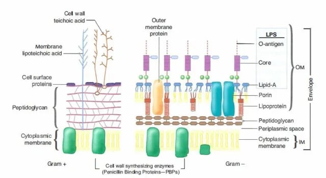 Image 2:Cell wall of Gram-positive and Gram-negative Bacteria
The differences in Gram-positive and Gram-negative bacteria cell wall composition account for the Gram staining differences. Gram-positive cell wall contains a thick layer of peptidoglycan with numerous teichoic acid cross-linking, which resists decolorization.
In aqueous solutions, crystal violet dissociates into CV+ and Cl – ions that penetrate through Gram-positive and Gram-negative cell walls. The CV+ interacts with negatively charged components of bacterial cells, staining the cells purple. When added, iodine (I- or I3-) interacts with CV+ to form large crystal violet-iodine (CV-I) complexes within the cytoplasm and outer layers of the cell.
The decolorizing agent (ethanol or an ethanol and acetone solution) interacts with the lipids of both gram-positive and gram-negative bacteria membranes.
- The alcohol dissolves the lipids of the Gram-negative outer membrane, disrupting it and leaving the thin peptidoglycan layer exposed. The cell wall becomes leaky, and the large CV-I complexes are washed out of the cell.
- The highly cross-linked and multi-layered peptidoglycan of the gram-positive cell dehydrates after the addition of ethanol. Thus ethanol treatment traps the large CV-I complexes within the cell.
After decolorization, the gram-positive cell remains purple. In contrast, the gram-negative cell has lost the purple color and becomes visible only after the counterstain, the positively charged dye safranin, is applied. Safranin stains the decolorized gram-negative cells red or pink. Because safranin is a paler counterstain, it does not mask the crystal violet-iodine complex already trapped in gram-positive cells, so those cells remain purple.
Steps of Gram Staining
Classic Gram staining techniques involve the following steps:
- Fixation of clinical materials to the surface of the microscope slide either by heating or by using methanol. (Methanol fixation is recommended rather than heat fixation. Methanol fixation preserves the morphology of host cells and bacteria. Heating the slide causes cell distortion, could increase cell debris, and may cause erroneous Gram stain results.).
- Application of the primary stain (crystal violet). Crystal violet is a dark blue to purple dye. It stains all cells blue/purple.
- Application of mordant: The iodine solution (mordant) is added to form a crystal violet-iodine (CV-I) complex; all cells continue to appear blue.
- Decolorization step: the critical step that distinguishes gram-positive from gram-negative cells. The solvent (acetone or ethanol) washes the CV-I complex out of gram-negative cells, which turn colorless, while gram-positive cells retain it and stay blue. The mechanism is explained in the Principle section below.
- Application of counterstain (safranin): The red dye safranin stains the decolorized gram-negative cells red/pink; the gram-positive bacteria remain purple.
Find information and process for the Preparation of Gram Staining Regent
Preparation of the smear
- Take a clean grease free slide.
- Transfer a loop of the sample (for example, sputum, CSF, or pus) to the microscope slide. If performing a Gram stain from a bacterial colony, first put a drop or a few loopful of water and emulsify the bacterial colony in the water drop.
- Spread the sample to an even-thin film over a circle of 15 mm diameter.
- Air dry the sample, and once the sample gets air dried, heat fix the smear by passing it through a bunsen burner three times. Heat application helps the cell adhesion (fixation) to the glass slide and prevents its loss during rinsing.
Allow the slide to cool to the touch before applying the stain. Alternatively, the smear can be fixed using methanol.
Methanol fixation
Place or hold the slide over a paper towel and flood the slide with absolute methanol for two minutes. Alternatively, dip the slide into a Coplin jar filled with methanol.
Once two minutes have passed, tilt the slide and drain off the excess methanol and let the slide air dry. Do not wipe or blot the slide, as this can remove cells.
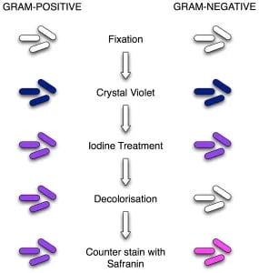 Image 3: Procedure of Gram Staining; note the color change after each step
Gram Staining Procedure
Before handling the specimen slide or culture material, review the microbiology laboratory safety rules for specimen handling and culture safety. Gram staining involves contact with live cultures or clinical specimens; so all the foundational safety practices mentioned there also apply for gram staining procedure.
The gram staining procedure involves four major steps; staining with crystal violet, fixing the dye, applying a decolorizer, and counter-staining.
- Flood air-dried, heat-fixed smear of cells for 1 minute with crystal violet staining reagent. Please note that the quality of the smear (too heavy or too light cell concentration) will affect the Gram Stain results.
- Wash slide in a gentle and indirect stream of tap water for 2 seconds.
- Flood slide with the mordant: Gram’s iodine. Wait 1 minute.
- Wash slide in a gentle and indirect stream of tap water for 2 seconds.
- Flood slide with decolorizing agent (acetone-alcohol decolorizer). Wait 10-15 seconds or add drop by drop to slide until the decolorizing agent running from the slide is clear.
- Flood slide with a counterstain, safranin. Wait 30 seconds to 1 minute.
- Wash slide in a gentle, indirect stream of tap water until no color appears in the effluent.
- Allow the slide to air dry by tilting it onto a paper towel or over a sink. Alternatively, gently dry the slide by blotting it using a lint-free bibulous paper. Please do not use a wiping motion, as it can remove the smear.
- The slide is now ready to view under the microscope. First, focus on the image using the high dry objective lens marked 40x. Then, without removing the slide, switch to the high-power oil immersion objective lens marked 100x. Use immersion oil and observe the results under oil immersion (100x) using a bright-field microscope. This will result in an overall magnification of 1,000x.
After staining, first check that decolorization was adequate (see Quality Control): on a smear with pus cells, neutrophil nuclei should stain red, not blue.
Results
- Gram-negative bacteria will stain pink/red and
- Gram-positive bacteria will stain blue/purple.
Reporting Gram smears
The report should include the following information:
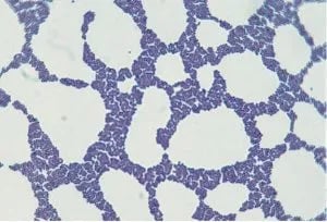 Figure: Staphylococcus in Gram Stain
- Numbers of bacteria present, whether many, moderate, few, or scanty
- Gram reaction of the bacteria, whether Gram-positive or Gram-negative
- Morphology of the bacteria, whether cocci, diplococci, streptococci, rods, or coccobacilli. Also, whether the organisms are intracellular.
- Presence and number of pus cells
- Presence of yeast cells and epithelial cells.
Example: A urethral smear report might read: ‘Moderate numbers Gram-negative intracellular diplococci and many pus cells.’
How to Remember: Gram Staining Steps
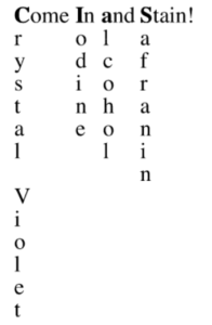 "Come In And Stain" the four reagents in order:
- Come = Crystal violet (primary stain, all cells turn purple)
- In = Iodine (mordant, fixes the dye)
- And = Alcohol/Acetone (decolorizer, the critical step)
- Stain = Safranin (counterstain, GN cells turn pink)
The decolorization step is the heart of the procedure. Everything before it is preparation; everything after it is visualization. The entire gram-positive/gram-negative distinction rests on those 10–15 seconds of decolorizer contact time.
Neutrophil nuclei as your built-in QC: on any smear with pus cells, red nuclei mean decolorization was correct, blue nuclei mean repeat the slide. (See Quality Control for detail.)
Gram Stain Appearances of Clinically Important Organisms
Gram-positive organisms
| Organism | Gram reaction | Morphology | Arrangement | Key identifying feature on gram stain |
|---|---|---|---|---|
| Staphylococcus aureus | Positive | Cocci | Irregular grape-like clusters | Clusters; golden-yellow pigment on culture |
| Staphylococcus epidermidis | Positive | Cocci | Clusters (smaller than S. aureus) | Similar to S. aureus — culture and coagulase differentiate |
| Streptococcus pyogenes (GAS) | Positive | Cocci | Long chains | Long chains; no clusters |
| Streptococcus agalactiae (GBS) | Positive | Cocci | Short chains or pairs | Shorter chains than GAS |
| Streptococcus pneumoniae | Positive | Lancet/bullet-shaped | Diplococci (pairs) | Lancet shape; may have capsule halo |
| Enterococcus faecalis | Positive | Cocci | Pairs and short chains | Similar to streptococci; distinguished biochemically |
| Bacillus anthracis | Positive | Large rods | Long Chains; square ends | Square-ended rods in long chains (bamboo/jointed appearance); central/subterminal spores |
| Bacillus cereus | Positive | Large rods | Chains | Similar to B. anthracis; spores prominent |
| Clostridium tetani | Positive | Rods | Single or pairs | Terminal spore gives "drumstick" or "tennis racket" appearance |
| Clostridium perfringens | Positive | Large rods | Single or pairs | "Boxcar" shape; subterminal spores; rarely sporulates in tissue |
| Clostridium difficile | Positive | Rods | Single | Subterminal spores; gram-variable in old cultures |
| Listeria monocytogenes | Positive | Short rods | V or L-shaped pairs | Resembles diphtheroids; may be mistaken for corynebacteria |
| Corynebacterium diphtheriae | Positive | Club-shaped rods | Palisades; V and L formations ("Chinese letters") | Club shape; metachromatic granules with methylene blue stain |
| Actinomyces spp. | Positive | Branching filaments | Tangled masses | Branching; sulfur granules in pus |
| Nocardia spp. | Positive (weakly) | Branching filaments | Tangled, beaded | Weakly gram-positive; acid-fast with modified ZN stain |
Gram-negative organisms
| Organism | Gram reaction | Morphology | Arrangement | Key identifying feature on gram stain |
|---|---|---|---|---|
| Neisseria gonorrhoeae | Negative | Kidney-bean shaped cocci | Diplococci, intracellular (within PMNs) | Intracellular diplococci in PMNs; specific for gonorrhea in urethral smears |
| Neisseria meningitidis | Negative | Kidney-bean shaped cocci | Diplococci, intra + extracellular | Both intracellular and extracellular diplococci in CSF |
| Moraxella catarrhalis | Negative | Cocci | Diplococci | Similar to Neisseria; distinguished by oxidase and DNase |
| Haemophilus influenzae | Negative | Very small coccobacilli | Single or pairs | Tiny, barely visible; can be missed on cursory examination |
| Escherichia coli | Negative | Rods | Single | Standard gram-negative rod; no distinctive feature |
| Klebsiella pneumoniae | Negative | Plump rods | Single; mucoid appearance | Mucoid surrounding (capsule); thick rods |
| Pseudomonas aeruginosa | Negative | Slender rods | Single or pairs | Slender; blue-green pigment on culture (not visible on gram stain) |
| Bacteroides fragilis | Negative | Pleomorphic rods | Single; pale staining | Pale, irregular; often vacuolated; anaerobic specimen |
| Fusobacterium nucleatum | Negative | Long rods with tapered pointed ends | Single | Spindle/fusiform shape with sharply tapered ends |
| Vibrio cholerae | Negative | Curved rods | Single; comma-shaped | Comma or S-shape; "fish in stream" appearance in stool |
| Campylobacter jejuni | Negative | Curved/S-shaped rods | Single | Seagull wing or S-shape; very slender |
| Helicobacter pylori | Negative | Curved/S-shaped rods | Single | Curved; found in gastric biopsy material |
Organisms not reliably seen on gram stain
| Organism | Why not seen | Alternative method |
|---|---|---|
| Mycobacterium spp. (incl. M. tuberculosis) | Waxy, high-lipid cell wall resists the stain | Ziehl-Neelsen acid-fast stain |
| Treponema pallidum | Too thin (0.1–0.2 µm) to be seen when Gram stained | Dark-field microscopy; fluorescent antibody |
| Leptospira spp. | Too thin to be seen | Dark-field microscopy |
| Mycoplasma spp. | No cell wall, so no peptidoglycan to retain stain | Culture on specialised media; PCR |
| Chlamydia trachomatis | Intracellular and very small | Giemsa stain (inclusion bodies); PCR |
| Rickettsia spp. | Intracellular and very small | Giemsa or other tissue stains; PCR |
| Legionella pneumophila | Poor uptake of the red counterstain | Silver stain; Dieterle stain; DFA; culture on BCYE |
Note: To be visible on a slide, organisms that stain by the Gram method must be present at roughly 10⁴ to 10⁵ organisms per milliliter of sample fluid.
The Gram stain is not just a morphological exercise; it is a clinical decision tool. The pattern seen on a direct smear from a clinical specimen can and should influence empiric antibiotic selection before any culture result is available.
| Gram Stain Pattern | Common Organisms | Immediate Clinical Implication |
|---|---|---|
| GP cocci in clusters | S. aureus, CoNS | Consider anti-staphylococcal coverage; screen for MRSA in high-risk settings |
| GP cocci in chains | Streptococcus spp., Enterococcus | Penicillin/ampicillin coverage; correlate with beta-hemolysis on blood agar |
| GP lancet-shaped diplococci | S. pneumoniae | High-dose penicillin or ceftriaxone; check for capsule (quellung reaction) |
| GN diplococci, intracellular | N. gonorrhoeae, N. meningitidis | In CSF: start ceftriaxone immediately without waiting for culture confirmation |
| GN rods (large, lactose fermenter) | E. coli, Klebsiella | Broad-spectrum GN coverage; risk-stratify for ESBL |
| GN rods (smaller, non-fermenter) | Pseudomonas, Acinetobacter | Anti-pseudomonal coverage in ICU/burns; consider carbapenem resistance |
| GN coccobacilli | Haemophilus, Brucella, Pasteurella | Context-dependent, check bite history, animal exposure, CSF |
| GP rods (boxcar-shaped) | Bacillus, Clostridium perfringens | Gas gangrene/wound: add anaerobic coverage urgently |
| GP branching filaments | Actinomyces, Nocardia | Prolonged penicillin (Actinomyces) or TMP-SMX (Nocardia) |
| Yeast cells (GP, round, budding) | Candida spp., Cryptococcus | Antifungal coverage; confirm with culture and India ink if Cryptococcus suspected |
| No organisms seen | Any | Culture; consider intracellular pathogens (Mycoplasma, Chlamydia, Rickettsiae) |
Key rule: A Gram stain from a normally sterile site (CSF, blood, joint fluid) showing any organisms at all is a serious finding requiring immediate action, do not wait for culture.
Limitations
The sensitivity of the Gram stain procedure is low. Sometimes, you may fail to see the organism in Gram Stain smear, but the same clinical specimen may yield organisms when cultured. To be visible on a slide, organisms that stain by the Gram method must be present at roughly 10⁴ to 10⁵ organisms per milliliter of centrifuged fluid.
Gram staining technique is not recommended for spirochetes and mycobacteria. Mycobacteria stain weakly with gram stain, and bacteria such as Mycoplasma, Rickettsiae, Chlamydiae do not take up the dyes used in Gram stain or are too small to be seen with light microscopy.
Not all bacteria are visible on a Gram stain. The organisms listed below are the medically important bacteria that cannot be seen, or cannot be reliably identified, using this technique.
Quality Control
Always check new batches of staining reagents using a smear containing known Gram-positive (S. aureus ATCC 25923) and Gram-negative (E. coli ATCC 25922) organisms before use on clinical specimens.
The neutrophil nuclei check is the fastest in-run QC available on any clinical smear containing pus cells: in a correctly decolorized smear, neutrophil nuclei stain red/pink. If they stain blue/purple, decolorization is insufficient and the entire slide must be repeated. This check takes two seconds and catches the single most common gram staining error before a wrong result is reported.
Common QC failures and causes:
| Problem | Likely Cause | Action |
|---|---|---|
| All bacteria staining GP (even known GN) | Under-decolorization | Increase decolorizer contact time; check reagent freshness |
| All bacteria staining GN (even known GP) | Over-decolorization | Reduce contact time; use drop-by-drop technique |
| Faint or no staining | Expired crystal violet or safranin | Replace reagents; check storage conditions |
| Pale counterstain | Old or diluted safranin | Replace safranin |
| Iodine solution yellow, not brown | Degraded iodine | Replace, always store in brown/opaque container |
| Smear washes off slide | Inadequate fixation | Ensure complete air-drying before heat fixation; use methanol fixation |
Troubleshooting Gram Stain Errors
| Problem | Likely cause | Solution |
|---|---|---|
| All organisms stain gram-negative (gram-positive appear pink) | Over-decolorization, most common error | Reduce decolorization time; use drop-by-drop acetone-alcohol rather than flooding |
| All organisms stain gram-positive (gram-negative appear purple) | Under-decolorization | Increase decolorization time; check acetone-alcohol concentration |
| Pale, weak staining overall | Stain reagents old or diluted; smear too thin | Prepare fresh reagents; use adequate bacterial suspension |
| Background staining/precipitate on slide | Dirty slide; stain not filtered; flooding with stain | Use clean grease-free slides; filter crystal violet before use |
| Loss of cells during washing | Smear not properly fixed | Fix adequately with methanol; avoid vigorous washing stream |
| Gram-variable results in same smear | Old culture used; antibiotic-treated specimen; excessive heat fixation | Use 18–24 hour culture; note antibiotic history; use methanol fixation |
| Nucleus of neutrophils appears blue not red | Under-decolorization, key quality check | Extend decolorization; repeat with fresh smear |
| Organisms not visible despite clinical suspicion | Below detection threshold (< 10⁴ organisms/mL) | Request culture; centrifuge specimen before smearing |
Variations in Gram Reaction
Various factors influence the results of Gram staining. Sometimes the result might be entirely different than you have anticipated.
- A thick smear will require more decoloration than a thin smear. When the smear is too thick, Gram-negative bacteria may not fully decolorize during decolorization steps and appear as Gram-positive.
Pitfalls in the Interpretation of Gram’s Stains
| Organism | Classic Presentation | Variant Presentation | Comments |
|---|---|---|---|
| Streptococcus pneumoniae | Gram-positive, lancet-shaped, diplococci | Elongated cocci, resembling short bacilli | May be misinterpreted as mixed organisms; over-decolorized cells may be mistaken for gram-negative coccobacilli. |
| Acinetobacter spp. | Gram-negative coccobacilli | Gram-negative cocci; gram-variable staining is common | May be mistaken for Neisseria spp. and reported as gram-negative cocci; search the smear to find some organisms that demonstrate elongated forms, which are not seen in Neisseria. |
| Clostridium perfringens | Boxcar-shaped gram-positive bacilli | Gram-positive cocci | May be mistaken for Streptococcus pneumoniae and reported as gram-positive cocci; in addition to a coccal form, cells retain crystal violet tenaciously during decolorization. |
| Clostridium perfringens | Boxcar-shaped gram-positive bacilli | Gram-variable or Gram-negative bacilli | Maybe mistaken for gram-negative bacilli; the boxcar shape is a clue that the organism is gram-positive; other Clostridia and Bacillus spp. May also appear similar. |
| Yeast, especially Cryptococcus neoformans | Gram-positive round or oval cells with budding | Gram-variable cells | May be mistaken for artifacts; size and shape distinguish them from bacteria. |
Frequently Asked Questions
What does it mean if neutrophil nuclei appear blue instead of red on a Gram stain?
What is the clinical significance of gram-negative intracellular diplococci in a urethral smear?
Why do gram-positive bacteria sometimes stain gram-negative?
References and further reading
- Tille PM. Bailey & Scott's Diagnostic Microbiology. 15th ed. Elsevier; 2022.
- Procop GW, Church DL, Hall GS, et al. Koneman's Color Atlas and Textbook of Diagnostic Microbiology. 7th ed. Wolters Kluwer; 2017.
- Murray PR, Rosenthal KS, Pfaller MA. Medical Microbiology. 9th ed. Elsevier; 2020.
- Garcia LS. Clinical Microbiology Procedures Handbook. 4th ed. ASM Press; 2016.
- Cowan ST. Cowan and Steel's Manual for the Identification of Medical Bacteria. 3rd ed. Cambridge University Press; 1993.

Tankeshwar Acharya, MSc (Medical Microbiology)
Tankeshwar Acharya is an Assistant Professor in the Department of Microbiology at Patan Academy of Health Sciences (PAHS), Nepal, where he has been teaching and practicing clinical microbiology for over 14 years. He is the founder of Microbe Online, one of the leading free microbiology education resources on the web, covering bacteriology, mycology, parasitology, immunology, and clinical laboratory diagnostics written from direct experience in both the classroom and the diagnostic laboratory.
Comments
No comments yet. Be the first to share your thoughts.
Leave a comment
All comments are reviewed before they appear.