
Normal Flora of the Skin: Types, Roles, and Culture Report Meaning
What normal skin flora is, the resident and transient organisms that live there, and what "growth of skin flora" means on a blood or urine culture report.

On this page
A blood culture comes back on a patient who spiked a fever last night. The report reads: Staphylococcus epidermidis isolated from one bottle. The intern wants to start vancomycin. The resident pauses. S. epidermidis is one of the most common bacteria on human skin, and it is also the single most common contaminant in blood cultures. So which is it here: a real bloodstream infection, or a bit of the patient's own skin that rode in on the needle?
You cannot answer that from the organism name alone. You answer it from where the skin flora lives, how it behaves, and how the culture was collected and reported. That is what this article is about.
Human skin is home to billions of diverse bacteria, archaea, fungi, and viruses, with bacteria making up the vast majority. Typically, a person has around 1,000 species of bacteria on their skin. The skin microbiome is seeded at birth. Most microorganisms live in the stratum corneum’s superficial layers and the hair follicles’ upper parts. Some microorganisms, however, reside in the deeper areas of the hair follicles. A variety of aerobic bacteria, anaerobic bacteria, and fungi inhabit these regions.
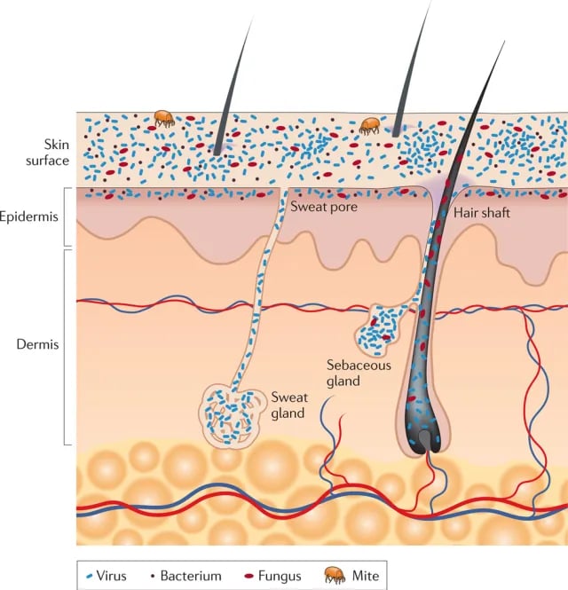 Figure: Schematic of skin histology viewed in cross-section with microorganisms and skin appendages (image source)
Skin surface varies greatly in chemical composition and moisture content. Generally, the skin surface is not a favorable place for microbial growth, as it is dry, salty, and poor in nutrients. The skin is also subject to periodic drying. But human skin is not uniform. It provides many different micro-environments. Someone has aptly compared it with geographic regions of the earth; the desert of the forearm, the cool woods of the scalp, and the tropical forest of the armpit.
Residential microbial populations are higher in moist and warm areas of the skin such as axilla, perineum, and interdigital spaces of the toes. Availability of nutrients is another major factor that alters the quantity and diversity of microflora in a particular niche. Several kinds of sweat glands (such as eccrine glands, apocrine glands, and sebaceous glands) present in the skin secrete fluids rich in microbial nutrients. The distribution of sweat glands on skin surfaces and differences in the composition of their secretions also affect the nature of skin flora.
Do you know?
Sweat itself does not smell, bacteria on your skin are the culprits for your body odor. Underarm odor develops as a result of bacterial activity on the secretions of the apocrine glands.
Types of Skin Flora
The microorganisms of the normal flora of the skin are either transient or residents.
- Transient microflora: Skin is the largest and most exposed organ so it is continually being inoculated with microorganisms. Most of these organisms can’t multiply and ultimately die either because of low moisture content or low pH on the skin surface.
- Resident microflora: Resident microorganisms are able to multiply, not merely survive, on the skin. The normal flora of the skin consists primarily of gram-positive bacteria.
Traditionally, skin flora were identified by use of culture-based methods, which used to favor easily culturable organisms and fail to capture the complete diversity of the microbiome. To circumvent this problem, nowadays, sequencing methods ( 16S ribosomal RNA gene sequencing for bacteria) are used to enumerate skin microbiota.
Gram-positive bacteria
Gram-positive bacteria are predominant members of the skin microbiome as they are better adapted to the dry conditions of the skin. These include Staphylococci, Micrococci, and a variety of both aerobic and anaerobic corynebacteria. Cutibacterium acnes (formerly Propionibacterium acnes) start colonizing after a child reaches the age of 10. It is ordinarily a harmless resident but certain strains can contribute to acne at puberty.
Gram-negative bacteria
Gram-negative bacteria are minor constituents of the skin flora as they can’t compete with gram-positive organisms. They are seen mostly in moist intertriginous areas, such as the toe webs and axilla.
The Gram-negative organism best adapted to moist skin is Acinetobacter. Other Gram-negatives such as Escherichia coli, Klebsiella, Enterobacter, and Proteus may be found in moist folds, but these are usually transient organisms from fecal or environmental sources rather than established residents.
Fungi (molds/yeasts)
The lipophilic yeast genus Malassezia is the dominant fungal resident of human skin. It concentrates in sebum-rich areas such as the scalp, face, and upper trunk. Candida albicans is a normal resident of mucosal surfaces (mouth, gut, and vagina), not of healthy dry skin. It can appear transiently on skin and can colonize moist or damaged skin, where it may cause opportunistic infection. Other fungi such as Aspergillus spp., Cryptococcus sp., and Rhodotorula sp. may be recovered from the skin, especially in moist regions such as the feet.
What "growth of skin flora" means on a culture report
When a laboratory reports "growth of skin flora," "mixed skin flora," or "normal skin flora," it is not naming a disease. It is telling you that the organisms that grew are the ordinary commensals that live on everyone's skin, most often coagulase-negative staphylococci (such as Staphylococcus epidermidis), Micrococcus, corynebacteria (diphtheroids), and Cutibacterium acnes. The lab is signaling one of two things: either these organisms entered the sample during collection, or, in the right clinical setting, one of them is causing a real infection. The report tells you what grew. It cannot tell you which of those two situations you are in. That judgment is yours to make, and it depends on the specimen type, how the sample was collected, and the patient.
The quantity words: scanty, light, moderate, heavy, rare, mixed
Many labs grade how much grew. The exact words vary between laboratories, but the ladder is consistent:
| Term on report | What it means | Usual reading for a normally sterile-site sample |
|---|---|---|
| Rare or scanty (or scant) growth | Very few colonies, often on one part of the plate only | Points toward contamination |
| Light growth | Small number of colonies | Often contamination, judged with clinical picture |
| Moderate growth | Intermediate number of colonies | Judged with clinical picture |
| Heavy growth | Colonies across the plate, often a pure culture | More concerning, but skin flora can still be a contaminant |
| Mixed growth | Two or more different organisms | For a sterile site, usually points toward contamination |
Two cautions. First, quantity alone does not prove significance. A heavy growth of a skin commensal from a poorly collected sample is still a contaminant. Second, "mixed" is a strong hint. A true bloodstream infection or a true urinary tract infection is usually caused by one organism. When a report from a normally sterile site lists several skin organisms together, collection contamination is the leading explanation.
Reading "skin flora" in a blood culture
Blood is normally sterile, so anything that grows demands an explanation. Skin flora, especially coagulase-negative staphylococci, are the most common blood-culture contaminants, because the needle passes through the patient's skin to reach the vein. If the skin is not disinfected well, a few resident organisms travel into the bottle. The interpretation logic uses several clues together:
- Number of positive bottles or sets. Blood cultures are usually drawn as sets from separate sites. When a skin organism grows in only one bottle of one set and the others stay negative, contamination is the likely answer. When the same organism grows in several separate sets, true infection becomes much more likely.
- The same species, repeatedly. One isolate of a skin commensal is weak evidence. The same species, from separate draws, is stronger evidence of real bacteremia.
- Time to positivity. Contaminants usually take longer to turn a bottle positive, because very few organisms were introduced. A bottle that flags positive quickly reflects a higher starting number of organisms, which fits true infection better.
- The patient and the setting. A skin commensal is far more likely to be a real pathogen in a patient with a central venous catheter, a prosthetic heart valve, a prosthetic joint, or a weakened immune system. In these patients, S. epidermidis is a genuine cause of device-associated infection, not a nuisance.
The single-bottle S. epidermidis in an otherwise well patient with no device is most likely a contaminant, and the right next step is often a repeat culture rather than immediate vancomycin. Change one fact, a central line and the same organism in two separate sets, and the reading flips to true catheter-associated bacteremia.
Reading "skin flora" in a urine culture
Urine collected clean-catch passes over skin and the outer genital area, so some skin and urogenital flora in the sample is expected. Reports such as "mixed skin flora," "mixed urogenital and/or skin flora," or "mixed growth" usually mean the sample picked up surface organisms during collection, not that the bladder is infected.
A true urinary tract infection is typically a single organism, most often Escherichia coli, at a significant colony count. When a urine report shows several skin or urogenital organisms mixed together, the usual reading is a collection problem, and the practical answer is a properly collected repeat sample rather than antibiotics.
Reading "skin flora" in a wound swab
Wounds are open to the air and sit on skin, so every wound is colonized by skin organisms whether or not it is infected. A surface swab recovers whatever is living on the wound surface, which usually includes normal skin flora, so a report of "mixed skin flora" or "mixed growth" from a superficial swab is common and, on its own, does not mean the wound is infected. This is the point that catches people out: colonization is not infection. Wound infection is diagnosed from the patient and the wound (spreading redness, warmth, increasing pain, purulent discharge, fever, stalled healing), not from the culture report alone. The culture's job is to identify the organism and guide antibiotic choice once infection has already been judged to be present clinically.
Two practical consequences follow. First, how the sample is collected changes what grows. A swab dragged across surface slough or pus picks up more skin and surface contaminants, which is why the wound is cleaned first and the sample is taken from clean viable tissue (see wound swab collection and the Levine technique and collecting a pus sample for the correct method). A poorly collected swab reads as "mixed skin flora" for the same reason a poorly collected blood culture does: it sampled the skin, not the lesion. Second, a single skin commensal reported from a wound with no clinical signs of infection is usually colonization, and treating it with antibiotics is often the wrong response. As with blood and urine, the report tells you what grew. Whether it matters is a clinical decision.
The one question that resolves almost every case
When you see skin flora on a report from a normally sterile site, ask: does the microbiology fit a single, consistent organism, and does the clinical picture support infection? If the answer is a single organism growing repeatedly in a patient who looks infected or who carries a device, treat it as real. If the answer is mixed organisms, a single stray isolate, or a well patient with no device, treat it as likely contamination and, where possible, repeat the sample. The organism name is where the question starts, not where it ends.
Factors Affecting Skin Flora
Although the resident microflora on an individual’s skin remains relatively constant, various factors can affect the nature and extent of the normal flora:
- Change in weather conditions and exposure to sunlight alters the temperature and humidity of the skin thus altering the number and diversity of skin microflora. Exposure to UV radiation kills the normal flora.
- Individual’s health, age, and gender determine the type and the density of skin flora. Age-related changes in skin structure and function alter the microflora of the skin. For example, young children have a more varied microflora and carry more gram-negative bacteria and potential pathogens than adults.
- Personal hygienic habits influence the resident microflora. Unclean individuals usually have higher microbial population densities on their skin. Bathing or showering cleans the skin by the mechanical removal of bacteria shed on corneocytes but the resident microflora are replenished immediately. Bacteria living in the deeper areas of the hair follicles act as a reservoir for recolonization after the surface bacteria are removed.
- Host genotype, lifestyle, and pathophysiology also alters the normal skin flora.
 Figure: Factors contributing to variation in the skin microbiome
Beneficial roles of Skin Flora
- A barrier to infection: Skin is colonized by beneficial microorganisms that suppress the growth of pathogens by outcompeting them for space and food. For example, when the normal flora is inhibited by antibiotics, overgrowth of fungi such as C. albicans can occur.
Cutibacterium acnes digest the oily substances from the sebaceous gland to create an acidic environment that makes it hard for the pathogen to colonize. S. epidermidis produces antimicrobial peptides and also stimulates antimicrobial peptide production by keratinocytes, which may provide protection against pathogenic bacteria. S. epidermidis produce chemicals that help reinforce the tight junctions between skin cells and protect the skin's physical integrity. - Immunological defenses: Shortly after birth, S. epidermidis communicates with regulatory T cells effectively training the immune system to tolerate commensal skin microbes while remaining alert against invaders. It boosts innate immune responses against intracellular bacteria.
When the balance is broken
- Opportunistic Infections: Skin normal flora can cause disease when they gain access to sterile sites such as tissues or blood.
- Surgical-wound infections: Surgical-wound infections are commonly associated with skin flora including various staphylococci and streptococci and Cutibacterium acnes.
- Osteomyelitis: Staphylococcus aureus is the most common bacterial cause of acute osteomyelitis in children and adults. Staphylococcus epidermidis and Cutibacterium acnes can cause osteomyelitis in patients with hip or knee prostheses as they enter the bloodstream at the site of skin wound or intravenous catheter.
- Endocarditis: S. aureus is the prominent cause of right-sided endocarditis in intravenous drug abusers. It is found that skin flora enter venous blood at the site of needle inoculation. Similarly, in people with prosthetic heart valve transplants, S. epidermidis is the major cause of endocarditis.
- Contaminants in blood culture: If disinfection is not done properly while drawing blood for culture, normal skin flora may grow in blood culture. S. epidermidis is one of such contaminants in diagnostic laboratories.
How to Remember
Skin as geography. Picture the skin as a map of climates. The forearm is desert: dry, few organisms. The scalp is cool woods. The armpit and groin are tropical: warm, moist, densely populated. Wherever it is warm and wet, expect more flora. This single image predicts where residents concentrate and why moist folds culture heavily.
Resident versus transient, in one line. Residents live there (they multiply and come back after washing). Transients are just visiting (they land, fail to multiply, and die off). Washing removes the visitors and thins the residents, but the residents return from deep in the hair follicles.
The contaminant rule of thumb: "one and mixed means doubt; same and again means real." One stray skin organism, or several mixed together, points to contamination. The same single species growing again in a separate draw points to real infection. This compresses the whole blood-culture logic into a phrase you can recall at the bench.
Key exam facts
| Fact | Detail and memory aid |
|---|---|
| Dominant skin bacteria | Gram-positive: coagulase-negative staphylococci (mainly Staphylococcus epidermidis), Micrococcus, corynebacteria (diphtheroids), Cutibacterium acnes. They tolerate dry, salty skin better than Gram-negatives. |
| Dominant skin fungus | Malassezia is the main resident yeast genus (lipophilic, likes sebum-rich areas: scalp, face, upper trunk). |
| Resident vs transient | Residents multiply and re-colonize after washing. Transients land and die off. Residents hide in hair follicles and return. |
| Where flora is densest | Warm, moist sites: axilla, groin, perineum, toe webs. "Tropical" skin regions. |
| "Skin flora" on a wound swab | Every open wound is colonized by skin organisms. Colonization is not infection. Infection is a clinical diagnosis; the culture guides drug choice, not the diagnosis. |
| Most common blood culture contaminant | Coagulase-negative staphylococci, especially S. epidermidis, introduced through skin at the venepuncture site. |
| When a skin commensal is a real pathogen | Devices and hosts: central lines, prosthetic valves, prosthetic joints, immunocompromised patients. Also osteomyelitis with prostheses, prosthetic-valve endocarditis. |
| "Mixed skin flora" on a sterile-site report | Usually collection contamination. True infection is usually a single organism. |
| Body odor origin | Sweat is odorless. Bacteria acting on apocrine gland secretions produce the smell. |
| Cutibacterium acnes | Formerly Propionibacterium acnes. Colonizes from around age 10. Harmless resident that can contribute to acne at puberty; also a cause of indolent device infection. |
Where Students Get Confused
"Growth of skin flora means the patient has a skin infection." No. It means the organisms that grew are ordinary skin commensals. Whether they matter depends on the specimen and the patient, not on the phrase itself. On a sterile-site sample it most often signals contamination.
"Candida albicans is normal flora of the skin." This is a common error. C. albicans is normal flora of mucosal surfaces (mouth, gut, vagina), not of normal dry skin. It can transiently appear on skin and colonize moist damaged skin, but it is not a resident of healthy skin the way Malassezia is.
"E. coli and Klebsiella are normal skin flora." They are listed on some sources, but as transient contaminants from fecal or environmental sources, not established residents. The established Gram-negative that survives on moist skin is Acinetobacter. Do not memorize the Enterobacterales as resident skin flora.
"S. epidermidis in a blood culture means sepsis." Not by itself. It is the commonest contaminant. It becomes a real diagnosis when the same species grows in more than one separate set, or in a patient with a device or immune compromise. A single positive bottle in a well patient with no device is usually contamination.
"Transient and resident are just two words for the same thing." They are not. Residents multiply on skin and return after washing. Transients cannot multiply there and die off. The distinction is the whole reason washing reduces but never sterilizes skin.
True or false: "Resident microorganisms live within the skin and are difficult to remove." True. Residents live in the superficial layers and, importantly, deep in hair follicles, which act as a reservoir. Surface washing removes shed organisms, but the follicular reservoir re-colonizes the surface, which is why residents are difficult to remove permanently.
References and further readings
- Byrd, A. L., Belkaid, Y., & Segre, J. A. (2018). The human skin microbiome. Nature Reviews Microbiology, 16(3), 143–155. https://doi.org/10.1038/nrmicro.2017.157
- Grice, E. A., & Segre, J. A. (2011). The skin microbiome. Nature Reviews Microbiology, 9(4), 244–253. https://doi.org/10.1038/nrmicro2537
- Davis, C. P. (1996). Normal Flora. In S. Baron (Ed.), Medical Microbiology (4th ed.). University of Texas Medical Branch at Galveston. http://www.ncbi.nlm.nih.gov/books/NBK7617/
- Eisenstein, M. (2020). The skin microbiome. Nature, 588(7838), S209. https://doi.org/10.1038/d41586-020-03523-7
- Pastar, I., O'Neill, K., Padula, L., et al. (2020). Staphylococcus epidermidis boosts innate immune response by activation of gamma delta T cells and induction of Perforin-2 in human skin. Frontiers in Immunology, 11, 550946. https://doi.org/10.3389/fimmu.2020.550946
- Tille, P. M. (2022). Bailey & Scott's Diagnostic Microbiology (15th ed.). Elsevier.
- Procop, G. W., et al. (2017). Koneman's Color Atlas and Textbook of Diagnostic Microbiology (7th ed.). Wolters Kluwer.
Frequently Asked Questions
What does "growth of skin flora" mean on a culture report?
What does "growth of skin flora" mean on a culture report?
It means the organisms that grew are the ordinary commensals that live on everyone's skin, most often coagulase-negative staphylococci such as Staphylococcus epidermidis, along with Micrococcus, corynebacteria, and Cutibacterium acnes. It is not a disease name. On a sample from a normally sterile site, such as blood, it most often means the organisms entered during collection rather than that they are causing infection.
What does "mixed skin flora" mean?
What does "mixed skin flora" mean?
It means two or more different skin organisms grew together. A true bloodstream or urinary infection is usually caused by a single organism, so several skin organisms mixed together on a sterile-site report usually point to contamination during collection rather than a real infection.
Does "scanty" or "light" growth of skin flora matter?
Does "scanty" or "light" growth of skin flora matter?
Usually not, on its own. Scanty, rare, or light growth of skin commensals from a normally sterile site points toward contamination. Quantity is only one clue, though. It is read together with the specimen type, how the sample was collected, and the patient's clinical picture.
Is Staphylococcus epidermidis in a blood culture always contamination?
Is Staphylococcus epidermidis in a blood culture always contamination?
No. It is the commonest blood-culture contaminant, but it can also cause real infection, especially in patients with a central line, a prosthetic heart valve, a prosthetic joint, or a weakened immune system. The reading depends on how many separate cultures grew the same organism and on the patient. The same species growing in more than one separate set is much more likely to be a real infection than a single positive bottle in a well patient.
What is the difference between resident and transient skin flora?
What is the difference between resident and transient skin flora?
Resident flora multiply on the skin and re-colonize it after washing, living in the surface layers and deep in hair follicles. Transient flora land on the skin from the environment, cannot multiply there, and die off. Washing removes transients and thins residents, but residents return from the follicular reservoir.
What are the main bacteria of normal skin flora?
What are the main bacteria of normal skin flora?
Mostly Gram-positive organisms: coagulase-negative staphylococci (mainly Staphylococcus epidermidis), Micrococcus, corynebacteria (diphtheroids), and Cutibacterium acnes. Gram-negative bacteria are minor and are found mainly in moist areas such as the toe webs and armpits.
What does "skin flora" or "mixed growth" mean on a wound swab?
What does "skin flora" or "mixed growth" mean on a wound swab?
It means the swab grew the ordinary organisms that live on skin, which every open wound carries. On its own it does not mean the wound is infected. Wound infection is diagnosed from the patient and the wound, such as spreading redness, pain, pus, or fever, not from the culture report alone. The culture identifies the organism and guides antibiotic choice once infection has been judged present.

Tankeshwar Acharya, MSc (Medical Microbiology)
Tankeshwar Acharya is an Assistant Professor in the Department of Microbiology at Patan Academy of Health Sciences (PAHS), Nepal, where he has been teaching and practicing clinical microbiology for over 14 years. He is the founder of Microbe Online, one of the leading free microbiology education resources on the web, covering bacteriology, mycology, parasitology, immunology, and clinical laboratory diagnostics written from direct experience in both the classroom and the diagnostic laboratory.
Comments
No comments yet. Be the first to share your thoughts.
Leave a comment
All comments are reviewed before they appear.