Sabouraud Dextrose Agar (SDA): Composition, Principle, Uses, and Colony Morphology
Sabouraud Dextrose Agar (SDA) is the standard medium for fungal isolation. Learn its composition, how its acidic pH selects for fungi, colony morphology of dermatophytes and yeasts, cycloheximide modification, and clinical uses.
A 35-year-old man presents with a slowly spreading, scaly, ring-shaped lesion on his inner thigh. His clinician suspects tinea cruris — a dermatophyte infection. Skin scrapings are sent to the microbiology laboratory. The technician inoculates two media: a KOH preparation for direct microscopy and a Sabouraud Dextrose Agar plate for culture. The KOH prep gives a result within minutes; the SDA plate will take 1 to 4 weeks, but it will provide the species-level identification needed to guide treatment and confirm whether the infection is from Trichophyton rubrum (the most common cause worldwide) or another dermatophyte.
That slow-growing, acid-tolerant, dextrose-rich environment is what Sabouraud Dextrose Agar was specifically designed to provide.
Sabouraud Agar or Sabouraud Dextrose Agar (SDA) is a selective medium primarily used for the isolation of dermatophytes. Other fungi, yeasts, and filamentous bacteria such as Nocardia can also grow in SDA. The acidic pH of this medium (pH about 5.0) inhibits the growth of bacteria but permits the growth of yeasts and most filamentous fungi. Antibacterial agents can also be added to augment the antibacterial effect.
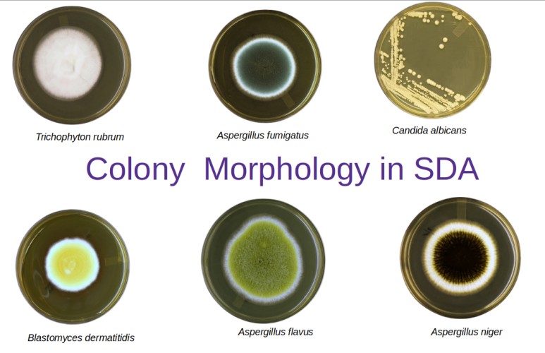 Figure: Fungal colonies (front-side) on Sabourad Dextrose Agar
This medium is also helpful to determine the mycological evaluation of food, contamination in cosmetics, and clinically to aid in the diagnosis of yeast and fungal infections.
Addition of antibiotics like chloramphenicol, gentamicin, and tetracycline as selective agents can inhibit the overgrowth of competing bacteria while permitting the successful isolation of fungi and yeasts. Various other modifications are also reported by using cycloheximide, penicillin, streptomycin, neomycin depending upon the intended use.
Principle
Sabouraud Dextrose Agar comprises of enzymatic digest of casein and animal tissues which provide a nutritious source of amino acids and nitrogenous compounds for the growth of fungi and yeasts.
Dextrose is a fermentable carbohydrate incorporated in high concentrations as a carbon and energy source. Agar is the solidifying agent. The addition of antibiotics like chloramphenicol and/or tetracycline acts as broad-spectrum antimicrobials to inhibit the growth of a wide range of gram-positive and gram-negative bacteria. Gentamicin is added to further inhibit the growth of gram-negative bacteria.
Why acidic pH selects for fungi:
Most clinically significant bacteria grow optimally at pH 7.2–7.4 and are significantly inhibited or killed at pH 5.0–5.6. Fungi, by contrast, have a much wider pH tolerance and grow well across a range of pH 2.0–9.0, with optimal growth around pH 5.0–6.0 for most species. Sabouraud Dextrose Agar exploits this difference: its acidic pH (approximately 5.6) creates an environment that favours fungal growth while suppressing most bacteria — particularly the rapidly growing Gram-negative organisms that would otherwise overgrow slow-growing fungal colonies in clinical specimens.
The high dextrose concentration (40 g/L — four times that of standard bacteriological media) provides an abundant carbon and energy source that particularly favours fungi adapted to carbohydrate-rich environments, including dermatophytes infecting keratinised skin, hair, and nails.
Important limitation: The acidic pH and absence of blood or serum means SDA does not support the mould phase of dimorphic fungi (Histoplasma capsulatum, Blastomyces dermatitidis, Coccidioides immitis) at 37°C for primary isolation. Brain Heart Infusion (BHI) agar is preferred for primary isolation of dimorphic fungi suspected from clinical specimens.
Composition of SDA
| Ingredients | Gm/L |
|---|---|
| Mycological peptone (enzymatic digest of casein and animal tissues) | 10 gm |
| Dextrose | 40 gm |
| Agar | 15 gm |
Preparation of SDA
- Suspend 65 gm of the medium in one liter of purified water.
- Heat with frequent agitation and boil for one minute to completely dissolve the medium.
- Autoclave at 121° C for 15 minutes.
- Cool to 45 to 50°C and pour into Petri dishes or tubes for slants.
- To process specimens, streak the specimen onto the medium with a sterile inoculating loop to obtain isolated colonies.
- Incubate the plates at 25 – 30°C in an inverted position (agar side up) with increased humidity.
- Cultures should be examined weekly for fungal growth and held for 4 – 6 weeks before being reported as negative.
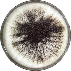 Figure: Mold colony with black pigmentation in SDA
Result and interpretation
After sufficient incubation, SDA plates should show isolated colonies in streaked areas and confluent growth in areas of heavy inoculation. Examine plates for fungal colonies exhibiting typical color and morphology. Additional procedures should be performed to confirm the findings.
Yeasts will grow as creamy to white colonies. Molds will grow as filamentous colonies of various colors.
Colony Morphology on SDA
Fungal colonies on SDA are examined for two sides: the obverse (front); the top surface showing colony color, texture, and surface morphology and the reverse (underside), which often shows pigmentation that is diagnostically important, particularly for dermatophytes.
Incubation temperature matters: dermatophytes are incubated at 25–28°C; Candida and other yeasts grow well at both 25°C and 37°C; dimorphic fungi grow in their mould phase at 25°C on SDA.
| Organism | Obverse color/texture | Reverse color | Incubation | Clinical significance |
|---|---|---|---|---|
| Candida albicans | Cream to white, smooth, pasty, yeast-like odour | Cream | 25–37°C, 24–48 hrs | Most common fungal pathogen; oral, vaginal, invasive candidiasis |
| Trichophyton rubrum | White to buff, flat or granular, powdery surface | Deep wine-red to cherry-red | 25–28°C, 1–3 weeks | Most common cause of tinea pedis, onychomycosis, tinea cruris |
| Trichophyton mentagrophytes | White to cream, flat or granular, may be powdery | Yellow-brown to tan | 25–28°C, 1–2 weeks | Tinea pedis, tinea unguium; zoophilic strains cause inflammatory ringworm |
| Microsporum canis | White to pale yellow, flat, fluffy | Bright lemon-yellow to golden-yellow | 25–28°C, 1–2 weeks | Tinea capitis and tinea corporis; most common dermatophyte from animals |
| Microsporum gypseum | Cinnamon-brown, granular, powdery | Pale yellow to tan | 25–28°C, 1–2 weeks | Geophilic; soil exposure; granular texture due to abundant macroconidia |
| Aspergillus fumigatus | Blue-green, powdery | Pale yellow | 37°C (grows well), 2–5 days | Invasive aspergillosis in immunocompromised patients |
| Aspergillus flavus | Yellow-green, powdery | Pale yellowish | 25–37°C, 3–5 days | Aflatoxin production; opportunistic infection |
| Aspergillus niger | Initially white, then black ("salt and pepper") | Pale yellow | 25–37°C, 3–5 days | Otomycosis; rarely invasive |
| Aspergillus nidulans | Greenish-blue with whitish edge | Yellow to brownish | 25–37°C | Less common Aspergillus species |
| Rhodotorula spp. | Pinkish-orange, creamy, salmon-coloured | Pink-orange | 25–37°C, 2–4 days | Emerging opportunist in immunocompromised patients; catheter-associated |
| Trichosporon mucoides | White to cream, yellowish, wrinkled | White to cream | 25–37°C | Superficial and invasive infections in immunocompromised |
| Geotrichum candidum | White to cream, flat with aerial mycelium | White | 25°C | Geotrichosis; dairy-associated contaminant |
Reverse pigmentation is the key to dermatophyte identification. The deep wine-red to cherry-red reverse of T. rubrum is one of the most reliable macroscopic identifiers in clinical mycology. When examining a slow-growing, white, powdery colony on SDA, always flip the plate and examine the underside before proceeding to microscopy.
Modifications of Sabouraud Agar
Cycloheximide Modification
Cycloheximide (actidione) is added to SDA at 0.4–0.5 g/L to inhibit the growth of saprophytic (environmental) fungi that would otherwise overgrow slow-growing pathogenic dermatophytes in clinical specimens. This modification is particularly useful for skin, hair, and nail cultures where environmental moulds are expected as contaminants.
Critical limitation of cycloheximide-containing SDA:
Cycloheximide is not selective only for saprophytes — it also inhibits several clinically important fungi:
| Fungi inhibited by cycloheximide | Clinical significance |
|---|---|
| Candida spp. (most) | Common cause of mucosal and invasive infections |
| Cryptococcus neoformans | Serious opportunistic meningitis in HIV patients |
| Aspergillus spp. | Leading cause of invasive mould infections |
| Trichosporon spp. | Systemic infections in immunocompromised |
| Pseudallescheria boydii | Mycetoma; keratitis |
Practical rule: When a specimen may contain Candida, Cryptococcus, or Aspergillus (e.g., BAL, blood, CSF), SDA without cycloheximide must be used alongside or instead of cycloheximide-containing SDA. Many laboratories therefore routinely inoculate two SDA plates — one with cycloheximide (for dermatophytes), one without (for all other fungi).
Exam mnemonic — "Cycloheximide CCAT": Cycloheximide inhibits Candida, Cryptococcus, Aspergillus, and Trichosporon. These are the four genera you must specifically consider when choosing whether to use the cycloheximide modification.
SabHI Agar (Sabouraud-Brain Heart Infusion Agar)
SabHI agar is formulated by combining equal parts Sabouraud Dextrose Agar and Brain Heart Infusion (BHI) agar. This hybrid medium retains some of SDA's selectivity against bacteria while gaining BHI's richness and ability to support fastidious organisms.
Why it was developed: Standard SDA does not reliably recover dimorphic fungi (Histoplasma, Blastomyces, Coccidioides, Sporothrix) from primary clinical specimens because these organisms require richer nutrient conditions for primary isolation, particularly at 37°C in their yeast phase. BHI alone supports them but also permits bacterial overgrowth. SabHI balances both requirements.
Key property: SabHI does not promote conidiation of filamentous fungi as effectively as SDA. This means that while recovery rates are higher on SabHI, characteristic sporulation patterns used for identification may be less pronounced — subcultural onto plain SDA is often required for definitive identification.
How to Remember
SDA is defined by three features — and each one is clinically meaningful:
- Low pH (~5.6) = selects for fungi over bacteria. Think: "fungi like acid, bacteria do not."
- High dextrose (40 g/L) = energy-rich environment that fuels slow-growing fungi. Four times the glucose of standard media.
- Incubation at 25–28°C for dermatophytes = room temperature, not body temperature. Dermatophytes grow in keratinised skin at the body surface (cooler than core temperature). Their lab growth temperature mirrors their natural niche.
The reverse side rule: Always examine the reverse of a dermatophyte colony on SDA. The pigment on the underside is often more diagnostic than the surface.
- Wine-red reverse = T. rubrum (most common worldwide)
- Lemon-yellow reverse = Microsporum canis (from cats and dogs)
- Tan/brown reverse = T. mentagrophytes
Cycloheximide = a trade-off:
- With cycloheximide: good for dermatophytes, bad for Candida, Cryptococcus, Aspergillus
- Without cycloheximide: grows everything including saprophytic moulds that contaminate
- When in doubt: use both plates
How SDA fits in the mycology media battery:
| Specimen | First choice | Why |
|---|---|---|
| Skin scraping, nail, hair | SDA + cycloheximide | Select for dermatophytes, inhibit saprophytes |
| BAL, CSF, blood | SDA without cycloheximide + BHI | Cannot miss Candida, Cryptococcus, Aspergillus |
| Vaginal swab | SDA without cycloheximide | Candida must grow |
| Suspected dimorphic fungi | BHI agar (primary), SDA (subculture) | BHI better for primary recovery |
Limitations of Sabouraud Agar
- It does not promote the conidiation of filamentous fungi.
- Antimicrobial agents added into a medium to inhibit bacteria may also inhibit certain pathogenic fungi.
- Avoid overheating a medium with an acidic pH; this may result in a soft medium.
Note: I am grateful to Yuri for permitting us to use his photos. For more images please visithttp://thunderhouse4-yuri.blogspot.com/
References and further readings
- Acharya T., Hare J. (2022) Sabouraud Agar and Other Fungal Growth Media. In: Gupta V.K., Tuohy M. (eds) Laboratory Protocols in Fungal Biology. Fungal Biology. Springer, Cham. https://doi.org/10.1007/978-3-030-83749-5_2

Tankeshwar Acharya, MSc (Medical Microbiology)
Tankeshwar Acharya is an Assistant Professor in the Department of Microbiology at Patan Academy of Health Sciences (PAHS), Nepal, where he has been teaching and practicing clinical microbiology for over 14 years. He is the founder of Microbe Online, one of the leading free microbiology education resources on the web, covering bacteriology, mycology, parasitology, immunology, and clinical laboratory diagnostics written from direct experience in both the classroom and the diagnostic laboratory.