
EMB Agar: Composition, Principle, and Colony Morphology
How eosin and methylene blue produce the metallic green sheen, why Klebsiella ferments lactose without one, how to read EMB plates in reflected versus transmitted light, and when EMB beats MacConkey.

On this page
A water quality inspector sends a sample from a municipal tap to the laboratory, concerned about fecal contamination following recent flooding. The technician plates the sample onto EMB agar. Within 24 hours, colonies with a striking metallic green sheen appear under reflected light: the unmistakable signature of Escherichia coli, a confirmed indicator of fecal contamination.
That metallic green sheen is one of the most distinctive colony appearances in all of bacteriology, and it is produced only by organisms that ferment lactose so vigorously that the acidified medium causes eosin and methylene blue dyes to combine into a dark precipitate that reflects green light.
EMB agar is used wherever you need to (1) select for Gram-negative enteric organisms, and (2) immediately distinguish the vigorous lactose fermenters (the fecal coliforms) from everything else.
Introduction
Eosin Methylene Blue (EMB) agar is a selective and differential culture medium. It selectively promotes the growth of Gram-negative bacteria and aids in the differentiation of lactose fermenter and non-lactose fermenting colonies.
EMB Agar is used for the isolation of fecal coliforms. EMB Agar can be streaked for isolation or used in the Membrane Filter Technique. EMB agar inoculated with Escherichia coli demonstrates growth with green-metallic sheen colonies.
 Figure: Escherichia coli colonies in Eosin Methylene Blue Agar (Note: Greenish Metallic Sheen)
EMB agar, first described by Holt-Harris and Teague, contained lactose and sucrose as sources of carbohydrates. Levine modified the medium by adding peptone and phosphate, removing sucrose from the formula, and increasing the lactose content. This aided in differentiating fecal and non-fecal types of coliforms. It also differentiates salmonellae and other non-lactose fermenters from the coliforms.
EMB agar serves two simultaneous purposes: selection and differentiation, making it one of the most informative primary plating media in clinical microbiology:
- Selective: Eosin Y and methylene blue dyes inhibit gram-positive organisms and most fastidious gram-negative bacteria, allowing gram-negative enteric bacilli to grow
- Differential: Lactose as the fermentable sugar, with eosin and methylene blue as pH-sensitive dyes, produces dramatically different colony appearances depending on how vigorously an organism ferments it, ranging from the metallic green sheen of vigorous fermenters to the colorless colonies of non-fermenters
Another commonly used media for selective isolation of Gram-negative rods and differentiation of the member of Enterobacteriaceae as lactose fermenter and non-lactose fermenter is MacConkey Agar.
Principle
EMB agar contains lactose as the fermentable carbohydrate. The original Holt-Harris and Teague formulation also contained sucrose, but the Levine modification used in most laboratories today removes sucrose and doubles the lactose. Differentiation depends on how vigorously an organism ferments the available sugar and therefore how much acid it produces at the colony surface.
- Lactose-fermenting gram-negative bacteria acidify the medium, which reduces the pH, and the dye produces a dark purple complex usually associated with a green metallic sheen. This metallic green sheen indicates the vigorous lactose fermentation typical of fecal coliforms.
- Organisms that are slow lactose-fermenters produce less acid, and the colonies appear brown-pink.
- Non-lactose fermenters increase the pH of the medium by deamination of proteins and produce colorless or light pink colonies.
Eosin Y and methylene blue are pH indicator dyes that combine to form a dark purple precipitate at low pH; they also inhibit the growth of most Gram-positive organisms. Peptic digest of animal tissue is a source of carbon, nitrogen, and other essential growth nutrients. Phosphate buffers the medium.
Why E. coli produces metallic green sheen
The characteristic metallic green sheen of E. coli on EMB agar is one of the most recognizable and diagnostically significant colony appearances in microbiology. Understanding why it occurs requires understanding the chemistry of the dye complex:
E. coli is a vigorous fermenter of lactose. This fermentation produces so much acid that the pH drops dramatically at the colony surface. At this very low pH:
- Eosin Y and methylene blue precipitate out of solution and are absorbed in large quantities by the colony
- The massive dye complex formed at very low pH produces a dark purple-black colony color
- The surface of the colony develops a metallic iridescence due to light interference from the densely packed dye crystals, similar to the iridescent sheen seen on a soap bubble or oil film on water
This metallic green sheen is therefore a direct indicator of vigorous acid production from lactose fermentation, a hallmark of E. coli among the Enterobacteriaceae. On the original sucrose-containing formulation, sucrose fermenters could also produce a sheen, which is precisely why Levine removed it. Organisms that ferment lactose less vigorously (like Klebsiella) may produce dark colonies but without the metallic sheen.
Composition of EMB Agar
The original EMB agar (Holt-Harris and Teague) contained both lactose and sucrose. Levine modified the formula by removing sucrose and doubling the lactose content: this modification reduces the number of false-positive metallic sheen colonies from sucrose-only fermenters and improves discrimination of true fecal coliforms. Levine EMB is the formulation used in most clinical and water microbiology laboratories today.
Ingredients | EMB agar (gm/L) | Levine EMB agar (gm/L) |
Peptone | 10 g | 10g |
Lactose | 5 g | 10g |
Sucrose | 5g | – |
Dipotassium phosphate (K₂HPO₄) | 2g | 2g |
13.5g | 13.5g | |
Eosin Y | 0.4g | 0.4g |
Methylene blue | 0.065g | 0.065g |
pH: 7.1 ± 0.2 at 25 °C.
Why two dyes? Eosin Y alone would give a reddish precipitate. Methylene blue alone would give a blue one. Together, at acidic pH, they form a dark purple-black complex that has the unique property of creating a metallic green sheen when the precipitate is dense enough (as in vigorous fermentation). The combination is synergistic: neither dye alone produces the metallic sheen.
Preparation of EMB agar
- Weigh and suspend 35.96 grams of dehydrated media in 1000 mL distilled water.
- Mix until the suspension is uniform and heat to boiling to dissolve the medium completely.
- Sterilize by autoclaving at 15 lbs pressure (121°C) for 15 minutes.
- Cool to 45-50°C, and with frequent gentle swirling, pour the media into sterile Petri plates.
Note: frequent swirling is recommended to restore the blue color of methylene blue and to suspend the flocculent precipitate, if any. - Label with initials of the name of the medium, and the date of preparation.
- Store the plates upside down (lids below) in the refrigerator until use.
Inoculation and Incubation
- Allow the plate to reach room temperature and dry the agar surface before use.
- Inoculate directly from the specimen. Suitable specimens include urine, stool, wound swabs, and, in water microbiology, the membrane filter after filtering a measured water volume.
- If culturing from a swab, roll the swab over a small area at the edge of the plate first, then streak out for isolation with a sterile loop.
- Incubate aerobically at 35 to 37°C. EMB does not require carbon dioxide.
- Read at 18 to 24 hours for the primary result, and again at 48 hours. Late lactose fermenters such as Citrobacter freundii and Shigella sonnei can appear colorless or pale at 24 hours and darken by 48 hours, so an early-only read can misclassify a fermenter as a non-fermenter.
- For the membrane filter technique in water testing, place the filter on the EMB surface and count metallic-sheen colonies at 24 to 48 hours as presumptive fecal coliforms.
Reading on EMB plate
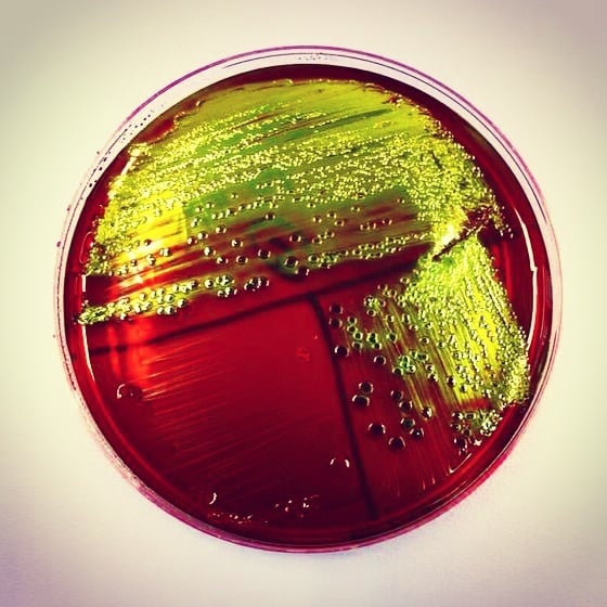 Figure: EMB agar inoculated with Escherichia coli demonstrating growth with green-metallic sheen colonies. (Image source: Carmen Moreno González)
Examine the plate in reflected light (room light shining down onto the plate surface) for metallic sheen. Then hold the plate up to a transmitted light source (light shining through from below) to see the dark centers. Missing the reflected light step is the most common cause of misread EMB plates: colonies that show metallic green sheen only in reflected light may appear simply as dark colonies in transmitted light.
Quality Control of EMB agar
Sterility testing can be performed by incubating 3 to 5 uninoculated plates from each batch at 35 to 37°C for 18-24 hours. Any growth in the media should be regarded as contamination, and the whole lot should be discarded.
Organism | Growth and colony characteristics |
E. coli ATCC 25922 | Good growth, blue-black colonies with a green metallic sheen |
Salmonella enterica subsp. enterica serovar Typhimurium ATCC 14028 | Luxuriant growth, colorless to amber colonies |
Enterococcus faecalis ATCC 29212 | Inhibition (partial) |
Shigella flexneri ATCC 12022 | Moderate to heavy growth, colorless to amber colonies |
Performance testing of prepared EMB agar plates can be done by inoculating known strains of bacteria into the medium and observing growth and colonial characteristics.
Colony Morphology on EMB Agar
Strong lactose fermenters (dark colonies with metallic sheen)
| Organism | Colony appearance | Key features |
|---|---|---|
| Escherichia coli | Small to medium (2–3 mm), dark purple-black with characteristic metallic green sheen; flat, dry, with irregular edge | Metallic green sheen is the hallmark of vigorous acid production from lactose fermentation; most reliable presumptive indicator of E. coli |
Moderate lactose fermenters (dark pink to brown-pink colonies, no sheen)
| Organism | Colony appearance | Key features |
|---|---|---|
| Klebsiella pneumoniae | Large (4–6 mm), dark pink to brown-pink, very mucoid, dome-shaped; no metallic sheen | Mucoid capsule produces glistening, dome-shaped appearance; may string when touched |
| Klebsiella aerogenes (formerly Enterobacter aerogenes) | Dark brown-pink center, large and mucoid, 3–4 mm; no metallic sheen | Ferments lactose but less vigorously than E. coli, so acid production is insufficient for the dense dye precipitate that produces the sheen |
| Klebsiella oxytoca | Similar to K. pneumoniae: large, mucoid, dark pink-brown | Distinguished from K. pneumoniae by indole positivity |
| Enterobacter cloacae | Dark pink, slightly mucoid; 2–3 mm | Smaller and less mucoid than Klebsiella; may have slightly darker center |
| Serratia marcescens | Pink-brown at 37°C; may show red/pink pigment (prodigiosin) at room temperature | Distinguished by DNase positivity and prodigiosin pigment at 25°C |
| Citrobacter freundii | Pale pink at 24 hours (late fermenter); darker at 48 hours | Late lactose fermentation, may appear NLF at 24 hours; H₂S positive on TSI |
Non-lactose fermenters (colorless or transparent colonies)
| Organism | Colony appearance | Key features |
|---|---|---|
| Salmonella Typhi | Colorless to pale pink, translucent, convex; 1–2 mm | Small, non-mucoid; no distinctive features on EMB, TSI needed for definitive direction |
| Salmonella spp. (non-typhoidal) | Colorless to pale pink, convex, smooth; 2–3 mm | Cannot distinguish from Shigella by EMB alone |
| Shigella spp. | Colorless, flat, translucent; 1–2 mm; slightly irregular edge | S. sonnei may show faint pink at 48 hours (late lactose fermenter) |
| Proteus mirabilis | Colorless, spreading; characteristic foul odor; reduced swarming on EMB compared to blood agar | Swarming is reduced on EMB, though less reliably than on MacConkey, which contains bile salts |
| Proteus vulgaris | Colorless, spreading; similar to P. mirabilis | Indole positive; distinguished biochemically |
| Pseudomonas aeruginosa | Colorless to pale, irregular, spreading; may show blue-green pigmentation (pyocyanin); characteristic grape-like odor | Non-fermenter; oxidase positive; characteristic sweet grape odor |
| Acinetobacter baumannii | Colorless to pale, opaque, convex; 1.5–2 mm | Non-fermenter; oxidase negative; important MDR nosocomial pathogen |
| Morganella morganii | Colorless, flat; 2–3 mm | Urease positive; phenylalanine deaminase positive |
Organisms inhibited: no growth on EMB agar
| Organism | Why inhibited |
|---|---|
| Staphylococcus aureus | Inhibited by eosin Y and methylene blue dyes |
| Streptococcus spp. | Inhibited by dyes |
| Enterococcus spp. | Inhibited by dyes |
| Neisseria gonorrhoeae | Fastidious gram-negative which is inhibited by dye concentration |
| Haemophilus influenzae | Fastidious, requires X and V factors not present |
| Campylobacter spp. | Fastidious, requires microaerophilic conditions and selective supplements |
Uses of EMB agar
1. Isolation and differentiation of Gram-negative enteric organisms EMB is used as a primary plating medium for clinical specimens where Gram-negative enteric organisms are expected: urine, stool, and wound specimens. It selects for Gram-negatives while simultaneously differentiating lactose fermenters (including E. coli and Klebsiella) from non-fermenters (Salmonella, Shigella).
2. Water quality testing (fecal coliform detection) EMB agar is the standard confirmatory medium for detecting fecal coliforms in drinking water, sewage, and food samples. Samples that produce metallic green sheen colonies on EMB after positive presumptive MPN (Most Probable Number) tests confirm the presence of E. coli, a definitive fecal contamination indicator.
3. Differentiation of E. coli from other coliforms Among lactose-fermenting Gram-negatives, the metallic green sheen specifically identifies E. coli (vigorous fermenter) from Enterobacter/Klebsiella (moderate fermenter, mucoid, no sheen). This distinction is important because E. coli in urine is always clinically significant, while Enterobacter may represent colonization rather than infection.
4. Colon-typhoid-dysentery group differentiation EMB is used alongside MacConkey agar for selective isolation of Salmonella and Shigella from stool specimens. The colorless colonies of these non-fermenters on EMB are picked for confirmatory TSI and serology.
5. Candida isolation (modified Levine EMB) Levine EMB with chlortetracycline (0.1 g/L added after autoclaving) suppresses bacterial flora and supports Candida albicans isolation, which produces characteristic "spidery" or "feathery" colonies under CO₂ conditions.
EMB Agar vs MacConkey Agar
Both EMB and MacConkey are selective and differential media for gram-negative enteric organisms, and they are frequently used together in a primary plating battery. Understanding their differences helps choose the right medium for each application:
| Feature | EMB Agar | MacConkey Agar |
|---|---|---|
| Selective agents | Eosin Y + methylene blue dyes | Crystal violet + bile salts |
| Sugars | Lactose only (Levine); lactose + sucrose (original formulation) | Lactose only |
| pH indicator | Eosin Y + methylene blue (change from clear to dark at low pH) | Neutral red (changes from colorless to pink at low pH) |
| E. coli appearance | Dark purple-black with metallic green sheen | Flat, dry, dark pink with bile precipitate halo |
| Klebsiella appearance | Large, dark pink-brown, very mucoid | Large, dark pink, mucoid |
| Non-fermenters | Colorless/transparent | Colorless/pale |
| Gram-positive inhibition | Yes (dyes) | Yes (crystal violet + bile) |
| Fastidious GNR inhibition | More inhibitory | Less inhibitory (no crystal violet) |
| Sucrose fermenters | Not detected on Levine EMB; detected on the original formulation | Not detected |
| Water quality testing | Preferred, E. coli metallic sheen = fecal coliform | Used but metallic sheen not produced |
| Historical use | Fecal coliform differentiation | Enteric pathogen isolation |
| FDA-BAM approved | Yes, for E. coli in food/water | Yes |
The key practical difference: MacConkey is more commonly used in clinical laboratories for routine stool and urine cultures because its neutral red indicator is more stable and the pink/colorless distinction is simpler to read.
EMB is essential in water microbiology where the metallic green sheen provides definitive identification of E. coli as a fecal indicator, and in settings where distinguishing E. coli from Klebsiella colony morphology is required.
When to use EMB over MacConkey:
- When the metallic green sheen of E. coli is specifically needed for presumptive fecal coliform identification (water testing, food microbiology)
When to use MacConkey over EMB:
- Routine clinical specimen plating: MacConkey is less inhibitory for some fastidious gram-negatives
- When observing swarming pattern of Proteus: bile salts in MacConkey better inhibit swarming
In clinical practice: Both are often inoculated simultaneously from the same specimen to maximize information: MacConkey for Enterobacteriaceae identification and Proteus assessment; EMB for presumptive E. coli confirmation by metallic sheen.
→ MacConkey Agar: Composition, Preparation, Uses and Colony Characteristics
How to Remember
EMB = dye-based selection and differentiation
Both eosin Y and methylene blue are dyes that:
- Inhibit Gram-positives (dye uptake disrupts cell membrane function)
- Act as pH indicators (combine at acid pH → dark purple precipitate → metallic green sheen when dense)
The sheen is physics, not chemistry. Remind yourself: the metallic green is light reflecting off a dense dark layer, not a green pigment. This is why it only appears in reflected light and disappears under transmitted light.
Three colony types, three clinical meanings:
| Colony on EMB | What it means | What to do next |
|---|---|---|
| Metallic green sheen | Vigorous LF, probably E. coli | Confirm with IMViC, TSI, or MALDI-TOF |
| Mucoid, dark center, no sheen | Moderate LF, probably Klebsiella/Enterobacter | Confirm with urease, indole, Voges-Proskauer |
| Colorless | Non-fermenter, possibly Salmonella or Shigella | Pick for TSI + serology immediately |
EMB vs MacConkey: EMB is for when you need to identify E. coli specifically (water testing, fecal coliform confirmation). MacConkey is for when you need a general enteric screen. In clinical practice, use MacConkey; in water microbiology, use EMB.
Where Students Get Confused
- The green sheen is physics, not a green pigment. The metallic green is light reflecting off a dense layer of precipitated dye, like the sheen on a soap bubble or oil film. It appears only in reflected light (light shining down onto the plate) and can vanish under transmitted light, where the same colony looks simply dark. Reading only in transmitted light is the most common way the sheen is missed.
- Dark colony does not always mean sheen-positive. Klebsiella and Enterobacter ferment lactose enough to make dark pink-brown colonies, but not vigorously enough for the dense dye layer that produces the sheen. Dark and mucoid without sheen points to Klebsiella; dark with a metallic green sheen points to E. coli.
- Colorless does not distinguish Salmonella from Shigella. Both are non-fermenters and both are colorless on EMB. EMB narrows them to the non-fermenter group but cannot separate them. Pick colorless colonies for TSI and serology.
- Late fermenters can read colorless at 24 hours. Citrobacter freundii and Shigella sonnei may look pale at 24 hours and darken by 48 hours. Confirm a non-fermenter appearance with a 48-hour read before reporting it.
- A sheen on the original EMB is not the same as on Levine EMB. On the older sucrose-containing formulation, a sucrose fermenter could also produce a sheen, which is a false positive for E. coli. Levine EMB (lactose only) is used precisely to avoid this. Know which formulation your laboratory uses.
References and further readings
- Tille PM. Bailey & Scott's Diagnostic Microbiology. 15th ed. St. Louis: Elsevier; 2022.
- Leber AL, editor. Clinical Microbiology Procedures Handbook. 4th ed. Washington, DC: ASM Press; 2016.
- Procop GW, Koneman EW. Koneman's Color Atlas and Textbook of Diagnostic Microbiology. 7th ed. Wolters Kluwer; 2017.
- US Food and Drug Administration. Bacteriological Analytical Manual (BAM). FDA; 2023.
- Holt-Harris JE, Teague O. A new culture medium for the isolation of Bacillus typhosus from stools. J Infect Dis. 1916;18(5):596–600.
- Cheesbrough M. District Laboratory Practice in Tropical Countries, Part 2. 2nd ed. Cambridge University Press; 2006.
Frequently Asked Questions
Why does E. coli produce a metallic green sheen on EMB agar?
Why does E. coli produce a metallic green sheen on EMB agar?
On the Levine EMB formulation used in most laboratories today, E. coli vigorously ferments lactose (the only sugar present), producing enough acid to sharply lower the pH at the colony surface. At this low pH, eosin Y and methylene blue precipitate in large quantities onto the colony. The densely packed dye crystals produce an iridescent metallic sheen, similar to a soap bubble, visible as green under reflected light. It is a direct indicator of vigorous acid production from lactose fermentation.
What is the difference between EMB and MacConkey agar?
EMB (Levine): eosin Y + methylene blue dyes, contains lactose only, E. coli shows metallic green sheen. The original Holt-Harris-Teague EMB also contained sucrose, but Levine EMB is standard today. MacConkey: crystal violet + bile salts, contains lactose only, E. coli shows dark pink with bile precipitate halo.
Why does Klebsiella produce mucoid colonies on EMB agar?
Why does Klebsiella produce mucoid colonies on EMB agar?
Klebsiella produces a thick polysaccharide capsule, using some fermented carbohydrate for capsule synthesis rather than acid production. Results in large, moist, dome-shaped mucoid colonies. On EMB they appear dark pink-brown and mucoid but without the metallic green sheen characteristic of E. coli.
Can gram-positive bacteria grow on EMB agar?
No, eosin Y and methylene blue dyes are toxic to gram-positive organisms, penetrating gram-positive cell walls and disrupting membrane function. Most Staphylococcus, Streptococcus, and Enterococcus species are completely inhibited. EMB is not suitable when gram-positive pathogens are suspected.
What is the significance of EMB agar in water quality testing?
EMB is FDA-BAM approved for E. coli detection in food and water. The metallic green sheen provides rapid, reliable presumptive fecal coliform identification. In the membrane filter technique, a measured water volume is filtered and the membrane placed on EMB, apperance of metallic green sheen colonies at 24–48 hours are counted as presumptive fecal coliforms.
Why are Salmonella and Shigella colorless on EMB agar?
Why are Salmonella and Shigella colorless on EMB agar?
Both are non-lactose fermenters. Without acid production, the pH does not fall and dyes remain soluble rather than precipitating onto colonies. Colorless colonies on EMB suggest non-lactose fermenters but cannot distinguish Salmonella/Shigella from Proteus or Pseudomonas, for which TSI and specific antisera required.
What does a dark pink colony without metallic sheen indicate on EMB?
Moderate lactose or sucrose fermentation, enough acid for dye precipitation but not enough for metallic sheen. Most typically Klebsiella pneumoniae (large, mucoid, dark pink-brown) and Enterobacter species. E. coli (metallic sheen) vs Klebsiella (dark, no sheen) is one of the most useful differential observations on EMB.

Tankeshwar Acharya, MSc (Medical Microbiology)
Tankeshwar Acharya is an Assistant Professor in the Department of Microbiology at Patan Academy of Health Sciences (PAHS), Nepal, where he has been teaching and practicing clinical microbiology for over 14 years. He is the founder of Microbe Online, one of the leading free microbiology education resources on the web, covering bacteriology, mycology, parasitology, immunology, and clinical laboratory diagnostics written from direct experience in both the classroom and the diagnostic laboratory.
Comments
No comments yet. Be the first to share your thoughts.
Leave a comment
All comments are reviewed before they appear.