
Streptococcus pneumoniae: Properties, Pathogenesis, and Diagnosis
Streptococcus pneumoniae: lancet-shaped diplococci, capsule-driven pathogenesis, alpha-hemolysis, optochin sensitivity and bile solubility for lab diagnosis.

On this page
A previously healthy 68-year-old man is recovering from a week of influenza when he suddenly worsens: high fever, shaking chills, sharp pleuritic chest pain, and a productive cough bringing up rust-colored sputum.
A chest film shows dense consolidation of one lower lobe. A Gram stain of the sputum settles the direction of the whole workup in a single field: lancet-shaped, Gram-positive diplococci. The story of how this organism moved from a harmless resident of his nose to the cause of lobar pneumonia, and how the laboratory confirms it, is the subject of this article.
Streptococcus pneumoniae (pneumococci) is a part of the normal nasopharyngeal and oropharyngeal flora. It is an important etiological agent of upper and lower respiratory tract infections (URTI and LRTI), bacteremia, and septicemia. Streptococcus pneumoniae is also associated with otitis media, sinusitis, meningitis, and endocarditis.
Characteristics of Streptococcus pneumoniae
Streptococcus pneumoniae is a Gram-positive coccus. It retains crystal violet and appears purple on Gram stain. The cells are lancet-shaped (pointed, like the tip of a spear) and typically lie in pairs, base to base, as diplococci. They may also occur singly or in short chains. Each coccus is roughly 0.5 to 1.25 micrometers across.

Key features at a glance:
- Gram-positive, lancet-shaped diplococci
- Non-motile and non-spore-forming
- Encapsulated (a polysaccharide capsule surrounds the cell; this is the single most important feature for both virulence and diagnosis)
- Catalase-negative and oxidase-negative
- Alpha-hemolytic on blood agar
- Fastidious and capnophilic (grows best with added CO₂)
The lancet shape and the paired arrangement are the first clues on a direct Gram stain of sputum or CSF, and they are worth committing to memory: two flame-shaped cells pointed at each other.
Diseases caused by Streptococcus pneumoniae
Streptococcus pneumoniae lives quietly in the nasopharynx of healthy carriers. Almost all the disease it causes can be understood as the organism spreading from that home base in one of two directions: outward into the air-filled spaces next door (local, contiguous spread), or inward into the bloodstream and the sites it seeds from there (invasive spread). The polysaccharide capsule is what makes the invasive route possible, because it resists phagocytosis and lets the organism survive in blood.
Local (contiguous) spread from the nasopharynx
- Otitis media: the most common bacterial cause of middle ear infection in children. The reason it is a childhood disease is anatomical: a child's Eustachian tube is short and horizontal, so organisms pass easily from the nasopharynx into the middle ear. As the tube lengthens and angles down with age, this route becomes harder.
- Sinusitis: spread into the paranasal sinuses, by the same contiguous route.
- Bronchitis: downward spread into the airways, often after a viral upper respiratory infection has damaged the mucosa.
Lower respiratory tract: pneumonia
Streptococcus pneumoniae is the most common bacterial cause of community-acquired pneumonia, classically a lobar pneumonia in infants and the elderly, and often following a viral infection such as influenza that has injured the respiratory lining.
In the alveoli, pneumococci trigger an outpouring of fluid, red cells, and white cells. The red cells breaking down in that exudate give the classic rust-colored (blood-tinged) sputum.
Invasive disease: bacteremia and meningitis
Once the capsule lets the organism into the bloodstream, it can seed distant sites.
- Bacteremia: pneumococcus in the blood, which can arise from pneumonia or from an apparently minor focus, and which is the gateway to the sites below.
- Meningitis: S. pneumoniae is one of the most common causes of bacterial meningitis (alongside Haemophilus influenzae and Neisseria meningitidis). The cerebrospinal fluid typically shows a very high white cell count, high protein, and low glucose. Pneumococcal meningitis is serious and carries a high rate of complications.
- Other invasive sites: endocarditis, septic arthritis, and peritonitis can follow bloodstream spread.
Who is at high risk, and why
The same capsule that drives invasive disease also explains who suffers most from it. Clearing encapsulated organisms from the blood depends heavily on the spleen and on opsonization by complement and antibody.
People without a working spleen, whether from surgical removal (asplenia) or from functional loss as in sickle cell disease, cannot clear encapsulated pneumococci efficiently and are at high risk of overwhelming pneumococcal bacteremia and sepsis.
This is the direct clinical reason pneumococcal vaccination is strongly recommended for asplenic and sickle cell patients: the defense they have lost is exactly the one this organism's capsule is built to defeat.
Virulence Factors
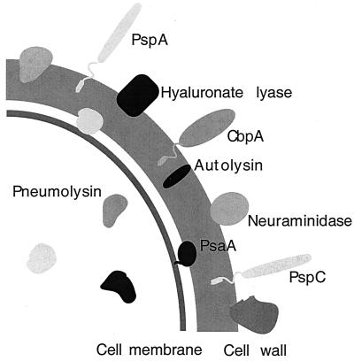 Figure: Schematic diagram of the virulence factors of Streptococcus pneumoniae
Four things have to happen, in roughly this order, for pneumococcus to go from harmless nasal colonizer to the organism causing meningitis: it sticks, it sneaks past the mucosal guard, it hides, and when conditions are right, it self-destructs to attack.
 - Teichoic acid anchors the organism to respiratory and endothelial epithelium. Clinical link: this is the first step in colonization, no adherence, no infection.
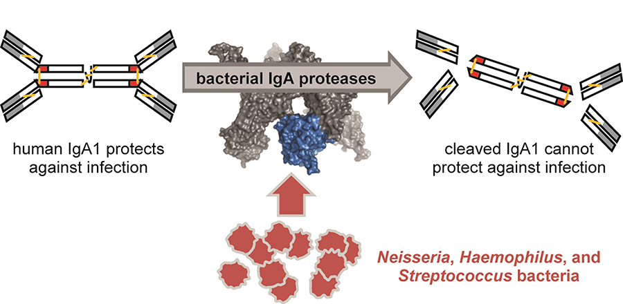
- IgA1 protease cleaves IgA1, the antibody that normally patrols and protects mucosal surfaces. Clinical link: this is specifically why pneumococcus, along with H. influenzae and N. meningitidis, can establish itself on a mucosal surface that should otherwise be defended.
- Polysaccharide capsule, the single most important virulence factor here. It blocks complement-mediated opsonization and phagocytosis, and physically resists mechanical clearance by mucus. Non-encapsulated mutants lose virulence almost entirely in animal models. Clinical link: more than 93 capsular serotypes exist, and the pneumococcal vaccines are built directly around this capsule, immunity is type-specific because the capsule itself is the target.
- Autolysin (LytA), a peptidoglycan-degrading enzyme that triggers the bacterium's own stationary-phase lysis. Clinical link: this is the mechanism, not a side effect, autolysin is what releases pneumolysin into surrounding tissue. It's also why old cultures show collapsed, flattened colonies (draughtsman colonies) by 24-48 hours.
- Pneumolysin, a pore-forming toxin that targets any host cell membrane containing cholesterol, essentially all human cells. It damages respiratory epithelium, inhibits the neutrophil respiratory burst, and interferes with classical complement fixation. Clinical link: this is the direct cause of tissue damage in pneumonia and meningitis, and the reason damage isn't confined to one tissue type.
- Other contributors: pneumococcal surface protein A (PspA, blocks complement), neuraminidase (aids colonization by acting on host glycans), peptidoglycan/teichoic acid fragments (highly inflammatory in the CNS, contributing to the damage seen in meningitis even after bacteria are cleared).
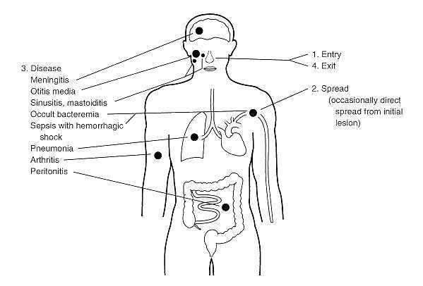 Figure: Types of disease caused by Streptococcus pneumoniae
How to Remember the Virulence Factors
Think of it as four steps: Stick, Sneak, Hide, Self-destruct.
Stick. Teichoic acid is Velcro, it gets the organism attached to the respiratory lining before anything else can happen.
Sneak. IgA1 protease is a key cut specifically for one lock. IgA1 is the guard posted at every mucosal surface in your body, and this enzyme is shaped to disable exactly that guard and no other antibody class. That specificity is the giveaway that this is a deliberately evolved mechanism, not incidental damage.
Hide. The capsule is an invisibility cloak. Strip it off in the lab and the organism becomes almost harmless, which tells you the capsule isn't just one feature among many, it's the main reason this organism survives contact with your immune system at all.
Self-destruct. This is the one students remember once they hear it right: autolysin is a soldier pulling the pin on a grenade. The bacterium triggers its own cell wall breakdown, and the explosion releases pneumolysin into the surrounding tissue. The bacterium doesn't survive that moment, but the toxin it released does the damage. This single mechanism explains two separate observations in the lab and the clinic at once: why old pneumococcal colonies collapse into a flattened "draughtsman" shape (the autolysin has fired), and why pneumonia and meningitis both involve direct tissue destruction rather than just inflammation (the pneumolysin that autolysin released is doing it).
One more hook that costs nothing: pneumolysin needs cholesterol to punch its pore. Nearly every human cell membrane has cholesterol. That's the whole reason this one toxin causes damage in lung, meninges, and elsewhere rather than being tissue-specific.
Reservoirs: Harmlessly inhabit the upper respiratory tract of humans.
Transmission: Respiratory droplets are not considered highly communicable. Often colonizes without causing infection.
Growth Requirements
Streptococcus pneumoniae is a fastidious organism: it will not grow on plain nutrient agar and needs an enriched medium. It is also capnophilic, meaning it grows best in an atmosphere with added carbon dioxide (5 to 10% CO₂, or a candle jar in resource-limited settings).
- Atmosphere: facultatively anaerobic, but recovery is far better with 5 to 10% CO₂. It is not a strict anaerobe and not an obligate aerobe.
- Temperature: optimum 35 to 37°C.
- Media: grows on enriched media such as blood agar and chocolate agar. On blood agar it shows alpha-hemolysis; on chocolate agar it grows as small grey mucoid colonies, but the alpha-hemolysis is not visible because the red cells have already been lysed to make the medium. To see the hemolysis, read the blood agar plate.
For the media themselves, see the dedicated guides on blood agar, chocolate agar, and types of bacteriological culture media.
Laboratory diagnosis
Laboratory diagnosis of Streptococcus pneumoniae infection is based on finding characteristics shape of the organism in the sample, characteristic colony morphology, biochemical reactions, susceptibility to certain diagnostic discs, and latex agglutination test.
Sample
Specimens used for the laboratory diagnosis of Streptococcus pneumoniae maybe one or more of the followings depending on the clinical case
- Specimens from respiratory tract: Sputum, lung aspirate, pleural fluid
- Body fluids, e.g., Blood/ cerebrospinal fluid
- Exudates from the joint, middle ear, and other sites
Sample Collection
- Sputum
Collect > 1.0 ml expectorated sputum in a sterile screw-capped container. - Lung aspirate/ pleural fluid
Collect > 1.0 ml by percutaneous needle aspiration in a sterile screw-capped tube. - Blood
Clean the venipuncture site with 70% alcohol and iodine, allow it to evaporate, and collect blood aseptically in a culture broth with an anticoagulant. In case of adults, collect 5-10 ml of blood in culture bottle. For children < 12-year-old, collect 1.5-2.0 ml of blood. Mix the blood and broth by rotating gently to avoid clotting. - Cerebrospinal fluid (CSF)
Clean the skin over L3-L4 inter-space with 70% alcohol and iodine. Collect > 1.0 ml CSF in a sterile screw-capped tube. Keep the CSF in an incubator at 35-37 degree centigrade, if not processed immediately. - Exudates from joints/middle ear
Collect > 1.0 ml by aspiration in a sterile screw-capped tube or add directly to a culture broth used for blood culture.
Note: DO NOT REFRIGERATE THE SAMPLE
Sample Transport
Streptococcus pneumoniae is a fastidious bacteria. Care must be taken during the transport of specimens. Specimens must be transported promptly to the laboratory within 1-2 hours.
Blood can only be transported after collecting in a culture broth containing appropriate anticoagulant. The inoculated medium can be held at room temperature (20°C– 25°C) for 4 – 6 hours before incubation at 37°C. The samples during transportation should be protected from extremes of temperature (less than 18°C, more than 30°C) and direct sunlight.
Culture and Identification
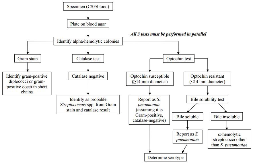 Figure: Flow chart for identification and characterization of a S. pneumoniae isolate
- Perform Gram staining of the sample (sputum/CSF)
- Gram staining shows Gram-positive, lanceolate-shaped diplococci, often in pairs. For the staining method itself, see Gram staining.
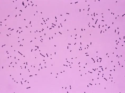 Figure: Gram positive diplococci: Streptococcus spp
Culture and Sensitivity
- Inoculate sample onto blood agar and chocolate agar plate.
- Incubate at 37°C with 5-10% CO₂ for 24 – 48 hours.
Colony morphology
- Colonies on blood agar plate are small (0.5 mm), round, translucent, or mucoid with alpha-hemolysis (A green discoloration of the agar around the colonies). Alpha-hemolytic property differentiates S. pneumoniae from many species, but not from the commensal alpha-hemolytic viridans streptococci.
- Young alpha-hemolytic colonies appear raised, and in 24 – 48 hours, colonies are flattened with a depressed center called the draughtsman colony. It is due to partial autolysis (these colonies are tentatively identified as Pneumococci).
- Streptococcus viridans also produces alpha-hemolytic colonies but does not produce draughtsman colony.
Colony characteristics: Alpha hemolytic (partial discoloration around the bacterial colony in blood agar), mucoid colony (if organism possesses polysaccharide capsule).
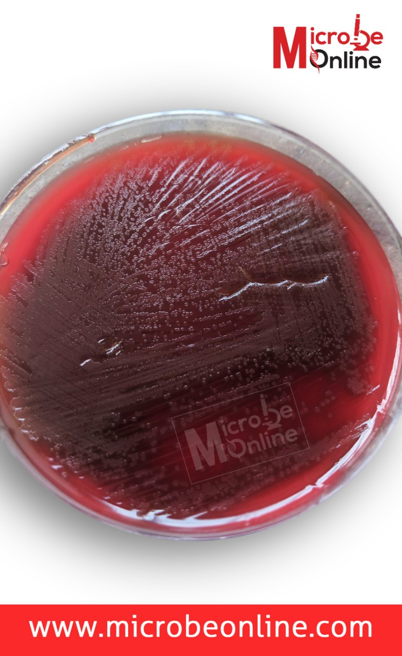 Figure: Draughtsman colonies of S. pneumoniae
The following confirmatory tests further identify alpha-hemolytic colonies.
Rapid test (catalase test): negative (no bubbling when colonies are emulsified in hydrogen peroxide). This separates streptococci from staphylococci, which are catalase-positive; it does not by itself identify pneumococcus. See Catalase test
Optochin test
Principle in one line: optochin (ethylhydrocupreine hydrochloride) inhibits S. pneumoniae at low concentrations by targeting its ATP synthase, while viridans streptococci are unaffected.
Expected result for pneumococcus: sensitive (zone of inhibition ≥14 mm with a 5 µg disk). Any zone under 14 mm, including no zone, is equivocal and must be confirmed by bile solubility, because some genuine pneumococci are optochin-resistant.
Full procedure, the 14 mm cutoff, resistance mechanism, and where the test fits among the Gram-positive disk tests: Optochin Sensitivity Test
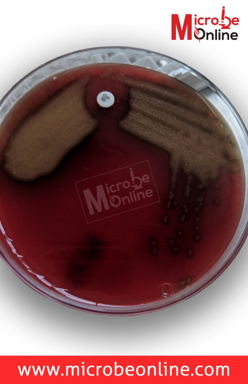 Figure: Alpha hemolysis in Blood Agar by Streptococcus pneumoniae, Observe the zone of inhibition around Optochin disk.
Bile solubility test
Bile salts (sodium deoxycholate) activate the pneumococcus's own autolysin, the same self-destruct enzyme described in the Virulence section above. This test doesn't use some separate lab trick, it simply triggers the organism's own lysis mechanism directly in a tube.
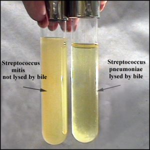 Figure: Bile solubility test: Streptococcus pneumoniae colonies are lysed by bile
Interpretation: clearing or loss of turbidity = soluble = positive = confirms S. pneumoniae. Persistent turbidity = insoluble = negative, organism is not S. pneumoniae, most likely viridans streptococci.
Read more: Full procedure: Bile Solubility Test
Biochemical Tests for S. pneumoniae
Name of the test | Result |
Catalase Test | Negative |
Optochin sensitivity test | Sensitive |
Bile solubility test | Soluble |
Hemolysis | Alpha-hemolysis |
Oxidase Test | Negative |
O/F Test | Fermentative |
Motility | Non-motile |
Detection of the antigen
C-carbohydrate antigen of the Streptococcus pneumoniae can be detected in the urine for the diagnosis of pneumonia and in CSF for the diagnosis of pneumococcal meningitis.
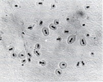 Figure: Streptococcus pneumoniae shows a quellung positive reaction.
Quellung Reaction: Positive
When Streptococcus pneumoniae of certain types are mixed with specific antipolysaccharide serum of the same type or with polyvalent antiserum on a microscope slide, the capsule swells markedly, and the organism agglutinates by cross-linking of the antibodies.
Where students get confused
- Any optochin zone under 14mm is not a negative result, it's an unfinished test. This is the single most common error. Whether the zone is 10mm, 5mm, or completely absent, the next step is identical: bile solubility. Don't try to read meaning into the exact sub-14mm size, treat all of them the same way until bile solubility settles it.
- Viridans streptococci will trip up an optochin-only workflow. Both are alpha-hemolytic on Blood Agar, and both can look superficially similar on Gram stain. The real differentiators are the draughtsman colony (pneumococcus only), capsule presence, optochin sensitivity, and bile solubility, not hemolysis pattern alone, since viridans is also alpha-hemolytic.
- Bile solubility and bile esculin are not the same test. Bile solubility lyses pneumococcus colonies in the presence of bile salts and confirms S. pneumoniae. Bile esculin tests for esculin hydrolysis in the presence of bile and is used for Enterococcus and Group D Streptococcus. Same word "bile," completely different organisms and completely different mechanisms.
- Catalase negative only rules out Staphylococcus, it doesn't confirm pneumococcus. All streptococci, including the harmless viridans group, are catalase-negative. Catalase tells you "not Staph," nothing more.
Antimicrobial Susceptibility
- Perform antimicrobial susceptibility test against a selected group of antimicrobials by a disk-diffusion method
- Reporting of results: Streptococcus pneumoniae isolated and resistance patterns with tested antibiotics
Initially, penicillin was the drug of choice to treat S. pneumoniae infections as all S. pneumoniae isolates were exquisitely susceptible to penicillin. Still, now drug-resistant S. pneumoniae (DRSP) poses a serious threat to global health as we are observing an alarming worldwide increase in the incidence.
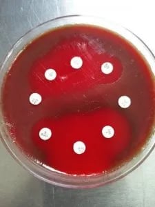 Figure: Antimicrobial sensitivity testing of Streptococcus pneumoniae
Key exam facts
One mnemonic ties the lab identification together. Streptococcus pneumoniae is the BOSS of community-acquired pneumonia: alpha-hemolytic on Blood agar, Bile Soluble, Optochin Sensitive. And even a BOSS does not show up unprotected: it wears a sugar-coated polysaccharide capsule as armor against the immune system, which is why it has more than 90 serotypes and why the vaccines target exactly that coat.
| Feature | S. pneumoniae | Memory hook |
|---|---|---|
| Gram stain | Lancet-shaped diplococci | Two flames pointed at each other |
| Gram reaction | Gram-positive | Purple, retains crystal violet |
| Catalase | Negative | Rules out Staphylococcus, nothing more |
| Optochin | Sensitive (zone ≥14 mm) | The "O/S" in BOSS |
| Bile solubility | Soluble | The "B" in BOSS; bile triggers the organism's own autolysin |
| Hemolysis | Alpha (on blood agar) | Same as viridans, so not a differentiator on its own |
| Atmosphere | Facultative anaerobe, capnophilic | Grows best with 5% CO₂ |
| Colony at 24–48 h | Draughtsman (flattened, depressed center) | Autolysin has fired, the soldier pulled the pin |
| Main virulence factor | Polysaccharide capsule | Remove it and virulence collapses |
| Tissue-damage toxin | Pneumolysin | Needs cholesterol, so it hits nearly any human cell |
| CSF in meningitis | High WBC, low glucose, high protein | Bacteria consume glucose; neutrophils pour in |
| Otitis media in children | Short horizontal Eustachian tube | Anatomy, not just "common in kids" |
| High-risk hosts | Asplenia, sickle cell disease | Capsule defeats the spleen-dependent clearance they've lost |
Frequently Asked Questions
Why is Streptococcus pneumoniae alpha-hemolytic like viridans streptococci, yet far more dangerous?
Why is Streptococcus pneumoniae alpha-hemolytic like viridans streptococci, yet far more dangerous?
Hemolysis pattern alone doesn't reflect virulence. S. pneumoniae's danger comes from its polysaccharide capsule (the major anti-phagocytic virulence factor) and pneumolysin, a pore-forming toxin that damages nearly any host cell containing cholesterol. Viridans streptococci lack a capsule and are far less invasive as a result.
What is the difference between bile solubility and bile esculin tests?
Bile solubility uses bile salts to lyse S. pneumoniae colonies by triggering the organism's own autolysin, confirming pneumococcus. Bile esculin tests whether an organism can hydrolyze esculin in the presence of bile, used to identify Enterococcus and Group D streptococci. Same word "bile," completely different organisms and mechanisms.
Why does the pneumococcal vaccine need to cover so many different serotypes?
S. pneumoniae has more than 90 distinct capsular serotypes, and immunity to the capsule is type-specific. Antibodies raised against one serotype's capsule don't protect against a different serotype, so vaccines must include multiple capsular polysaccharides to provide broad coverage.
Can Streptococcus pneumoniae be part of normal flora without causing disease?
Can Streptococcus pneumoniae be part of normal flora without causing disease?
Yes. S. pneumoniae commonly colonizes the upper respiratory tract harmlessly. Disease occurs when the organism spreads beyond its normal niche, such as into the lungs, bloodstream, or meninges.
Why does CSF show low glucose in pneumococcal meningitis?
Is Streptococcus pneumoniae aerobic or anaerobic?
Is Streptococcus pneumoniae aerobic or anaerobic?
It is a facultative anaerobe, so it can grow with or without oxygen, but in the laboratory it grows best in an atmosphere enriched with 5 to 10% carbon dioxide (it is capnophilic). It is neither an obligate aerobe nor an obligate anaerobe.
Why is the sputum rust-colored in pneumococcal pneumonia?
Why is the sputum rust-colored in pneumococcal pneumonia?
The rust or brick-red color comes from red blood cells that leak into the alveoli during infection and are broken down. Pneumococci multiplying in the alveoli trigger an outpouring of fluid and inflammatory cells, and the altered blood gives the sputum its characteristic rusty tint.
What is a draughtsman colony?
What is a draughtsman colony?
It is the flattened, coin-like colony with a depressed center that pneumococcus forms by 24 to 48 hours. It is caused by the organism's own autolysin partially breaking down the colony from the center outward. It is not a different organism; it is simply an older pneumococcal colony.
Why does the capsule matter so much in Streptococcus pneumoniae?
Why does the capsule matter so much in Streptococcus pneumoniae?
The polysaccharide capsule blocks complement-mediated opsonization and phagocytosis, letting the organism survive in the bloodstream and tissues. Non-encapsulated strains lose almost all their virulence. Because immunity is directed against the capsule and there are more than 90 capsular types, the pneumococcal vaccines are built around capsular polysaccharides.
Why are people without a spleen at high risk from Streptococcus pneumoniae?
Why are people without a spleen at high risk from Streptococcus pneumoniae?
The spleen is central to clearing encapsulated bacteria from the blood. Pneumococcus's polysaccharide capsule resists phagocytosis, so removing that organism depends on splenic filtering and opsonization. People without a functioning spleen, after surgical removal or from sickle cell disease, lose that defense and can develop overwhelming pneumococcal bacteremia and sepsis. This is why pneumococcal vaccination is strongly recommended for them.
References
- Bogaert, D., De Groot, R., & Hermans, P. W. (2004). Streptococcus pneumoniae colonisation: the key to pneumococcal disease. The Lancet Infectious Diseases, 4(3), 144-154. https://doi.org/10.1016/S1473-3099(04)00938-7
- Mitchell, A. M., & Mitchell, T. J. (2010). Streptococcus pneumoniae: virulence factors and variation. Clinical Microbiology and Infection, 16(5), 411-418. https://doi.org/10.1111/j.1469-0691.2010.03183.x
- O'Brien, K. L., Wolfson, L. J., Watt, J. P., et al. (2009). Burden of disease caused by Streptococcus pneumoniae in children younger than 5 years: global estimates. The Lancet, 374(9693), 893-902. https://doi.org/10.1016/S0140-6736(09)61204-6
- Jacobs, M. R. (2004). Streptococcus pneumoniae: epidemiology and patterns of resistance. The American Journal of Medicine, 117(Suppl 3A), 3S-15S. https://doi.org/10.1016/j.amjmed.2004.07.003
- Jedrzejas, M. J. (2001). Pneumococcal virulence factors: structure and function. Microbiology and Molecular Biology Reviews, 65(2), 187-207. https://doi.org/10.1128/MMBR.65.2.187-207.2001
- Mellroth, P., Daniels, R., Eberhardt, A., et al. (2012). LytA, major autolysin of Streptococcus pneumoniae, requires access to nascent peptidoglycan. Journal of Biological Chemistry, 287(14), 11018-11029. https://pmc.ncbi.nlm.nih.gov/articles/PMC3322828/
- Hyams, C., Camberlein, E., Cohen, J. M., Bax, K., & Brown, J. S. (2010). The Streptococcus pneumoniae capsule inhibits complement activity and neutrophil phagocytosis by multiple mechanisms. Infection and Immunity, 78(2), 704-715. https://doi.org/10.1128/IAI.00881-09

Tankeshwar Acharya, MSc (Medical Microbiology)
Tankeshwar Acharya is an Assistant Professor in the Department of Microbiology at Patan Academy of Health Sciences (PAHS), Nepal, where he has been teaching and practicing clinical microbiology for over 14 years. He is the founder of Microbe Online, one of the leading free microbiology education resources on the web, covering bacteriology, mycology, parasitology, immunology, and clinical laboratory diagnostics written from direct experience in both the classroom and the diagnostic laboratory.
Comments
No comments yet. Be the first to share your thoughts.
Leave a comment
All comments are reviewed before they appear.