
Quellung Reaction: Principle, Procedure, Serotyping, and Clinical Applications

On this page
A 68-year-old man is admitted with community-acquired pneumonia and bacteremia. Blood cultures grow gram-positive lancet-shaped diplococci, confirmed as Streptococcus pneumoniae. The reference laboratory wants to know the serotype: is this a vaccine-preventable serotype covered by PCV13? Was this a vaccine failure? Is there a community cluster of the same serotype?
The Quellung reaction answers all of these questions. By mixing the isolate with type-specific antisera and observing capsular swelling under the microscope, the exact serotype can be determined, one of over 90 different capsular types. The result determines whether the patient's infection should have been prevented by available vaccines and contributes to national surveillance data on pneumococcal serotype distribution.
The Quellung reaction, described by Friedrich Neufeld in 1902, remains the reference standard for pneumococcal serotyping more than a century after its discovery.
The Quellung (or Neufeld) reaction is the gold standard technique for serotyping Streptococcus pneumoniae (pneumococcus). This microscopic “precipitin test” can be used to identify pneumococci or to determine the capsular serotype of individual pneumococcal isolates.
There are over 90 different capsular serotypes of S. pneumoniae. This technique utilizes a high-quality microscope and specific pneumococcal antisera (commercially available as pooled, group, or serotype-specific) and is commonly used in reference and research laboratories worldwide.
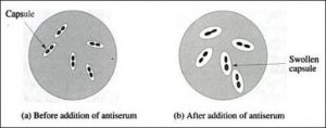 Figure: Swollen pneumococci capsule
The Quellung reaction (swelling of the capsule) is reasonably simple to perform and can be applied wherever a suitable microscope and antisera are available.
This method involves testing a pneumococcal cell suspension with pooled and specific antisera directed against the capsular polysaccharide. The antigen-antibody reactions are observed microscopically. A positive quellung reaction is the result of the binding of the capsular polysaccharide of pneumococci with type-specific antibodies contained in the typing antiserum.
The protocol has three main steps:
- preparation of a bacterial cell suspension,
- mixing of cells and antisera on a glass slide, and reading the Quellung reaction using a microscope.
It is recommended to initially test with pooled antisera in succession until a positive reaction is observed. Typing should then proceed by testing with the individual group and serotype-specific antisera included in the antisera pool that gave a positive reaction to determine the serogroup and serotype.
Some strains of H. influenzae produce a polysaccharide capsule, which is demonstrable by capsule stains and a Quellung reaction with type-specific antisera.
Principle
Anticapsular antibodies present in the serum react with the carbohydrate material of the pneumococcal capsule, causing a microprecipitin reaction on the surface of the Streptococcus pneumoniae. This antigen-antibody reaction causes a change in the refractive index of the capsule so that it appears “swollen” and more visible.
After the addition of a counterstain (methylene blue), the pneumococcal cells stain dark blue and are surrounded by a sharply demarcated halo which represents the outer edge of the capsule. The light transmitted through the capsule appears brighter than either the pneumococcal cell or the background. Single cells, pairs, chains, and even clumps of cells may have positive quellung reactions.
Why the capsule appears to "swell": The dominant explanation is that the swelling is largely an optical effect, not true physical enlargement. When type-specific antibody binds to the capsular polysaccharide, it forms a microprecipitin layer on the capsule surface that changes the capsule's refractive index, thus making it refract light differently from the surrounding medium. The capsule becomes sharply demarcated and brightly visible as a halo around the dark blue cell body. The name Quellung (German: swelling) describes the visual appearance, not the underlying mechanism.
Procedure of the quellung reaction
A. Preparation of a bacterial cell suspension
- Grow the isolate(s) to be tested for 18-24 hours on a blood agar plate (BAP) at 35-37°C with ~5% CO2 (or in a candle-jar).
- From overnight growth on the BAP, use a sterile loop to prepare a light to moderate cell suspension (approximately equal to a 5.0 McFarland density standard) in 0.5 ml of 0.85% saline.
Optimum quellung reactions can be observed when there are 25-50 cells visible in a microscopic field at 1000X magnification.
B. Mixing of cells and antisera on a glass slide
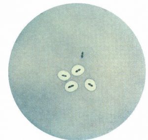 Figure: Result of Quellung Reaction
- Dispense equal amounts of antiserum (5 µl) and methylene blue (5 µl) onto a microscope slide.
- Add approximately 0.2-1.0 µl of the diluted cell suspension and mix all three with a pipette tip.
- Cover the suspension with a 22 mm square cover-slip and incubate at room temperature (25°C) for 10-15 minutes.
- Do not allow the fluid on the slide to dry.
C. Reading the Quellung reaction using a microscope
- Examine the slide at 1000X using an oil immersion lens.
- Begin testing with pooled antisera. Once a positive reaction is obtained, proceed with individual group and serotype-specific antisera included in the pooled antisera that gave the positive reaction to determine the serogroup and serotype.
Results
- A positive quellung reaction is observed when the capsule appears as a sharply demarcated halo around the dark blue stained cell
- A negative quellung reaction is observed when there is no appearance of a clear, enlarged halo surrounding the stained cell.
The Pooled Antiserum Hierarchy: How Serotyping Works
S. pneumoniae has over 90 capsular serotypes, which are organized into serogroups (based on structural similarity) and individual serotypes within each group. Reference laboratories use a hierarchical panel of antisera to determine the exact serotype efficiently rather than testing 90+ individual antisera on every isolate.
The standard approach uses antisera from Statens Serum Institut (SSI, Denmark), the global reference laboratory for pneumococcal typing:
Step 1. Omniserum (pool of all serotypes): Test with omniserum first. This contains antibodies against all known pneumococcal capsular types. A positive reaction confirms the isolate is pneumococcal and is encapsulated. A negative omniserum reaction excludes pneumococcus (or indicates a very rare untypeable strain).
Step 2. Pool sera (A–O or similar groupings): Several pool sera are available, each containing antibodies against a subset of serotypes. Test the isolate against each pool until a positive reaction is obtained. This narrows the serotype to one pool's subset.
Step 3. Group sera: Within the positive pool, test with group-specific sera to identify the serogroup (e.g., group 19 contains serotypes 19A, 19B, 19C, 19F).
Step 4. Type-specific sera: Finally, test with individual type-specific sera within the positive group to determine the exact serotype (e.g., 19F vs 19A, clinically important because 19A is a common multidrug-resistant serotype not covered by earlier PCV7 vaccines).
Practical note: This hierarchical approach typically requires 10–15 individual antiserum tests per isolate rather than 90+, making routine typing feasible in a reference laboratory.
Clinical and Epidemiological Applications
Pneumococcal Serotyping for Vaccine Surveillance
The primary contemporary use of the Quellung reaction is determining whether pneumococcal disease strains are covered by available vaccines:
- PCV7 (7-valent, older): Serotypes 4, 6B, 9V, 14, 18C, 19F, 23F
- PCV13 (13-valent, widely used): PCV7 serotypes + 1, 3, 5, 6A, 7F, 19A
- PCV15/PCV20 (newer): Extended coverage including additional serotypes
When a vaccinated child develops invasive pneumococcal disease, Quellung serotyping determines whether this is:
- A vaccine failure (infection with a covered serotype despite vaccination)
- Serotype replacement (infection with a non-covered serotype, increasingly common post-vaccination)
- Vaccine escape (rare: serotype switching)
This surveillance data drives national immunization policy decisions.
Outbreak Investigation
When clusters of pneumococcal disease occur: in care homes, hospitals, schools, serotyping by Quellung confirms whether isolates from different patients are the same serotype, supporting or refuting a common source outbreak.
Direct CSF Testing (Bedside Application)
The Quellung reaction can be performed directly on CSF in suspected pneumococcal meningitis:
- Mix a drop of CSF with pneumococcal omniserum and methylene blue
- Examine under oil immersion
- A positive reaction provides presumptive pneumococcal identification within minutes
Sensitivity limitation: Direct CSF testing is less sensitive than culture-based Quellung because CSF may contain few organisms. A negative direct Quellung does not exclude pneumococcal meningitis.
Haemophilus influenzae Typing
Type b H. influenzae (Hib) is the cause of meningitis and epiglottitis in unvaccinated children. It also has a polysaccharide capsule demonstrable by Quellung with type-specific Hib antisera. Quellung remains a reference method for Hib confirmation, although agglutination tests have largely replaced it in routine use.
Limitations of the Quellung Reaction
| Limitation | Detail |
|---|---|
| Requires specific antisera | Commercial antisera (SSI) are expensive and require careful storage and validation; availability is limited outside reference laboratories |
| Requires expertise | Reading the reaction correctly requires experience: false positives (granular background) and false negatives (weak reactions) require an experienced microscopist |
| Limited sensitivity on direct specimens | On CSF or other direct specimens, low organism numbers may give weak or negative reactions even with genuine pneumococcal infection |
| Cannot detect all serotypes | Rare serotypes or novel variants may not react with available antisera; truly untypeable strains exist |
| Time-consuming for large numbers | The hierarchical testing approach requires multiple antiserum tests per isolate; high-throughput laboratories prefer molecular methods |
| Capsule loss during storage | Isolates should be tested fresh or stored carefully; repeated subculturing can lead to capsule loss and false-negative Quellung |
Modern Alternatives to the Quellung Reaction
The Quellung reaction remains the reference standard but is increasingly supplemented or replaced by molecular methods in high-throughput settings:
| Method | Principle | Advantages | Limitations |
|---|---|---|---|
| Quellung reaction | Antigen-antibody capsular swelling | Gold standard; works on live culture; identifies serotype directly | Requires antisera, expertise, microscope; slow for large numbers |
| Latex agglutination | Antibody-coated latex beads agglutinate with capsular antigen | Faster than Quellung; usable on CSF directly | Less discriminating for serotype (group level only for some) |
| Sequential multiplex PCR (seqPCR) | PCR amplification of capsular biosynthesis genes | High throughput; no antisera required; works on culture and direct specimens | Cannot distinguish intact capsule expression from gene presence |
| Whole genome sequencing (WGS) | In silico serotyping from genomic data | Simultaneous serotyping + resistance profiling + outbreak investigation | Expensive; requires bioinformatics; not yet routine outside reference labs |
| Microarray | Hybridization to serotype-specific probes | High throughput | Research use only |
Current practice in most reference laboratories: Quellung for confirmation and reference serotyping; seqPCR for high-throughput surveillance; WGS increasingly replacing both for comprehensive strain characterization.
How to Remember: Quellung Reaction
"Quellung = Swelling, but it is not really swelling": The German word Quellung means swelling, and the capsule appears to swell visually. But the mechanism is a change in refractive index caused by antibody binding, not true enlargement. This distinction is frequently tested.
The three-component reaction: Quellung = Organism + Type-specific antiserum + Methylene blue All three must be present. Without the specific antiserum, no reaction. Without methylene blue, the cell is invisible. Without the correct serotype antiserum, the capsule doesn't change appearance.
The hierarchy mnemonic: "Omni → Pool → Group → Type": Start broad (omniserum confirms pneumococcus), narrow progressively (pool → group → type) until you reach the exact serotype. Like narrowing a diagnosis from "infection" to "gram-positive bacteremia" to "pneumococcal bacteremia" to "pneumococcal serotype 19A."
Vaccine coverage hook: 19A is the serotype you need to know which is not covered by PCV7, added in PCV13, a common cause of treatment failure in the era of penicillin-resistant pneumococcal disease.
Key Exam Facts
| Feature | Detail |
|---|---|
| Full name | Quellung reaction (Neufeld reaction / capsular swelling reaction) |
| Named after | Friedrich Neufeld (1902) |
| Principle | Type-specific anticapsular antibody + methylene blue → capsule changes refractive index → appears as bright refractile halo |
| Positive result | Sharply demarcated bright halo around dark blue cell body |
| Negative result | No halo, or identical to non-specific antiserum control |
| Primary organism | Streptococcus pneumoniae (>90 serotypes) |
| Other organisms | Haemophilus influenzae type b; other encapsulated bacteria |
| Antiserum hierarchy | Omniserum → Pool sera → Group sera → Type-specific sera |
| Clinical applications | Pneumococcal serotyping; vaccine surveillance; outbreak investigation; direct CSF testing |
| McFarland density | ~5 McFarland for optimal cell concentration (25-50 cells per field at 1000x) |
| Reading magnification | 1000x oil immersion |
| Gold standard status | Yes, reference method for pneumococcal serotyping |
| Modern alternative | Sequential multiplex PCR (seqPCR); whole genome sequencing (WGS) |
| Key limitation | Requires specific antisera; expertise; not applicable in most routine labs |
References
- Henrichsen J. Six newly recognized types of Streptococcus pneumoniae. J Clin Microbiol. 1995;33(10):2759–2762.
- World Health Organization. Laboratory Methods for the Diagnosis of Meningitis caused by Neisseria meningitidis, Streptococcus pneumoniae, and Haemophilus influenzae. 2nd ed. Geneva: WHO; 2011.
- Statens Serum Institut. Pneumococcal Typing. https://www.ssi.dk/en/vaccines/pneumococcal-vaccine
- Brueggemann AB, Peto TE, Crook DW, et al. Temporal and geographic stability of the serogroup-specific invasive disease potential of Streptococcus pneumoniae in children. J Infect Dis. 2004;190(7):1203–1211.
- Murray PR, Rosenthal KS, Pfaller MA. Medical Microbiology. 9th ed. Elsevier; 2020.
Frequently Asked Questions
What is the Quellung reaction and what does it detect?
The Quellung reaction (also called the Neufeld reaction or capsular swelling reaction) is performed by mixing a capsulated bacterium with its type-specific anti-capsular antiserum and methylene blue. When the antibody binds to the capsule, the capsule appears swollen and highly refractile under the microscope, though this is an optical effect from a change in refractive index rather than true physical swelling. It is the gold standard for serotyping Streptococcus pneumoniae (which has over 90 capsular serotypes) and is also used for Haemophilus influenzae type b confirmation.
How does the pooled antiserum hierarchy work in pneumococcal serotyping?
What is the clinical use of the Quellung reaction in meningitis?
The Quellung reaction can be performed directly on CSF in suspected pneumococcal meningitis by mixing a drop of CSF with pneumococcal omniserum and methylene blue. A positive reaction provides presumptive pneumococcal identification within minutes, before culture or PCR results are available, allowing antibiotic selection to be confirmed. Sensitivity on direct CSF is limited by organism density, so a negative direct Quellung reaction does not exclude pneumococcal meningitis. The reaction also has application in Haemophilus influenzae type b confirmation and outbreak investigation to identify whether cases share the same serotype.

Tankeshwar Acharya, MSc (Medical Microbiology)
Tankeshwar Acharya is an Assistant Professor in the Department of Microbiology at Patan Academy of Health Sciences (PAHS), Nepal, where he has been teaching and practicing clinical microbiology for over 14 years. He is the founder of Microbe Online, one of the leading free microbiology education resources on the web, covering bacteriology, mycology, parasitology, immunology, and clinical laboratory diagnostics written from direct experience in both the classroom and the diagnostic laboratory.
Comments
No comments yet. Be the first to share your thoughts.
Leave a comment
All comments are reviewed before they appear.