
Dermatophytes: Lab Diagnosis and How to Tell the Three Genera Apart
How the laboratory identifies dermatophytes: KOH microscopy, SDA culture, Wood's lamp, and the differential tests that separate look-alike Trichophyton, Microsporum, and Epidermophyton species.

On this page
A young boy is brought to the clinic with a round, scaly, itchy patch on his scalp. Some hairs in the patch have broken off close to the skin, leaving small black dots.
Is this a bacterial infection, eczema, or a fungus? And if it is a fungus, which genus, and often which species? That single answer is what the laboratory is asked to provide, because the specimen to collect, the tests that follow, and the clinician's next step all depend on the name. The whole diagnostic problem is that Trichophyton, Microsporum, and Epidermophyton can look nearly identical, and separating them is the work this article walks through.
This is the everyday problem dermatophytes present. They are fungi that live on keratin, the tough protein in skin, hair, and nails. They cause the group of infections called tinea, better known as ringworm. The job of the laboratory is not only to confirm that a fungus is present, but to name the genus and often the species.
This article walks through the three genera, the shared laboratory methods used on every dermatophyte specimen, and the differential tests that tell very similar-looking fungi apart.
Dermatophytes are keratinophilic and keratinolytic fungi. They can affect all keratinized body areas (hair, skin, and nails) and cause cutaneous mycosis (dermatophyte infection or dermatophytosis).
Dermatophytosis spreads by direct or indirect contact with the infected animal or humans. Indirect transfer can occur from the swimming pool floor, showers, brushes, etc. Direct transfer can occur by the viable fungus in the keratin fragments of skin, hair, and nails. The three clinically significant dermatophytes are Trichophyton, Microsporum, and Epidermophyton.
- The genus Trichophyton is capable of invading the hair, skin, and nails.
- The genus Epidermophyton involves the skin and nails only (not hairs).
- The genus Microsporum involves only the hair and the skin (not nails).
Mnemonics to remember which dermatophyte does not affect what: Trichophyton: Infects all three (so Tri)– i.e. Skin, Nail and Hair Epidermophyton does not affect Hair (REMEMBER “E” is close to ‘H’ alphabetically and it does not infect near ones) Microsporum does not affect Nails as ‘M’ is close to ‘N’(similar scenario of not infecting neighbors)
Classification of Dermatophytes
Dermatophytes are classified into three genera based on the conidia’s morphology and formation: Trichophyton (T), Microsporum (M), and Epidermophyton (E). Conidia is the structure of asexual reproduction.
Many dermatophyte species produce two types of asexual spores: large, multi-celled macroconidia and small, single-celled microconidia. Arthroconidia are a separate structure. They are spores formed by the fragmentation of a hypha into segments, and they are the infective form found in shed skin, hair, and nails.
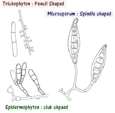 Figure: Conidia of dermatophytes
Dermatophytes are classified into three groups based on their habitat: soil (geophilic species), animals (zoophilic species), and man ( anthropophilic species).
- Anthropophilic organisms: T. rubrum, T.interdigitale, T. tonsurans, T. violaceum, M.audouinii, M. ferrugineum.
- Zoophilic organisms: M. canis ( originating from cats and dogs), M. nanum (originating from pigs), T. equinum (originating from horses), T. verrucosum( originating from cattle)
- Geophilic organisms: M. gypseum, M. fulvum
Which genus infects what: a quick way to reason it out
The three genera differ in which tissues they invade, and this is the first thing to fix in your mind because it narrows the possibilities before any test is done.
Trichophyton infects all three tissues: skin, hair, and nails. Epidermophyton infects skin and nails but never hair. Microsporum infects skin and hair but never nails.
So the tissue involved is already a clue. A nail infection rules out Microsporum. A hair or scalp infection rules out Epidermophyton. A pure skin infection could be any of the three, and you move to microscopy and culture to separate them. This is why the specimen you collect matters: a scalp case needs plucked hairs, a nail case needs deep nail scrapings, and sending the wrong specimen can miss the diagnosis.
Pathogenesis of Dermatophytes
Virulence factors of dermatophytes are arthroconidium (asexual spores), enzymes (keratinase, protease, lipase), and mannan of the fungal cell wall.
Adherence
Arthroconidium (asexual spores produced by dermatophytes) adheres to the surface of the tissue. Trichophyton rubrum adheres to the epithelial cells by the carbohydrate-specific adhesions, which are expressed on the surface of the spores. Fibrillar projections have been observed in the T. mentagrophytes during the adherence phase. At the skin surface, long and sparse (thin) fibrils connect fungal arthroconidia to keratinocytes.
The arthroconidium then germinates into hypha, and hypha enters into the stratum corneum (the outermost layer of the epidermis). In the pathogenesis of dermatophytoses, the initial interaction between the arthroconidia and the stratum corneum occurs 3-4 hours after contact.
Penetration
After the adherence, dermatophytes need to obtain the nutrients for their survival. To use the nutrients, the proteins, starch, cellulose, and lipids are degraded into smaller compounds. Dermatophytes then secrete various enzymes like proteases, lipases, elastases, collagenases, phosphatases, and esterases to degrade the complex compounds into simple ones. Keratinases degrade the keratin present in the host tissues into oligopeptides or amino acids. Proteolytic enzymes degrade the proteins of the skin. It helps in the penetration of the stratum corneum.
Evading the host response
Dermatophytes stay in the dead, keratinized outer layer of the skin and usually do not invade living tissue. This location already keeps them away from many immune defenses. The cell wall component mannan adds to this by suppressing the local inflammatory response and by slowing the turnover of keratinocytes. Normally, skin cells divide and shed steadily, which would carry the fungus away. By slowing this shedding, mannan helps the fungus stay in place and establish a persistent infection.
Putting it together
The sequence of a dermatophyte infection is: an arthroconidium lands on skin and adheres to keratinocytes, it germinates into a hypha within about 3 to 4 hours, the hypha grows into the stratum corneum, secreted keratinases and other proteases break down keratin for nutrients and open a path through the tissue, and mannan then dampens the host response so the infection persists and slowly spreads outward. This outward spread from a central point is what produces the ring-shaped lesion that gives ringworm its name.
The three genera at a glance
The three genera are separated by which conidia they make and which tissues they invade. Full species rosters, colony and microscopic morphology, labelled conidia diagrams, and genus-specific tests are on the individual pages.
| Genus | Infects | Conidia produced | Genus-specific confirmatory test | Full article |
|---|---|---|---|---|
| Trichophyton | Skin, hair, nail | Microconidia (many) + macroconidia (few, thin-walled) | Hair perforation, urease | Trichophyton species |
| Microsporum | Skin, hair (not nail) | Microconidia + macroconidia (many, thick-walled, spindle) | Rice grain test | Microsporum species |
| Epidermophyton | Skin, nail (not hair) | Macroconidia only (club-shaped, in clusters); no microconidia | — | Epidermophyton floccosum |
The single most useful discriminator to memorize: Epidermophyton is the only one of the three that makes no microconidia. If you see microconidia on the slide, it is Trichophyton or Microsporum, never Epidermophyton.
The infections dermatophytes cause: tinea (ringworm)
Dermatophytes cause a group of infections named for the body site: tinea capitis (scalp), tinea corporis (body), tinea pedis (foot, "athlete's foot"), tinea cruris (groin), tinea barbae (beard), tinea manuum (hand), and tinea unguium (nail). The same species, most often Trichophyton rubrum, can cause several of these. The site tells you where the infection is, not which organism caused it, so identification still depends on the laboratory workup below.
For the clinical presentation of each infection, the inflammatory patterns (kerion, favus, black dot), and treatment by site, see the dedicated article: Types of Tinea (Ringworm) Infections.
Laboratory diagnosis of Dermatophytes
Clinical observation and laboratory investigation play a crucial role in diagnosing fungal infection. The sample of choice are the skin, nail, and plucked hairs of short length from the involved sites.
1. Collection of samples
Hair, nail, and skin samples are taken in a folded square of black paper or the card. When paper is used, it helps to dry out the specimens preventing them from bacterial contamination. It helps to store the fungus for 12 months without losing its viability.
Hairs
For the hair sample collection, instead of cutting, hair is plucked from the edge of the lesion.
Skin
For the skin sample collection, it needs to be washed well and then scraped from the margin of the lesion onto the folded black paper.
Nail
Nails scrapping is taken from the nail bed or infected areas after discarding the outer layers.
Scales
It includes the hair stubs, the contents of plugged follicles, and skin scales.
Hairbrush sampling
Use the sterile plastic brush and brush it on the scalp. Then inoculate the plastic brush by pressing it in the agar suitable culture media.
2. Wood’s Lamp Examination
It is a device used to diagnose and manage superficial cutaneous fungal infections. It is used for the examination of the scalp and ringworm infection. Most of the infected hair fluorescence when exposed to ultraviolet light. Microsporum audouinii and Microsporum canis fluorescens bright green, whereas Microsporum gypseum does not fluorescens. Non-fluorescent fungi like T. tonsurans and T. verrucosum are difficult to detect. The fluorescence seen under Wood's lamp comes from a pteridine compound produced by the fungus in infected hairs. Sometimes false positives may occur if other substances also consist of pteridine. An error may occur when the ointments containing petroleum, jelly, scales, serum, exudates, lint, and dried soaps fluorescens bluish or purple.
| Microorganisms | Fluorescence Color |
|---|---|
| Microsporum audouinii | Bright-green |
| Microsporum canis | Bright-green |
| Microsporum ferrugineum | Blue-green |
| Microsporum distortum | Blue-green |
| Microsporum gypseum | Dull-yellow |
| Trichophyton schoenleinii | Dull-green |
| Malassezia furfur | Golden-yellow |
| Corynebacterium minutissimum | Coral-red |
3. Direct microscopy
Place the specimen on a clean, grease-free slide and add 10-20% KOH. Cover it with the cover slip and examine it after 20 minutes. In the skin or nails, branching arthrospores are seen. In the hair, Trichophyton spp. forms parallel rows of spores outside (ectothrix) or inside (endothrix) of the hair shaft. T. tonsurans and T. violaceum show the endothrix type of hair invasion. T. mentagrophytes show ectothrix type of hair invasion. Hairs infected with T. schoenleinii show hyphae and air spaces within the shaft. For the full method, see KOH wet mount preparation.
4. Fungal Culture
The suitable media for the fungal culture is Potato Dextrose Agar (PDA) or Sabouraud’s dextrose agar (SDA) used for culture. Incorporating antibiotics like gentamicin and chloramphenicol in Sabouraud’s media will inhibit the overgrowth of bacteria. Incorporating the cycloheximide will inhibit the overgrowth of the rapidly growing saprophytic fungi. Sabouraud peptone-glucose agar (Emmons’ modification) mixed with cycloheximide and chloramphenicol is commonly used. Its commercial names are Mycobiotic and Mycosel agars. Inoculation is done at 25-30℃ for 1-3 weeks. The fungi are identified based on colonial appearance, color, pigment production, and microscopic observation. For the microscopic observation, lactophenol cotton blue staining is performed.
Colony appearance and microscopic morphology are species-specific and are covered, with labelled diagrams, on each genus page linked above.
5. Dermatophyte Test Medium (DTM)
Dermatophyte Test Medium is used for the presumptive identification of dermatophytes from the other fungal or bacterial contaminants. It helps to distinguish whether the cutaneous lesion is caused by dermatophytes, other fungi, or bacteria. When incubated at 25℃, the dermatophytes turn the medium red, whereas bacteria and other fungi cannot change it. To avoid the false positive result caused by DTM, dermatophyte identification medium (DIM) is also used.
Telling the common look-alikes apart
Several dermatophytes look similar on culture, so a few differential tests carry most of the identification workload. The two questions that come up most often in the laboratory are separating the two most common Trichophyton species, and confirming Microsporum audouinii.
Hair perforation is an in vitro test that separates two look-alike pairs (T. rubrum vs T. mentagrophytes, and M. canis vs M. equinum). Its principle, method, and reading are covered in Hair Perforation Test; the interpretation for the common Trichophyton pair is in the comparison table below.
Urea hydrolysis (urease) also separates T. rubrum from T. mentagrophytes: T. mentagrophytes is urease positive, T. rubrum negative. Method and interpretation are in the Urease Test article.
Trichophyton rubrum versus Trichophyton mentagrophytes. These are the two most frequently isolated dermatophytes, and they are separated by a small set of tests that agree with each other:
| Feature | T. rubrum | T. mentagrophytes |
|---|---|---|
| Reverse pigment on SDA | Deep red | Absent or tan, variable |
| Microconidia shape | Tear-drop, along the hyphae | Round, in grape-like clusters |
| Macroconidia | Few, long, pencil-shaped | Cigar-shaped, sometimes with a tail |
| Hair perforation test | Negative (no perforation) | Positive (wedge-shaped perforation) |
| Urease (urea hydrolysis) | Negative | Positive within 2 to 4 days |
A useful memory anchor: T. mentagrophytes is the "active" one. It perforates hair (positive hair perforation test) and it hydrolyzes urea (positive urease test). T. rubrum does neither, and it shows the deep red reverse pigment instead. "Mentagrophytes moves, rubrum rests and reddens."
Confirming Microsporum audouinii. This species does not grow on sterile rice grains, while M. canis and most other Microsporum species grow well and sporulate. The rice grain test is the specific confirmatory test for M. audouinii; its method and reading are on the Microsporum species article.
A note on how the tests fit together. No single test names a dermatophyte. The workflow is layered: direct KOH microscopy confirms a fungus is present and shows the pattern of hair invasion (endothrix versus ectothrix), culture on SDA gives colony color and growth rate, lactophenol cotton blue microscopy of the colony shows macroconidia and microconidia shape, and only then do the differential tests (hair perforation, urease, rice grain) settle species that still look alike. Each step narrows the field; the final name comes from the combination, not from any one result.
Treatment depends on the infection site (topical for most skin disease, oral for nail and scalp disease) and is covered in the Types of Tinea (Ringworm) Infections article, along with prevention and control.
How to Remember
Which genus skips which tissue. Trichophyton takes all three (skin, hair, nail). For the other two, use the neighbor trick already on the page: Epidermophyton skips Hair ("E" sits near "H"), and Microsporum skips Nail ("M" sits near "N"). Each one fails to infect its alphabet neighbor.
Endothrix versus ectothrix, and why the black dots. Endo = inside the hair shaft. When the fungus grows inside the shaft, the hair is weak and snaps off right at the scalp, leaving the black-dot pattern (T. tonsurans, T. violaceum). Ecto = outside; the fungus coats the outside of the shaft (many Microsporum).
The two "active" tests point the same way. For the T. rubrum versus T. mentagrophytes problem, remember that mentagrophytes is the busy one: it perforates hair and splits urea (urease positive). T. rubrum stays quiet on both and shows off its red reverse pigment instead. "Mentagrophytes moves, rubrum rests and reddens."
Key exam facts in one table
| Fact | Detail |
|---|---|
| What dermatophytes eat | Keratin (keratinophilic and keratinolytic fungi) |
| Three genera | Trichophyton, Microsporum, Epidermophyton |
| Only genus with no microconidia | Epidermophyton floccosum (macroconidia only, club-shaped) |
| Trichophyton infects | Skin, hair, nail (all three) |
| Epidermophyton infects | Skin and nail (not hair) |
| Microsporum infects | Skin and hair (not nail) |
| Infective form | Arthroconidia (from fragmented hyphae) |
| Disease name | Dermatophytosis = tinea = ringworm |
| First-line microscopy | KOH wet mount |
| Culture media | Sabouraud dextrose agar (SDA) or PDA, with cycloheximide + chloramphenicol; incubate 25 to 30°C, 1 to 3 weeks |
| Wood's lamp positive | M. audouinii, M. canis (bright green); most Trichophyton do not fluoresce |
| T. rubrum vs T. mentagrophytes | Hair perforation and urease: mentagrophytes positive, rubrum negative |
| Rice grain test | Confirms M. audouinii (does not grow on rice) |
Where Students Get Confused
Microconidia, macroconidia, and arthroconidia are three different things. Macroconidia are large and multi-celled; microconidia are small and single-celled; both are used to identify the genus on culture. Arthroconidia are different again: they form when a hypha breaks into segments, and they are the infective spores found in shed skin and hair. Do not treat "microconidia" and "arthroconidia" as the same word.
Endothrix versus ectothrix is about location, not species. Endothrix means the fungus grows inside the hair shaft; ectothrix means outside. Some species tend toward one pattern, but the terms describe where the spores sit, not the name of the fungus. On KOH microscopy you report the pattern; culture gives the name.
Ringworm has no worm. It is a fungal infection. The name comes from the ring-shaped lesion with a raised, advancing border and central clearing, not from any parasite.
Wood's lamp does not detect every dermatophyte. A negative Wood's lamp does not rule out tinea. Many important species, including most Trichophyton, do not fluoresce, so the lamp is a useful screen for certain Microsporum scalp infections but never a substitute for KOH and culture.
References
- Chander J. Textbook of Medical Mycology. 4th ed. Jaypee Brothers Medical Publishers; 2018.
- Carroll KC, Pfaller MA, et al. Manual of Clinical Microbiology. 12th ed. ASM Press; 2019.
- Procop GW, Church DL, Hall GS, et al. Koneman's Color Atlas and Textbook of Diagnostic Microbiology. 7th ed. Wolters Kluwer; 2017.
- Tille PM. Bailey and Scott's Diagnostic Microbiology. 15th ed. Elsevier; 2022.
- Su H, Packeu A, Ahmed SA, et al. Species distinction in the Trichophyton rubrum complex. J Clin Microbiol. 2019;57(9):e00352-19. https://doi.org/10.1128/JCM.00352-19
Frequently Asked Questions
What are dermatophytes?
What are dermatophytes?
Dermatophytes are a group of fungi that feed on keratin, the tough protein found in skin, hair, and nails. They cause the skin infections known as tinea or ringworm. The three medically important genera are Trichophyton, Microsporum, and Epidermophyton.
What is the difference between dermatophytes and dermatophytosis?
What is the difference between dermatophytes and dermatophytosis?
Dermatophytes are the fungi. Dermatophytosis is the infection they cause. Dermatophytosis is also called tinea or ringworm, and it is named by body site, such as tinea capitis on the scalp or tinea pedis on the feet.
How are the three dermatophyte genera different?
How are the three dermatophyte genera different?
They differ in which tissue they invade. Trichophyton infects skin, hair, and nails. Epidermophyton infects skin and nails but not hair. Microsporum infects skin and hair but not nails. The tissue involved is often the first clue to the genus.
How are dermatophyte infections diagnosed in the laboratory?
How are dermatophyte infections diagnosed in the laboratory?
The main steps are a KOH wet mount of skin, hair, or nail to look for fungal elements, followed by culture on Sabouraud dextrose agar to grow and identify the fungus. Colony appearance and the shape of macroconidia and microconidia under lactophenol cotton blue identify the genus, and differential tests separate similar species.
How do you tell Trichophyton rubrum from Trichophyton mentagrophytes?
How do you tell Trichophyton rubrum from Trichophyton mentagrophytes?
By a small set of tests that agree with each other. T. mentagrophytes perforates hair in the hair perforation test and hydrolyzes urea (urease positive). T. rubrum does neither and instead shows a deep red pigment on the reverse of the colony.
Why does ringworm form a ring?
Why does ringworm form a ring?
The fungus grows outward from the point where it started, spreading in a circle through the outer skin. The active edge advances while the center begins to clear, which produces the ring shape with a raised border. There is no worm involved.
Does a negative Wood's lamp rule out a fungal infection?
Does a negative Wood's lamp rule out a fungal infection?
No. Only some dermatophytes fluoresce under Wood's lamp, mainly certain Microsporum species on the scalp. Most Trichophyton species do not fluoresce, so a negative result does not exclude tinea. KOH microscopy and culture are still needed.

Tankeshwar Acharya, MSc (Medical Microbiology)
Tankeshwar Acharya is an Assistant Professor in the Department of Microbiology at Patan Academy of Health Sciences (PAHS), Nepal, where he has been teaching and practicing clinical microbiology for over 14 years. He is the founder of Microbe Online, one of the leading free microbiology education resources on the web, covering bacteriology, mycology, parasitology, immunology, and clinical laboratory diagnostics written from direct experience in both the classroom and the diagnostic laboratory.
Comments
No comments yet. Be the first to share your thoughts.
Leave a comment
All comments are reviewed before they appear.