
Viridans Streptococci: Pathogenesis and Lab Diagnosis
Viridans streptococci morphology, dental and endocarditis pathogenesis, and how optochin resistance and bile insolubility distinguish them from S. pneumoniae.

On this page
The viridans streptococci are important Gram-positive cocci comprising a large group of bacteria. These are found as mouth commensals, causing minor to life-threatening diseases. The word viridans has been derived from the Latin word Viridis as most species of this group gives green color due to alpha hemolysis (partial hemolysis) on blood agar.
General Characteristics
Viridans streptococci show general characteristics similar to other streptococcal species. Like streptococcal species, these organisms are Gram-positive cocci arranged in chains and are facultative anaerobes and catalase-negative.
Like S. pneumoniae, the group is alpha-hemolytic streptococci. These bacteria can be differentiated from S. pneumoniae by optochin resistance, bile solubility, absence of capsule, and arrangement, i.e., cocci in a chain (whereas S. pneumoniae are diplococci).
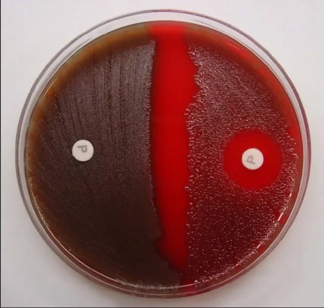 Figure: Viridans streptococci (right) are optochin resistant. Optochin-containing paper disc is added on blood agar previously inoculated with viridans streptococci. On incubation, it shows no zone of inhibition around the disc.
Viridans streptococci include five groups based on phenotype and each group contains at least 30 species. These groups are;
- S. anginosus group,
- S. mitis group,
- S. sanguinis group,
- S. salivarius group, and
- S. mutans group.
Viridans streptococci have no Lancefield antigen on the cell wall. Hence they are not classified serologically.
Habitat and Epidemiology
The organisms are generally present as harmless inhabitants of the mouth and oral cavity; gingiva, oropharynx, and gastrointestinal tract. They remain without causing harm unless there is a change in their environment like the host immune status is low or bad oral hygiene or the presence of an extra nutritional substrate. Hence infection caused by viridans streptococci is endogenous. The infection is low pathogenic in immunocompetent individuals. However, it can be severe and invasive, like endocarditis and septicemia, when low immune status.
Pathogenesis and Clinical Manifestations
Two virulence factors of viridans streptococci play vital roles in pathogenesis. The first is dextran which facilitates binding to fibrin platelet aggregates on damaged heart valves. Another is that these can form biofilms that assist in forming dental plaques.
Diseases caused by viridans streptococci are dental infections, sub-acute endocarditis, and others that may be life-threatening.
Dental infections
Viridans streptococci can bind to oral mucosa and tooth surfaces colonizing the oral cavity. Most common dental infections include dental caries, abscesses, and periodontal disease. If trapped in an oral cavity, a sucrose-rich diet supplies the substrate that bacteria convert into dextrans, creating a favorable environment for bacteria for demineralization and decay of teeth.
The common agent of dental caries is S. mutans. The S. anginosus group is a frequent cause of abscesses in the mouth, brain, and abdomen. S. sanguinis and S. mitis are the species most often isolated after dental procedures, entering the bloodstream and seeding damaged heart valves to cause subacute endocarditis. Hence patients with an existing heart problem need to use prophylactic antibiotics before surgery or dental procedure.
Subacute endocarditis
Viridans streptococci bind to fibrin platelet aggregates on damaged heart valves with the help of dextrans after entering the bloodstream. It leads to subacute endocarditis. A fibrin layer covers and protects the bacteria in the heart, which then causes bacterial endocarditis.
Others
Viridans streptococci can cause several other infections. Some of those are brain abscesses, abscesses on abdominal viscera, septicemia, and rarely meningitis.
Lab Diagnosis
Sample of choice
Depending on the type of symptoms/infection, the specimen for lab diagnosis of viridans streptococci can range from pharyngeal swabs, supragingival dental plaques, to blood.
Sample processing
The following methods are used for isolation and identification of the organism:
Microscopy
Under the microscope, viridans streptococci are seen as Gram-positive cocci in a chain when observed after Gram staining. These bacteria resemble S. pyogenes morphologically but may be more ovoid and are usually in short chains.
Biochemical tests
Viridans Streptococci are facultative anaerobes, catalase negative, and bile-insoluble, i.e., bile salts cannot lyse the bacteria.
Culture
Most of the viridans streptococci grow in nutrient agar or blood agar but fail to grow in MacConkey agar. The growth of some strains is enhanced if incubated in 5-10 % carbon dioxide incubators. These organisms produce small, colorless colonies in blood agar. Most cause partial (alpha) hemolysis resulting in green discoloration of blood agar, whereas others do not cause any hemolysis on the blood agar (gamma hemolysis).
Sensitivity test
Viridans streptococci are optochin resistant. Optochin-containing paper disc is added on blood agar previously inoculated with viridans streptococci. On incubation, it shows no zone of inhibition around the disc.
Similarly, molecular techniques are also used to identify different species of viridans streptococci.
Preventive Measures
Viridans streptococci are harmless human normal flora. The organisms cause opportunistic and endogenous infections like dental caries when the host environment is changed, which ultimately may lead to endocarditis, septicemia, or brain abscesses. One should maintain oral hygiene properly by brushing teeth regularly and cleaning the mouth after having sweet foods to avoid such infections. Also, patients with existing heart problems need prophylactic treatment with antibiotics, like penicillin, before surgery or a dental procedure.
Where students get confused
- Alpha-hemolysis alone never identifies an organism. Both S. pneumoniae and viridans streptococci share it, optochin and bile solubility are what actually separate them, not colony color.
- Viridans streptococci also resemble S. pyogenes on Gram stain, both form chains. Hemolysis pattern is the fast differentiator: GAS is beta-hemolytic (complete clearing), viridans is alpha or gamma (partial or none).
- "Harmless oral flora" doesn't mean "ignore in a blood culture." In a patient with a prosthetic or damaged heart valve, the same organism that's perfectly normal in the mouth is the textbook cause of subacute bacterial endocarditis, context (recent dental work, existing valve disease) changes the significance of the same lab result.
Key Exam Facts in One Table
| Feature | Viridans streptococci | Memory hook |
|---|---|---|
| Hemolysis | Alpha (or gamma) | Same green as pneumococcus |
| Optochin | Resistant | Opposite of pneumococcus |
| Bile solubility | Insoluble | No autolysin trigger |
| Capsule | Absent | Unlike pneumococcus |
| Lancefield antigen | None | Not serologically grouped |
| Key adhesion factor | Dextran | Binds damaged heart valves |
| Caries species | S. mutans | Mutans = mouth decay |
| Abscess species | S. anginosus group | Anginosus = abscess |
| Endocarditis species | S. sanguinis, S. mitis | Sanguinis = sanguine = blood |
How to Remember
Same color, opposite answers. Both S. pneumoniae and viridans streptococci turn the blood agar green (alpha-hemolysis), but every confirmatory test flips them apart:
| S. pneumoniae | Viridans streptococci | |
|---|---|---|
| Optochin | Sensitive | Resistant |
| Bile solubility | Soluble | Insoluble |
| Capsule | Present | Absent |
| Arrangement | Pairs (diplococci) | Chains |
Bile salts trigger autolysin of pneumococcus to self-destruct, that's what "bile soluble" actually means mechanically. Viridans streptococci don't carry that enzyme, so the bile solubility test comes back negative, this is the organism failing to do what pneumococcus does to itself.
Skip memorizing all five group names flat. Sort the important species by what they actually do:
- S. mutans = the caries organism. It ferments sucrose, makes dextran, and demineralizes enamel. Think "mutans = mouth decay."
- S. anginosus group = the abscess organisms. They seed pus collections in the mouth, brain, and abdomen. Think "anginosus = abscess."
- S. sanguinis and S. mitis = the bloodstream/valve organisms. They enter during dental procedures and stick to damaged heart valves, causing subacute endocarditis. Think "sanguinis = sanguine = blood."
References
- Doern, C. B., & Burnham, C.-A. D. (2010). It's not easy being green: The viridans group streptococci, with a focus on pediatric clinical manifestations. Journal of Clinical Microbiology, 48(11), 3829–3835. https://doi.org/10.1128/jcm.01563-10
- Sinner, S. W., & Tunkel, A. R. (2015). Viridans streptococci, nutritionally variant streptococci, and groups C and G streptococci. In Mandell, Douglas, and Bennett's Principles and Practice of Infectious Diseases (8th ed., pp. 2349–2361). Elsevier.
- Tille, P. M. (2022). Bailey & Scott's Diagnostic Microbiology (15th ed.). Elsevier..
Frequently Asked Questions
Why are viridans streptococci called "viridans"?
Can viridans streptococci cause infective endocarditis in a healthy heart?

Tankeshwar Acharya, MSc (Medical Microbiology)
Tankeshwar Acharya is an Assistant Professor in the Department of Microbiology at Patan Academy of Health Sciences (PAHS), Nepal, where he has been teaching and practicing clinical microbiology for over 14 years. He is the founder of Microbe Online, one of the leading free microbiology education resources on the web, covering bacteriology, mycology, parasitology, immunology, and clinical laboratory diagnostics written from direct experience in both the classroom and the diagnostic laboratory.
Comments
No comments yet. Be the first to share your thoughts.
Leave a comment
All comments are reviewed before they appear.