
Serial Dilution Method: Principle, Procedure, Uses, and Bacterial Count Calculation

On this page
A water sample collected from a municipal supply following flood damage arrives in the public health laboratory. The technician needs to report how many bacteria are present per milliliter, not just whether bacteria are present, but how many. The sample cannot be plated directly: if it contains 10⁷ bacteria/mL, plating 1 mL would produce 10 million colonies, which is uninterpretable. The sample must be diluted — but by how much?
Serial dilution is the technique that answers this question: a systematic sequence of 10-fold dilutions that reduces an unknown concentration to a countable range, then uses colony counts from the plate to calculate back to the original concentration.
Why Serial Dilution Is Necessary
Direct plating of clinical or environmental specimens is not possible for quantitative bacterial counting because most real-world samples contain far too many organisms to produce countable plates. A plate with more than 300 colonies has overlapping colonies that merge and cannot be counted individually. A plate with fewer than 30 colonies produces statistically unreliable counts.
Serial dilution solves this by creating multiple plates at different concentrations — ensuring that at least one dilution falls within the 30–300 countable range. The concentration from the countable plate is then multiplied by the dilution factor to calculate the original sample concentration.
When quantitative counts are clinically required:
- Urine culture: ≥10⁵ CFU/mL = significant bacteriuria; <10⁴ = likely contamination
- Food safety testing: S. aureus >10⁵ CFU/g = public health risk threshold; E. coli counts in dairy, meat, water
- Water quality: Total viable count per mL or per 100 mL
- Pharmaceutical sterility: Colony counts on surfaces and in air
- Research: Determining antibiotic kill rates over time
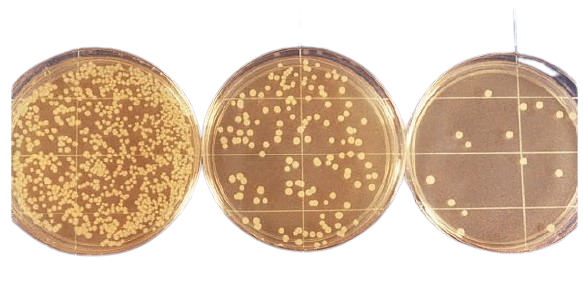 Figure: Agar plates plated with serial dilution of the test sample
Understanding Basics of Dilution
(In this section, I am discussing very basic information about dilution and terminologies. If you are already familiar with the basics, you can directly jump to preparing serial dilution).
Let’s say you have a glass of sugar solution at 6.0 M concentration and another glass with water. If you took 1 mL of sugar solution and dispensed it in a new tube, then added 9 mL of water and mixed. You just made a dilution.
To calculate the dilution factor (df), you need to divide the solution’s total volume by the sample volume (i.e., sugar solution in this case).
Total volume = (Volume of sample + Volume of diluent), i.e., 1 mL sugar solution + 9 mL water = 10 mL

Dilution Factor = Total volume/Sample volume = 10/1= 10
This dilution can be expressed by various terms like
- The dilution factor is 10 (dilution factor is the reciprocal of the dilution)
- It was a 10-fold dilution
- It was diluted by 1/10.
Calculating the concentration
To calculate the concentration of this diluted sample, multiply by the inverse of the dilution factor.
Final concentration = (Initial concentration X 1/ dilution factor), i.e., 0.6 M

Sometimes, you may wish to calculate backward. Let’s say you have a sample that has been diluted 1/10 with a concentration of 0.6 M and wants to calculate its undiluted concentration.
Undiluted concentration = (Diluted concentration X dilution factor)
= 0.6 M x10 = 6M.
In this example, we have used sugar solution as a sample and water as a diluent. In a real-world example, the sample can be an antibiotic solution, a patient specimen, food items, or drinking water. The diluent may be water, a buffer solution, or an actual growth medium.
Multiple or Serial Dilution
Let’s say you again diluted the sugar solution by taking 1 ml of diluted sugar solution in another 9 mL of water. Your final dilution ends up being 1/10 X 1/10 = 1/100 dilution. In other words, your sample has been diluted 100-fold (d.f. =100).
| Dilution Fold | Volume of Sample or Stock Solution | Volume of Diluent | Final Volume |
|---|---|---|---|
| 2 Fold Dilution | 5 mL | 5 mL | 10 mL |
| 10 Fold Dilution | 1 mL | 9 mL | 10 mL |
| 100 Fold Dilution | 0.1 mL | 9.9 mL | 10 mL |
If you want to measure the concentration of the final solution after two successive 10-fold dilutions, follow the same formula: (6 M X1/10 X 1/10) = 6/100 = 0.06 M
So, If you need 100-fold (10-2) dilution, you can either add 0.1 mL sample with 9.9 mL of diluent or make two successive 10-fold dilutions.
Similarly, if you need 10-6(1/106) dilution, you can make three successive 10-2(1/102) dilutions or six successive 10-1 dilutions.
Before preparing a dilution, prepare a table like this to avoid confusion or mistakes in diluting the sample.
Equipment
- Petri dishes, glass or plastic (at least 15 × 90 mm)
- Pipets with pipet aids or pipettors, 1, 5, and 10 ml, graduated in 0.1 ml units
- Erlenmeyer flask
- Dilution bottles
- Pipet and petri dish containers, adequate for protection
- Circulating water bath
- Incubator
- Colony counter
- Tally register
- Dilution blanks
- Plate count agar (PCA)
- Refrigerator
- Test tubes
- Vortex
Preparing Serial Dilution
In this example, 1 mL of the original sample is taken from a broth culture and added to 9 mL of sterile water, thereby diluting the culture by a factor of 10. This process is repeated until the desired concentration is reached.

Preparation of diluent
- Prepare six test tubes that can hold 20 mL or more in a rack and label them T1–T6. Each tube corresponds to one dilution step (for example, T3 is a 10⁻³ dilution, i.e., 0.001 of the stock concentration; its dilution factor, the reciprocal, is 10³).
- Pipet 9 mL of sterile water, saline, or broth into each of the six test tubes.
- Sterilize the diluent blanks by autoclave. Use aluminum foil to cover each of the six test tubes and then transfer them to an autoclave-compatible test tube rack. Sterilize for a minimum of 15 minutes at 121°C, 15 PSI.
- Remove blanks using heat-resistant gloves and allow to cool. When tubes reach room temperature, cover and store at 4°C until needed.
Serial Dilution Method
- Obtain the sample flask from the incubator and shake it vigorously.
- Pipet 1 mL of “solution” into the test tube labeled T1. Vortex T1.
- Remove 1 mL from test tube T1 and add it to test tube T2. Vortex T2.
- Remove 1 mL from test tube T2 and add it to test tube T3. Vortex T3.
- Remove 1 mL from test tube T3 and add it to test tube T4. Vortex T4.
- Remove 1 mL from test tube T4 and add it to test tube T5. Vortex T5.
- Remove 1 mL from test tube T5 and add it to test tube T6. Vortex T6.
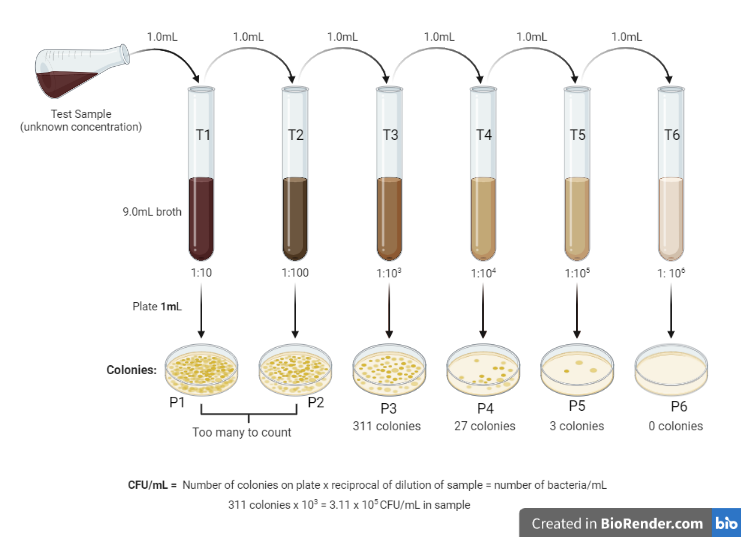 Figure: Serial dilution method for estimating viable count of bacteria
Plating into the Media
Thus prepared samples can be plated to culture media via the pour plate or spread plate technique. The procedure of the spread plate technique is mentioned here.
- Pipet 0.1 mL of a diluted sample from T1 directly onto a petri dish labeled P1. Repeat this step with each tube and plate to the corresponding petri dish (T2 to P2, T3 to P3, and so on).
- Obtain a sterile, disposable spreading rod or flame sterilize a glass spreading rod.
- In a clockwise or counterclockwise motion, glide the horizontal portion of the spreading rod to equally distribute the sample through the petri dish’s surface.
- Incubate the plates for 24 hours at the temperature appropriate to the sample (30–35°C for water and food counts per APHA/ISO methods; 35–37°C for clinical isolates).
Find more about the spread plate technique in this blog: Spread Plate Technique: Principle, Procedure, Results
If you want to try the pour plate procedure and also want to know more about this technique, visit this blog: Pour Plate Method: Procedure, Uses, (Dis) Advantages
Counting Number of Organisms
After 24 hours of incubation, remove the plate from the incubator and count the number of colonies using colony counter. The usual practice is to count colonies only on plates that have between 30 to 300 colonies.
Calculate the number of bacteria in the original sample using this formula:
CFU/mL = colonies counted × (reciprocal of the dilution) ÷ volume plated (mL)
The "reciprocal of the dilution" is the number you multiply back by to undo the dilution: for a 10⁻⁴ dilution it is 10⁴. The volume plated matters because only part of the diluted sample is transferred to the plate (0.1 mL for a spread plate).
Worked example: suppose the plate made from the 10⁻⁴ dilution had 45 colonies, and 0.1 mL was plated.
CFU/mL = 45 × 10⁴ ÷ 0.1 = 4.5 × 10⁶ CFU/mL in the original sample.
(Dividing by 0.1 mL is the same as multiplying by 10, which is why plating only 0.1 mL adds a factor of ten.)
Notes
- Refrain from estimating colonies count by counting >300 colonies or <30 colonies on the agar plate, as it leads to a high degree of error. A high count can be confounded by the error in counting too many small colonies or difficulty in counting overlapping colonies.
- Use separate sterile pipettes for each dilution. To do otherwise will increase the chances of inaccuracy because of the carry-over of organisms.
- Accuracy in quantitation is determined by accurate pipette use and adequate agitation of dilution tubes.
Troubleshooting: What to Do When Counts Are TNTC or TFTC
TNTC (Too Numerous To Count — >300 colonies/plate): Cause: The sample concentration was higher than expected; the dilutions were insufficient. Solution: Perform additional 10-fold dilutions (extend the dilution series) and re-plate. If the 10⁻⁴ plate was TNTC, try 10⁻⁵ and 10⁻⁶.
TFTC (Too Few To Count — <30 colonies/plate): Cause: The sample was over-diluted; the organism concentration was lower than expected. Solution: Plate a less dilute sample (e.g., if 10⁻³ was TFTC, try 10⁻² or undiluted).
No growth on any plate: Possible causes: Organisms died during transport (maintain cold chain); inhibitory substances in the sample not removed by dilution; organisms require special media not used; the sample was genuinely sterile.
Plates at adjacent dilutions inconsistent (e.g., 10⁻³ plate: 240 colonies; 10⁻⁴ plate: 5 colonies): Cause: Error in dilution preparation — a 10-fold dilution step was performed incorrectly. Solution: Re-dilute carefully, ensuring accurate pipetting volumes and complete mixing at each step. The 10-fold rule predicts that each successive dilution should give 10-fold fewer colonies; large departures from this indicate dilution error.
The 30–300 rule applies to both pour plate and spread plate methods. For spread plates using 0.1 mL inoculum, the effective countable range is 30–300 colonies per plate, corresponding to a colony concentration of 300–3,000 CFU per 0.1 mL applied.
Uses of Serial Dilution Method
Serial dilution is a core laboratory skill with wide application across microbiology. It is used to isolate and quantify microorganisms in samples such as water, food, and clinical specimens, and it underpins several other quantitative assays described below.
Serial dilution skill is required for bioburden testing, minimum inhibitory concentration (MIC), most probable number method (MPN), determination of antibody titer, and determination of minimum lethal dose.
To obtain a pure culture of microorganisms
Serial dilution method is routinely used to obtain a pure culture of microorganisms from mixed cultures. A series of dilutions are made from the sample and plated in the culture medium either by spread plate or by pour plate method.
Some of the resulting plates will contain a countable number of bacterial colonies on the agar. Each colony represents a pure growth, also known as a colony forming unit (CFU), that arises from a single bacterium.
Estimation of viable cell numbers
Serial dilution technique is widely used to estimate viable cell numbers in the standard plate count method and most probable number (MPN) technique. MPN methods are used for estimating the number of microorganisms in foods, wastewater, and other samples in which cell numbers need to be assessed routinely.
Standard plate count is used for estimating the number of viable organisms present in a sample. Because one may not know the approximate viable count ahead of time, multiple 10-fold dilutions of the sample are made and plated into the agar medium.
MIC and MBC Determination
The MIC and MBC determination procedure employs an antibiotic dilution assay in agar, culture tubes, or microtiter plate wells. Wells containing serial dilutions of antimicrobial agents are inoculated with a standard inoculum of a test organism. After which, the concentration of the drug that inhibits growth is determined by visual inspection or measuring turbidity.

The highest dilution (lowest concentration) of an antibiotic that completely inhibits growth is the MIC value for the test organism.
Determination of Antibody Titers
Antibody titer is the highest dilution (lowest concentration) of serum at which antigen-antibody reaction is observed. To determine antibody titer, serial dilutions of patient serum are prepared and assayed by various serological methods, such as ELISA.
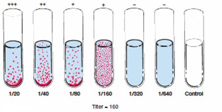 Figure: Determination of antibody titer
In cases wherein high concentrations of antigen or antibody are anticipated, false-negative prozone or postzone phenomena, respectively, can be avoided by repeating the test using serial dilutions of the specimen.
Phage Plaque Assay
Bacteriophage plaque assay is done to grow isolated plaques of phage particles within a lawn of bacteria. To achieve a plaque count on plates of 100-250 pfu (plaque forming units), phage stock should be serially diluted.
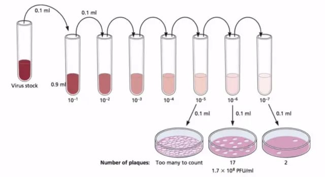 Figure: Phage plaque assay
Calculation of LD50
To calculate the lethal dose of viruses that do not cause recognizable effects in cell culture yet cause death in animals. Generally, 10-fold dilutions of viruses are made, and each dilution sample is injected into several sensitive animals.
After a suitable incubation period, the fraction of dead and live animals at each dilution is tabulated, and an end-point dilution is calculated. This is the dilution at which, for example, half of the injected animals die (the lethal dose for 50% or LD50).
Limitations
- Serial dilution is a labor-intensive process that is prone to multiple errors. To overcome these issues, bio-science companies are marketing various automated dilution equipment.
- The accuracy of the serial dilution and estimation of the viable count of bacteria depends on the homogenous dispersal of organisms in each dilution. Lack of proper pipetting or mixing of organisms may cause errors. Those errors can be minimized by giving hands-on training on proper pipetting, shaking each culture before sampling, and making several plates from each dilution.
- The viable count of bacteria may not represent the entire living bacterial population. It does not include organisms that may have died by the time plating was done; nor does it include organisms that cannot grow on the chosen medium. For example, anaerobic bacteria such as Clostridium perfringens, microaerophilic bacteria such as Campylobacter, and halophilic bacteria such as Vibrio parahaemolyticus (which requires salt) cannot grow on standard methods agar.
Advantages of Serial Dilution
Serial dilutions are much easier to make as they are made by repeating the same dilution step over and over, using the previous dilution as the input for the next dilution. By plotting the dilution and the relative number of organisms (or concentration), the number of organisms (or concentrations) in any dilution can be estimated.
Thus serial dilution avoids the problems associated with cumbersome calculation and dilution required to make dilutions of various concentrations.
How to Remember
Each dilution step multiplies the dilution by another factor of 10. A series of tubes, each receiving 1 mL of the previous tube into 9 mL of diluent, gives 10⁻¹, 10⁻², 10⁻³, and so on. The formula CFU/mL = colonies counted × (reciprocal of the dilution) ÷ volume plated always returns the original concentration.
The 30–300 rule — why these numbers: Below 30 colonies, the count is statistically unreliable (too much sampling error). Above 300 colonies, colonies overlap and merge (too crowded to count). The countable window of 30–300 represents the reliable range. Always plate at least 3 dilutions to ensure one falls in this range.
Serial dilution in clinical practice:
- Urine culture: calibrated 1 µL or 10 µL loop delivers a known volume — the colony count on the plate directly gives CFU/mL without needing a dilution series
- MIC testing: serial 2-fold dilutions of antibiotic in broth (not 10-fold) — same principle, different dilution factor
Where Students Get Confused
- Dilution vs dilution factor (the big one). A 10⁻⁴ dilution and a dilution factor of 10⁴ describe the same tube. The dilution (10⁻⁴, a fraction) is how much is left; the dilution factor (10⁴, its reciprocal) is what you multiply back by. Students mix these up constantly. Rule: you dilute by 10⁻⁴, you multiply back by 10⁴.
- Forgetting the volume-plated step. The formula is not just colonies × dilution factor. You also divide by the volume plated. Plating 0.1 mL (not 1 mL) adds a factor of ten. Leaving this out understates the count tenfold, and it is the most common calculation slip in the cluster.
- "10³ means a 1000-fold more concentrated sample." No. 10⁻³ (or "1/1000") is the dilution. Writing a diluted tube as "10³" instead of "10⁻³" flips the meaning. The positive-exponent number is the factor you multiply by at the end, not the state of the tube.
- Counting the wrong plate. Only a plate with 30–300 colonies is used for the calculation. Students sometimes count a TNTC or TFTC plate because it is the only one they plated. Always plate at least three dilutions so one lands in range.
- Reusing the same pipette down the series. Carrying the same tip from tube to tube drags organisms forward and inflates later counts. Use a fresh sterile tip at each step.
- Not mixing between steps. If a tube is not vortexed before the next transfer, the sample drawn is not representative, and adjacent dilutions stop following the 10-fold pattern. Uneven results between neighboring plates usually mean a mixing or pipetting error, not real biology.
- Assuming viable count = total count. The plate count only counts organisms that are alive and can grow on the chosen medium. Dead cells, injured cells, and organisms needing other conditions (anaerobes, microaerophiles, halophiles) are missed.
Key exam facts in one table
| Concept | Key exam fact and why it holds |
|---|---|
| What it is | A stepwise sequence of (usually 10-fold) dilutions that brings an unknown, too-high concentration down into a countable range, so the original count can be calculated back. |
| Why needed | Undiluted samples often hold far more than 300 colonies' worth of organisms, giving uncountable plates. Dilution guarantees at least one plate lands in the countable window. |
| Standard step | 1 mL sample into 9 mL diluent = a 10-fold (10⁻¹) dilution. Repeating multiplies the dilution by ten each time (10⁻¹, 10⁻², 10⁻³ …). |
| Dilution vs dilution factor | Dilution = the fraction remaining (10⁻⁴). Dilution factor = its reciprocal (10⁴), the number you multiply back by. Same tube, two numbers. |
| Countable range | 30–300 colonies per plate. Under 30 is statistically unreliable; over 300, colonies merge and undercount. |
| CFU/mL formula | CFU/mL = colonies counted × (reciprocal of dilution) ÷ volume plated. For 45 colonies at 10⁻⁴, 0.1 mL: 45 × 10⁴ ÷ 0.1 = 4.5 × 10⁶. |
| Volume-plated correction | Plating 0.1 mL (not 1 mL) means dividing by 0.1, i.e., multiplying by 10. Omitting it understates the count tenfold. |
| TNTC / TFTC | TNTC (>300): dilute further and re-plate. TFTC (<30): use a less dilute sample. |
| Consistency check | Each successive 10-fold plate should show about one-tenth the colonies of the previous. A large departure signals a dilution or mixing error. |
| Key applications | Viable plate counts, MPN, MIC/MBC (2-fold, not 10-fold), antibody titer, phage plaque assay, LD50. |
| Main limitation | Counts only viable, culturable organisms; misses dead, injured, and organisms needing other media or atmospheres. |
References and further readings
- Sanders, E. R. (2012). Aseptic laboratory techniques: plating methods. Journal of Visualized Experiments, (63), e3064. https://doi.org/10.3791/3064
- Madigan, M. T., Bender, K. S., Buckley, D. H., Sattley, W. M., & Stahl, D. A. (2021). Brock Biology of Microorganisms (16th ed.). Pearson.
- Sutton, S. (2011). Accuracy of plate counts. Journal of Validation Technology, 17(3), 42–46.
- American Public Health Association (APHA). (2017). Standard Methods for the Examination of Water and Wastewater (23rd ed.). APHA Press.
Frequently Asked Questions
Why must bacterial counts fall between 30 and 300 colonies per plate for a valid result?
How is the CFU/mL calculation performed after serial dilution and plating?
What are the main sources of error in serial dilution that can cause inconsistent results between dilution levels?

Tankeshwar Acharya, MSc (Medical Microbiology)
Tankeshwar Acharya is an Assistant Professor in the Department of Microbiology at Patan Academy of Health Sciences (PAHS), Nepal, where he has been teaching and practicing clinical microbiology for over 14 years. He is the founder of Microbe Online, one of the leading free microbiology education resources on the web, covering bacteriology, mycology, parasitology, immunology, and clinical laboratory diagnostics written from direct experience in both the classroom and the diagnostic laboratory.
Comments
No comments yet. Be the first to share your thoughts.
Leave a comment
All comments are reviewed before they appear.