Bacteriophage Plaque Assay: Principle, Procedure, Results
How the plaque assay counts infectious phage particles, why a "plaque" is the opposite of a bacterial colony, and where the dilution math trips students up.
A student hands their supervisor an incubated plate from a diagnostic virology bench and says, "nothing grew, the plate's basically clear." The supervisor looks at it and says the opposite: the sample is loaded with active phage.
Both of them are looking at the same plate. The difference is what "clear" means here. On a routine bacterial culture plate, growth shows up as colonies, visible mounds where bacteria multiplied. On a plaque assay plate, it's reversed: the background lawn of bacteria is the normal, expected growth, and a plaque is a hole punched into that lawn, a spot where phage killed every bacterium in the area. No growth is the positive result. Confluent, unbroken growth is the negative one.
This inversion trips up exactly the students who've just gotten comfortable with counting bacterial colonies (CFU) and then meet plaque-forming units (PFU) for the first time. The math for both techniques, serial dilution and a plate count, is nearly identical. What flips is what a "hit" looks like on the plate.
Bacteriophages fall into two broad groups based on how they replicate: virulent (lytic) phages, which always destroy their host cell, and temperate phages, which can also enter a dormant, non-lytic prophage state. Plaque assays specifically measure lytic activity, so only virulent phages, or temperate phages actively undergoing the lytic cycle, will form plaques.
For the full mechanism, see Bacteriophage Structure and Life Cycle.
As the world is struggling with the problems of increase in antimicrobial resistance, various research are underway to evaluate applications of phages to treat bacterial infections (as a replacement of antibiotic therapy).
Using phages to treat bacterial infections was developed back in the 1920s and 1930s in Eastern Europe and the Soviet Union.
Plaque assay is one of the widely used approaches for determining the quantity of infectious virus in a sample. Only viruses that cause visible damage to cells can be assayed in this way. Plaque assay was first developed to calculate the titers of bacteriophage stocks. Currently, its modified procedure is being used for the determination of titer of many different animal viruses too.
Principle of Phage Plaque Assay
When a suspension of an infective phage (e.g. T4 phage) is spread over the lawn of susceptible bacterial cells (e.g. Escherichia coli), the phage attaches the bacterial cell, replicate inside it, and kills it during its lytic release. Lysis of the bacteriophage is indicated by the formation of a zone of clearing or plaque within the lawn of bacteria. In the absence of lytic phage, the bacteria form a confluent lawn of growth.
Each plaque corresponds to the site where a single bacteriophage acted as an infectious unit and initiated its lytic cycle. The spread of infectious phage from the initially infected bacterial cell to the surrounding cells results in the lysis of the bacteria in the vicinity, eventually forming the plaque that is large enough to be visible to the naked eye. Plaques do not continue to spread indefinitely. The size of the plaque formed depends on the virus, the host, and the conditions of culture. The number of plaques that develop and the appropriate dilution factors can be used to calculate the number of bacteriophages i.e. plaque-forming units (PFU) in a sample.
 The medium used in phage plaque assays has a relatively low percentage of agar and therefore is called soft agar; it permits diffusion of phage to nearby uninfected cells but does not permit new phages to move to remote parts of the plate.
This is a useful contrast with bacterial viable counting: a plaque assay and a viable bacterial count both rely on serial dilution and counting discrete units on a plate, but the "unit" being counted is opposite in nature. A colony-forming unit (CFU) is visible growth; a plaque-forming unit (PFU) is visible destruction of growth. Reading a plaque assay plate with a bacterial-colony mindset is the single most common source of confusion for students new to virology technique.
Procedure for Bacteriophage Plaque Assay
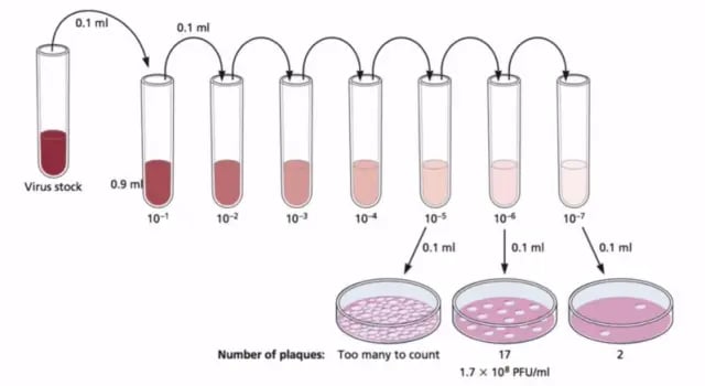 Figure: Bacteriophage Plaque Assay
Preparation of Stock Solution by serial dilution
This dilution scheme follows the same serial dilution logic used for bacterial viable counts; if that technique is already familiar, the setup here will feel identical. What differs is only what you're diluting and what you'll be counting afterward.
- Place six sterile saline tubes (0.9 ml each) in your test-tube rack
- Label one tube “control” and label the remaining five tubes consecutively from 10-1 through 10-5.
- Label six nutrient agar plates the same as the tubes.
- Using a sterile 1 ml pipette, aseptically transfer 0.1 ml of the bacteriophage suspension provided to the saline tube labeled 10-1.
- Mix the tube well by rolling it between the palms of your hands.
- With another 1 ml pipette, transfer 0.1 ml from the 10-1 tube to 10-2 tube. Mix the tube.
- Using a fresh pipette for each transfer, transfer 0.1 ml of the suspension from the 10-2 tube to the 10-3 tube, and continue this diluting procedure consecutively for the remaining saline tubes. Do not forget to mix each tube well before and after diluting.
Overlaying Plate with Phage-Agar Mixture
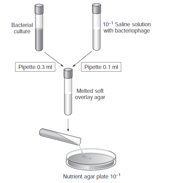 1. Obtain six tubes of melted soft overlay agar from the water bath. Pipette 0.3 ml of a broth culture of E.coli into each of the soft agar tubes. Mix each tube well by rolling it between your palms. Label each tube with your initials and return them to the water bath as soon as possible. Do not allow the agar to solidify. Note: you must work quickly here2. (Again work quickly) Remove one inoculated tube of soft agar from the water bath. Wipe off all the water from the surface of the tube. Using a 1 ml pipette, aseptically transfer 0.1 ml of the 10-1 saline phage dilution into the soft agar tube. Mix the agar tube by rolling it between your hands. 3. Immediately, aseptically pour the soft agar onto the surface of the nutrient agar plate correspondingly labeled as 10-1. Replace the lid and without picking up the plate, rotate it gently in a 6-to 8-inch circle on the surface of the table to evenly distribute the agar. 4. Using a fresh 1 ml pipette each time and working quickly, repeat steps 1 and 2 for the remaining saline phage dilution tubes and for the saline control tube. 5. For each dilution tube, use its correspondingly labeled nutrient agar plate. 6. Allow the soft agar to solidify. 7. Invert and incubate plates at 35°C to 37°C for 24 hours.
Results
- After incubation, examine each plate and count the number of plaques on each plate that has clearly differentiated plaques.
- Record your counts. Plates where plaques have covered the entire plate and where plaques are not clearly discernible from each other (more than 300 plaques) should be recorded as TNTC (too numerous to count).
This 300-plaque ceiling plays the same role as the 30–300 colony rule used for bacterial viable counts: above it, plaques begin to merge and become unreliable to count individually. The underlying reason is identical in both techniques, past a certain density, individual units on the plate are no longer statistically distinguishable from each other. - Calculate the number of lytic phages per milliliter that were in the original bacteriophage suspension using the formula mentioned above.
In your practical, you can count the plaque-forming units, calculate and tabulate it as follows:
| Dilution of phage | 10 -1 | 10 -2 | 10 -3 | 10 -4 | 10 -5 |
|---|---|---|---|---|---|
| Number of plaques | |||||
| Calculations of plaque units/ml |
Results of Bacteriophage Plaque Assay
If 48 plaques are observed in 10-5 dilution factor, as the 0.1 ml virus is added, plaque-forming units/ml will be 4.8 X 10⁷.
How to Remember
- PFU vs. CFU, in one line: CFU counts hills (bacterial growth); PFU counts holes (bacterial death). If the "unit" you're counting is something growing, you're doing a viable count. If it's a clear gap in something that should otherwise be solid growth, you're doing a plaque assay.
- Why the agar is "soft": Soft agar is loose enough to let phage particles diffuse a short distance to infect neighboring cells, which is what makes a plaque grow to a visible size, but not so loose that phage can travel across the whole plate. Think of it as a virus being able to knock on the neighbor's door, but not drive across town.
- The 300-plaque ceiling isn't arbitrary: It's the same logic as the 30–300 colony rule for bacterial counts. Past a certain density, adjacent units start merging into each other, and you're no longer counting discrete events, you're just looking at damage.
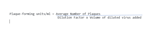
- Checking your PFU/mL formula the fast way: PFU/mL = plaques counted ÷ (dilution factor × volume plated). If your final number looks smaller than the number of plaques you actually counted, you've made an arithmetic direction error, since diluting the original sample should always inflate the calculated concentration, not shrink it.
Key exam facts in one table
| Concept | Detail | Memory aid |
|---|---|---|
| What a plaque represents | A zone of bacterial lysis caused by one infectious phage's lytic cycle spreading locally | The opposite of a colony: absence of growth, not presence of it. |
| Confluent lawn | Uniform bacterial growth with no phage activity present | The "negative" result, exactly the reverse of what a negative bacterial culture plate looks like. |
| Soft agar's function | Low agar percentage, permits local diffusion to nearby cells only | Keeps plaques discrete and countable instead of one giant zone of clearing. |
| Plaque size determinants | Virus, host, and culture conditions | Size is not a measure of phage concentration; number of plaques is. |
| PFU/mL formula | Plaques counted ÷ (dilution factor × volume plated) | Worked example: 48 plaques at 10⁻⁵ dilution, 0.1 mL plated → 4.8 × 10⁷ PFU/mL. |
| TNTC cutoff | More than 300 plaques per plate | Mirrors the 30–300 CFU rule for bacterial viable counts; both exist to keep counts statistically reliable. |
| Only lytic activity is measured | Temperate phages only form plaques while actively undergoing the lytic cycle | A lysogenized cell producing no active phage will not contribute to a plaque. |
Where Students Get Confused
- Reading a plaque as if it were a colony. A plaque is a clear zone of destroyed bacteria; a colony is a visible mound of bacterial growth. Mixing these up means reading a result as its exact opposite.
- Forgetting PFU and CFU measure fundamentally different events. Both use serial dilution and a plate count, and the math is nearly identical, but a CFU counts something alive and growing, while a PFU counts something a virus has already killed.
- Treating the TNTC cutoff as unique to bacteriology. The same reasoning behind the 30–300 CFU rule for bacterial viable counts applies here: above roughly 300 plaques, they begin to merge and can no longer be counted as discrete, reliable events.
- Confusing what determines plaque size vs. plaque number. Size depends on the virus, host, and incubation conditions; number of plaques (at a given dilution) is what's actually used to calculate phage concentration. A question describing unusually large plaques is not asking about phage concentration.
- Assuming a fixed number of dilution tubes is a universal rule. Different lab manuals and protocols use different numbers of dilution steps (5, 6, or 7) depending on the expected phage concentration in the sample. The number of tubes is a practical choice based on expected titer, not a fixed requirement of the technique itself.
References and Further Reading
- Racaniello, V. Virology Blog: Detecting viruses: the plaque assay. Retrieved from virology.ws.
- Madigan, M. T., et al. Brock Biology of Microorganisms. Pearson.
- Atlas, R. M. Principles of Microbiology.
- Pollack, R. A., et al. Laboratory Exercises in Microbiology.

Tankeshwar Acharya, MSc (Medical Microbiology)
Tankeshwar Acharya is an Assistant Professor in the Department of Microbiology at Patan Academy of Health Sciences (PAHS), Nepal, where he has been teaching and practicing clinical microbiology for over 14 years. He is the founder of Microbe Online, one of the leading free microbiology education resources on the web, covering bacteriology, mycology, parasitology, immunology, and clinical laboratory diagnostics written from direct experience in both the classroom and the diagnostic laboratory.