
Spread Plate Technique: Principle, Procedure, Uses, and Common Errors

On this page
During an investigation of a waterborne disease outbreak, the environmental health team collects water samples from a suspect borehole. Laboratory analysis requires a total coliform count (not just presence or absence) but how many per 100 mL.
Unlike the pour plate method, which mixes the inoculum into agar that may obscure slow-growing coliforms, the spread plate deposits every organism on the agar surface where colonies develop in full-size, accessible form — ideal for picking and identifying individual isolates alongside counting.
The spread plate technique is a viable-count method in which a liquid sample is spread across an agar surface to count or isolate the bacteria it contains. A perfect spread plate technique will result in visible and countable colonies of bacteria evenly distributed on the plate.
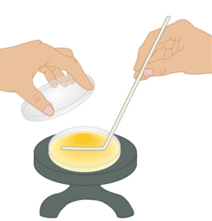 Figure: Spread-plate technique with a turntable and glass spreader.(Image source: Ref-1)
To get optimum results from the spread plate technique, one must be careful;
- to make accurate dilutions using pipettes (master serial dilution technique).
- to apply a balanced spread technique using a glass spreader to spread the inoculum evenly on the agar surface.
- to respect the necessary “short” time interval between agar inoculation and spreading
Each viable organism grows and divides to yield one colony, known as colony forming units (CFU).
Principle
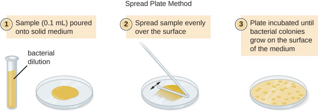
A liquid sample is diluted in a series of tubes containing sterile water or physiological saline. A fixed volume of sample, usually 0.1 mL, is removed from each tube and placed onto the agar surface. The sample is then spread evenly over the agar surface using a sterile, bent glass rod.
Following incubation, the dispersed cells develop into isolated colonies. The number of colonies is counted, then converted to the count in the original sample by multiplying by the reciprocal of the dilution and dividing by the volume plated (0.1 mL). The volume step matters: because only 0.1 mL is plated, leaving it out understates the true count by tenfold.
For solid or semi-solid samples (soil, food), the material is first homogenized in a suitable diluent, then processed through the same 10-fold serial dilutions before plating.
Requirements
- Glassware
- Screw-capped test tubes
- Sterile pipettes
- Sterile bent glass rods (bent in the shape of a hockey stick), or commercially available sterile spreaders
- Medium: Plate count agar or nutrient agar. The surface of the plate must not be too moist because the added liquid must soak in, so the cells remain stationary.
- Alcohol (ethanol)
Procedure for Spread Plate Technique
A: Serial dilution
- Prepare a series of at least 6 test tubes containing 9 ml of sterile distilled water.
- Using a sterile pipette, add 1ml of sample to the first tube of the set. Label it as 10-1.
- Mix the contents well by inverting the tube a few times.
- Take 1ml of the sample from the first tube and transfer it to the second tube. Label it as 10-2.
- Repeat the procedure with all the remaining tubes labeled until 10-6.
B. Plating
- Pipette out 0.1 ml from the appropriate desired dilution series onto the center of the surface of an agar plate.
- Dip the L-shaped glass spreader (hockey stick) into ethanol. Ethanol is used to sterilize the glass spreader.
- Pass the glass spreader through a bunsen burner, flame to ignite the ethanol, then let the flame burn off completely.
- Let the spreader cool for a few seconds (touch it to an unused edge of the agar first; if the agar hisses, wait longer). A spreader that is too hot will kill the cells and burn the agar surface.
- Spread the sample evenly over the surface using the cooled spreader, rotating the Petri dish underneath at the same time.
- Incubate the plate for 24–48 hours at the temperature appropriate to the sample (30–35°C for water, food, and environmental counts per APHA/ISO methods; 35–37°C for clinical isolates).
Note: The standard volume is 0.1 mL. Volumes above 0.1 mL are generally avoided because the excess liquid does not soak in and may cause colonies to coalesce, making them difficult to count. On very well dried plates, up to 0.2 mL can be plated when a larger volume is needed for a low-count sample, but 0.1 mL remains the routine volume.
 Calculation of Result
If your spread plate is successful, after incubation, you will get isolated countable colonies evenly spread across the surface of the agar. Count the number of colonies and multiply it by the appropriate dilution factor to determine the colony-forming units (CFU) present per ml in the original sample.
CFU/mL = colonies counted × (reciprocal of the dilution) ÷ volume plated (mL)
The "reciprocal of the dilution" is the number you multiply back by to undo the dilution. For a 10⁻⁴ dilution, the reciprocal is 10⁴. Because the spread plate uses only 0.1 mL, you also divide by 0.1 (which is the same as multiplying by 10).
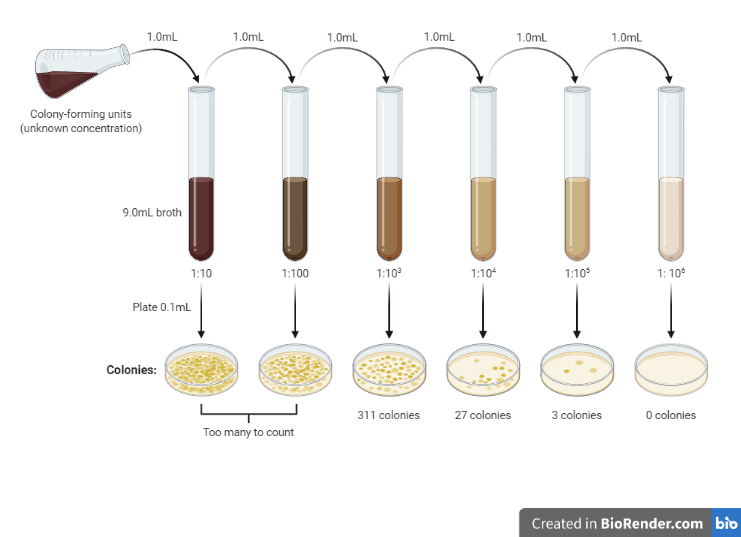 Figure: Serial dilution and number of colonies formed (Image source Biorender.com)
Worked example: suppose the plate made from the 10⁻⁴ dilution yielded 27 colonies, and 0.1 mL was plated. The reciprocal of a 10⁻⁴ dilution is 10⁴, so:
CFU/mL = 27 × 10⁴ ÷ 0.1 = 2.7 × 10⁶ CFU/mL in the original sample.
Common Errors and Troubleshooting
| Problem | Cause | Solution |
|---|---|---|
| Uneven colony distribution (heavy center, sparse edges) | Spreader not moved to the plate edges; spreading motion too small | Use the full plate surface in an overlapping spiral or back-and-forth pattern reaching to within 5 mm of the plate edge |
| Sample pools in the center without spreading | Plate surface not pre-dried; liquid does not absorb into agar | Pre-dry plates (lid slightly ajar, inverted) in a 35 to 37°C incubator for 15 to 30 minutes before use |
| Spreading marks visible as streaks of confluent growth | Spreader pressed too hard; agar surface damaged | Touch the spreader as lightly as possible — it should glide, not drag |
| >300 colonies, TNTC (too numerous to count) | Insufficient dilution | Extend serial dilution to higher dilutions; re-plate |
| <30 colonies, TFTC (too few to count) | Over-diluted or low-count sample | Plate a less dilute sample or plate a larger volume (up to 0.2 mL on pre-dried plates) |
| Satellite colonies around main colonies | Contamination from a spreading rod not sterilized between plates | Re-sterilize glass spreader by immersing in alcohol and flaming before each plate; allow to cool before spreading |
| Spreading rod too hot when applied | Agar surface burns; colonies fail to grow in the burned track | After flaming, cool the spreader on the edge of the agar plate before spreading — if the agar hisses, wait longer |
Benefits compared to pour plate technique
- All colonies grow on the surface, so they are full-size, well-separated, and easy to pick for identification or subculture.
- The inoculum never touches hot molten agar, so heat-sensitive and heat-stressed organisms survive and are counted accurately.
- Colonies are not obscured within the agar depth, making counting and morphology assessment easier than with the pour plate.
For a full side-by-side comparison of the two methods, see the pour plate method
Uses
- Viable counts of water, food, and beverages, where a precise number of organisms per volume is required.
- Enumeration and isolation of environmental microflora, for example from soil or surface swabs.
- Recovering heat-sensitive or heat-stressed organisms that might not survive the pour plate.
- Isolating individual colonies for identification, since all colonies are on the surface and easy to pick.
- Unlike the streak-plate technique, which is qualitative, the spread plate is quantitative: it gives an actual count, not just presence or absence.
How to Remember
Spread plate vs pour plate: Spread plate: inoculum spread on solid agar surface (0.1 mL, surface colonies only). Pour plate: inoculum mixed into molten agar before solidification (1.0 mL, surface and subsurface colonies).
Pre-drying is not optional: The spread plate uses 0.1 mL on a pre-dried surface. If the plate has any surface moisture, the 0.1 mL will not spread and will puddle in the center. Pre-drying is the single most commonly skipped step and the most common cause of failed spread plates in teaching laboratories.
The hockey stick spreader: The glass spreading rod bent into an L-shape (hockey stick) is used in a specific motion: place the horizontal portion on the agar, spin the plate slowly while keeping the spreader still (or move the spreader in a back-and-forth pattern). The goal is to distribute every microliter of inoculum as evenly as possible across the entire surface.
Where Students Get Confused
- Skipping the pre-dry step. This is the number-one cause of failed spread plates. If the agar surface holds any moisture, the 0.1 mL will not soak in and puddles in the center. Pre-dry plates (lid ajar, inverted) at 35 to 37°C for 15 to 30 minutes before plating.
- 10⁻⁴ vs 10⁴, the notation trap. The dilution is written 10⁻⁴ (a ten-thousand-fold dilution). The number you multiply back by is its reciprocal, 10⁴. Dilute by 10⁻⁴, multiply by 10⁴.
- Forgetting the 0.1 mL volume factor. Because only 0.1 mL is plated (not 1 mL), you divide by 0.1, which multiplies the count by ten. Leaving this out understates the result tenfold. This is the single most common calculation error on spread plates.
- Touching a hot spreader to the agar. After flaming off the alcohol, the spreader must cool before it touches the plate, or it kills the cells and scars the agar. Test on an unused edge; if the agar hisses, wait.
- Not spreading to the edges. Stopping short leaves a heavy center and sparse rim, so the count is uneven and unreliable. Carry the spread to within a few millimeters of the plate edge.
- Plating more than 0.1 mL to "get more colonies." Volumes above 0.1 mL do not soak in, so colonies coalesce and cannot be counted. If counts are too low, plate a less dilute sample rather than a larger volume.
- Reading plates outside the 30–300 window. Over 300 is TNTC (dilute further); under 30 is TFTC (use a less dilute sample). Counting a crowded plate always undercounts because merged colonies read as one.
Key exam facts in one table
| Concept | Key exam fact and why it holds |
|---|---|
| What it is | A viable-count method where a small volume of diluted sample is spread across a pre-dried agar surface, producing surface-only colonies for counting and isolation. |
| Volume plated | 0.1 mL (about one tenth of the pour plate's 1.0 mL). This is why the calculation includes a ÷0.1 (×10) step. |
| Pre-drying | Essential. The surface must be dry enough to absorb 0.1 mL so cells stay put; a moist plate causes pooling and failure. |
| Spreader | A bent glass rod (hockey stick), sterilized by dipping in ethanol and flaming, then cooled before use. |
| Colony location | Surface only, so all colonies are full-size, separated, and easy to pick, unlike the pour plate's small subsurface colonies. |
| Countable range | 30–300 colonies per plate. Over 300 = TNTC (dilute more); under 30 = TFTC (use a less dilute sample). |
| CFU/mL formula | CFU/mL = colonies counted × (reciprocal of dilution) ÷ volume plated. For a 10⁻⁴ dilution and 0.1 mL: × 10⁴ ÷ 0.1. |
| Heat effect | None. The inoculum never contacts molten agar, so heat-sensitive organisms survive, an advantage over the pour plate. |
| Incubation | 24–48 hours; 30–35°C for water/food/environmental counts, 35–37°C for clinical isolates. |
| Best used for | Water, food, and environmental (soil) viable counts, and isolating colonies for identification. |
| Vs streak plate | The streak plate is qualitative (isolation only); the spread plate is quantitative (gives a count). |
Reference and further readings
- Sanders, E. R. (2012). Aseptic laboratory techniques: plating methods. Journal of Visualized Experiments, (63), e3064. https://doi.org/10.3791/3064
- Madigan, M. T., Bender, K. S., Buckley, D. H., Sattley, W. M., & Stahl, D. A. (2021). Brock Biology of Microorganisms (16th ed.). Pearson.
- Sutton, S. (2011). Accuracy of plate counts. Journal of Validation Technology, 17(3), 42–46.
Frequently Asked Questions
Why must spread plates be pre-dried before use?
Why is only 0.1 mL plated, and how does it affect the calculation?
When should I choose a spread plate over a pour plate?
What do TNTC and TFTC mean on a spread plate?

Tankeshwar Acharya, MSc (Medical Microbiology)
Tankeshwar Acharya is an Assistant Professor in the Department of Microbiology at Patan Academy of Health Sciences (PAHS), Nepal, where he has been teaching and practicing clinical microbiology for over 14 years. He is the founder of Microbe Online, one of the leading free microbiology education resources on the web, covering bacteriology, mycology, parasitology, immunology, and clinical laboratory diagnostics written from direct experience in both the classroom and the diagnostic laboratory.
Comments
No comments yet. Be the first to share your thoughts.
Leave a comment
All comments are reviewed before they appear.