
Antigen Detection Tests for Disease Diagnosis: Methods, Examples, and Clinical Uses

On this page
A 32-year-old man presents on day 2 of fever, severe headache, and neck stiffness. CSF is collected by lumbar puncture. A Gram stain shows no organisms. Culture will take 48–72 hours. The patient is critically ill and cannot wait.
In the laboratory, a latex agglutination test for capsular polysaccharide antigens is performed on the CSF in 15 minutes and returns positive for Cryptococcus neoformans. Antifungal treatment begins immediately. The culture confirms Cryptococcus three days later.
This case captures the defining advantage of antigen detection: it identifies the pathogen directly, without waiting for the immune system to respond. In early infection, in immunocompromised patients who cannot mount adequate antibody responses, and in any situation requiring a same-visit result, antigen detection frequently provides diagnostic answers that antibody-based serology cannot.
This article and its companion: This article covers antigen detection — identifying pathogen components directly in clinical specimens. For antibody-based serological tests (ELISA, RPR, TPHA, Widal, ASO, Monospot, and more), see the companion article: Serological Tests for Disease Diagnosis.
Together these two articles cover the full spectrum of immunodiagnostic testing. The fundamental difference: antigen tests ask "Is the pathogen here right now?"; antibody tests ask "Has the body responded to this pathogen?"
Detection of specific antigens in a sample (blood, urine, CSF, or stool) using immunologic methods; precipitin tests, particle agglutination, immunofluorescence, immunochromatography, or enzyme immunoassay is the mainstay of rapid infection diagnosis. These test methods use polyclonal and monoclonal antibodies to detect specific antigens of the pathogen of interest.
Antigens are foreign substances, usually high molecular weight proteins or glycoproteins, that elicit the production of antibodies. One organism may contain different antigens that the host will recognize as foreign.
Why Antigen Detection? The Clinical Case
In any infection, antigen precedes antibody. The pathogen (or its proteins) enters the host before the immune system has time to generate a detectable antibody response. This creates a window period during which antibody tests are negative even though infection is present.
Antigen detection closes this window. It also solves three other diagnostic challenges that antibody testing cannot:
| Clinical situation | Why antibody testing fails | Why antigen testing works |
|---|---|---|
| Early acute infection (days 1–7) | Antibodies not yet detectable (window period) | Pathogen antigens are present and detectable immediately |
| Immunocompromised patients (HIV, transplant, chemotherapy) | Cannot mount adequate antibody response; serology unreliable | Detects pathogen directly regardless of immune status |
| Point-of-care / rapid diagnosis needed | ELISA and treponemal tests take hours to days | Lateral flow RDTs give results in 15–20 minutes |
| Monitoring active infection | IgG persists for life — cannot distinguish active from past | Antigen clearance with treatment confirms therapeutic response |
The practical rule: Use antigen detection first when the patient is acutely ill and early in the illness. Switch to or add antibody detection when the clinical picture is subacute, retrospective, or when immunity status is the question (post-vaccination, epidemiology).
Methods Used in Antigen Detection
The following methods are used to detect pathogen antigens in clinical specimens. Each has different sensitivity, specificity, speed, and infrastructure requirements:
| Method | Principle | Speed | Setting | Examples |
|---|---|---|---|---|
| Lateral flow / RDT (Rapid Diagnostic Test) | Antigen binds antibody-labelled particles; migrates along membrane; colour line appears | 10–20 minutes | Point-of-care, field, district laboratory | Malaria RDT, dengue NS1, HIV combo, COVID-19 Ag, Strep A |
| ELISA (EIA) | Antigen captured by antibody on plate; detected by enzyme-labelled second antibody; colour reaction | 2–4 hours | Laboratory | HBsAg, HBeAg, HIV p24, dengue NS1, H. pylori stool antigen |
| Latex agglutination | Antigen agglutinates antibody-coated latex beads — visible clumping | 15–30 minutes | Laboratory | Cryptococcal antigen (CSF/serum), meningococcal polysaccharide, Strep A, GBS |
| Immunofluorescence (DFA) | Fluorescent-labelled antibody applied directly to specimen; organism glows under fluorescence microscope | 1–2 hours | Reference laboratory | Legionella, Bordetella pertussis, HSV, RSV, Giardia |
| Co-agglutination | Antibody bound to Protein A of S. aureus Cowan I strain; antigen causes agglutination | 15–30 minutes | Laboratory | Lancefield group identification, Neisseria |
| Immunochromatography (ICT) | Antibody-labelled coloured particles; lateral flow format | 10–20 minutes | Point-of-care | Malaria, dengue, filariasis |
| Ouchterlony double diffusion | Antigen and antibody diffuse toward each other in agar; precipitin line at equivalence | 24–48 hours | Reference laboratory | Exoantigen confirmation of systemic fungi (Histoplasma, Blastomyces, Coccidioides) |
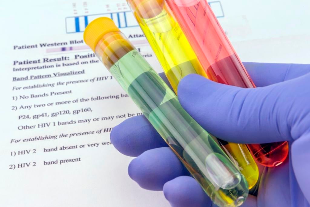 Figure: Blood sample and lab request for anti-HIV testing
Stool Antigen Test
Stool antigen tests (SATs) are noninvasive diagnostic tools for Helicobacter pylori infection, introduced after the urea breath test (UBT). This test was introduced after urea breath test (UBT). The currently used stool antigen test for diagnosing H. pylori infection is based on enzyme immunoassay (EIA) or immunochromatography (ICA).
Stool antigen test (SAT) is recommended by WHO for both initial diagnosis and test-of-cure after H. pylori eradication therapy. For test-of-cure, SAT should be performed ≥4 weeks after completing antibiotic treatment. Serology (anti-H. pylori IgG) is not suitable for test-of-cure because IgG remains elevated for months to years after successful eradication.
Urine Antigen Test
Urinary antigen testing has grown in popularity for several significant respiratory infections, particularly Legionella pneumophila (legionellosis), Streptococcus pneumoniae (pneumococcal pneumonia), and Histoplasma capsulatum (histoplasmosis). Though these organisms generally cannot be cultured from urine, antigens shed by them are concentrated in the kidney and excreted in urine. Soluble antigens from Streptococcus agalactiae and Haemophilus influenzae may concentrate in urine.
Urine antigens are then detected via an immunoassay such as an enzyme-linked immunosorbent assay (ELISA) or an immunochromatographic or lateral flow assay (LFA).
When urine antigen testing changes clinical management: The Legionella urinary antigen test (UAT) detects Legionella serogroup 1 responsible for ~80% of Legionnaires' disease within 3 hours. It allows targeted antibiotic therapy (levofloxacin or azithromycin instead of broad-spectrum) at a stage when culture results are not yet available. Similarly, the pneumococcal UAT can guide de-escalation from broad-spectrum coverage in severe community-acquired pneumonia within the first 24 hours.
Antigens present in Cerebrospinal fluid (CSF)
Soluble capsular polysaccharide antigens produced by the most common bacterial agents of meningitis including Streptococcus pneumoniae, Neisseria meningitidis, Haemophilus influenzae type b, and group B Streptococcus can be detected in CSF using latex agglutination, coagglutination, or commercial agglutination systems.
Cryptococcal antigen- the clinical priority: Cryptococcal meningitis is the most common cause of meningitis in HIV-positive adults in sub-Saharan Africa, responsible for 15–20% of AIDS-related deaths. WHO recommends Cryptococcal antigen (CrAg) lateral flow assay screening in all HIV-positive patients with CD4 <100 cells/µL before they develop symptoms. The CrAg LFA is >99% sensitive and specific, costs less than $5, and requires no laboratory equipment. Early detection at the asymptomatic stage dramatically reduces mortality.
Antigen testing in other samples
- Detection of group A beta-hemolytic antigen from throat swabs
- Detection of group B streptococcal antigen in vaginal secretions
Commonly Tested Antigens in Disease Diagnosis
| Antigen | Disease | Specimen | Test method | Clinical significance |
|---|---|---|---|---|
| p24 | HIV infection | Blood | ELISA; 4th-gen combo Ag/Ab test | Detectable 14–20 days post-infection — before anti-HIV antibodies appear; used in window period; neonatal diagnosis |
| HBsAg (Australia antigen) | Hepatitis B | Blood | ELISA; RDT | Presence = current HBV infection (acute or chronic); first serological marker to appear |
| HBeAg | Hepatitis B | Blood | ELISA | Indicates active viral replication and high infectivity |
| NS1 antigen | Dengue | Blood | ELISA; RDT | Detectable days 1–7 of illness — earlier than IgM; loses sensitivity after day 7; does not provide serotype |
| HRP2 (Histidine-rich protein 2) | Falciparum malaria | Blood | RDT | Expressed only by P. falciparum; most common malaria RDT target; may persist after parasite clearance (false positive) |
| pLDH (Parasite lactate dehydrogenase) | All malaria species | Blood | RDT | Detects all four Plasmodium species; clears within 24–48 hrs of treatment — better for test-of-cure than HRP2 |
| Aldolase | All malaria species | Blood | RDT (pan-malarial) | Pan-species marker; used alongside HRP2 and pLDH in combination RDTs |
| Cryptococcal polysaccharide antigen | Cryptococcal meningitis | CSF; serum | Latex agglutination; lateral flow assay | >99% sensitivity/specificity; critical for early diagnosis in HIV patients; CSF preferred |
| Pneumococcal polysaccharide | Pneumococcal pneumonia; meningitis | Urine; CSF | ICT; latex agglutination | Urine UAT: sensitivity 70–80% for pneumococcal pneumonia; remains positive days after treatment starts |
| Legionella serogroup 1 antigen | Legionnaires' disease | Urine | ICT; ELISA | 70–90% sensitivity; only detects serogroup 1 (~80% of cases); remains positive weeks after treatment |
| Histoplasma antigen | Histoplasmosis | Urine; BAL | EIA | Most sensitive test in disseminated histoplasmosis; essential in immunocompromised patients |
| H. pylori antigens | H. pylori gastritis/peptic ulcer | Stool | EIA; ICT | Preferred over serology for diagnosis and test-of-cure; positive result indicates active infection |
| Group A Streptococcal antigen | Strep throat; scarlet fever | Throat swab | Lateral flow RDT | Results in 5–10 minutes; negative rapid test should be backed up by throat culture in children |
| Group B Streptococcal (GBS) antigen | Neonatal GBS sepsis/meningitis | Vaginal/rectal; CSF; urine | Latex agglutination | Antenatal GBS screening guides intrapartum prophylaxis |
| Rotavirus antigen | Viral gastroenteritis | Stool | EIA; lateral flow | Most common cause of severe diarrhoea in children under 5; rapid antigen test widely used |
| COVID-19 (SARS-CoV-2) antigen | COVID-19 | Nasopharyngeal swab | Lateral flow RDT | High specificity (~99%); lower sensitivity than PCR especially in asymptomatic; most useful in symptomatic patients days 1–5 |
P24 Antigen
p24 is a capsid structural protein that makes up a protein ‘shell’ on the surface of the HIV virus. p24 test is generally only positive from about two to three weeks after infection with HIV.
Histidine rich protein 2 (HRP2)
HRP-II is an abundant protein expressed only by Plasmodium falciparum and is the target for the most commonly used RDTs.
Parasite lactate dehydrogenase (pLDH)
It is produced by the asexual and sexual stages (gametocytes) of malaria parasites. Malarial test kits that are currently available detect pLDH from all four species of Plasmodium. They can distinguish P. falciparum from the non-falciparum species, but cannot distinguish between P. malariae, P. ovale, and P. vivax.
 Figure: Microbiologist standing at desk and holding test tube rack while preparing blood samples for centrifuge
HBeAg
HBeAg is a hepatitis B viral protein. It is an indicator of active viral replication; this means the person infected with Hepatitis B can likely transmit the virus to another person (i.e. the person is infectious).
HBsAg (Australia antigen)
HBsAg is the surface antigen of the hepatitis B virus (HBV). It indicates the current hepatitis B infection.
NS1 Antigen
NS1 tests detect the non-structural protein NS1 of dengue virus in the serum using synthetically labeled antibodies. NS1 is detectable during the acute phase of dengue virus infections.
NS1 tests can be as sensitive as molecular tests during the first 0-7 days of symptoms. After day 7, NS1 tests are not recommended. A positive NS1 test result is indicative of a dengue infection but does not provide serotype information.
How to Remember
Antigen = the pathogen's ID card; antibody = the body's response to seeing the ID card. Antigen tests look for the ID card directly. Antibody tests look for proof the body has seen it. The ID card appears first — which is why antigen tests work earlier in infection.
The three specimen types and what they tell you:
| Specimen | What antigen testing finds | Classic example |
|---|---|---|
| Blood | Systemic infection / viraemia | HIV p24, dengue NS1, HBsAg, malaria RDT |
| Urine | Antigens concentrated in kidney and excreted | Legionella UAT, Pneumococcal UAT, Histoplasma EIA |
| CSF | CNS infection — organism in CSF or antigens shed into it | Cryptococcal CrAg, bacterial capsular antigens |
| Stool | GI infection or organisms shed in faeces | H. pylori SAT, rotavirus, Giardia, Cryptosporidium |
The rapid test revolution in LMIC settings: Malaria RDTs, dengue NS1 RDTs, HIV combo tests, Cryptococcal LFAs, and COVID-19 antigen tests all share one characteristic — they are lateral flow immunochromatographic tests that any trained health worker can perform in 15 minutes without laboratory equipment. These RDTs have transformed diagnostics in district hospitals and health posts where culture and ELISA are not available. Understanding the antigen each detects and its clinical window is the core knowledge for any healthcare worker in tropical settings.
The window period visual:
DAY 0         DAY 3–5          DAY 7–14         WEEKS–MONTHS
  |               |                |                  |
Infection    ANTIGEN peaks    IgM appears        IgG persists
              (detectable)    (seroconversion)   (for life)

Use: ← Antigen tests here → ← IgM tests here → ← IgG tests here →
The earlier the presentation, the more you need antigen detection. The later or more retrospective the diagnosis, the more antibody tests help.
Key exam facts in one table
| Antigen | Disease | First detectable | Memory aid |
|---|---|---|---|
| p24 | HIV | 14–20 days post-infection, before antibodies | The capsid shows up to the party before the antibody guest list is even printed |
| HBsAg | Hepatitis B | First marker to appear in acute infection | "s" for "surface" and "s" for "seen first" |
| HBeAg | Hepatitis B | Present during active replication | Think "e" = "excreting virus" high infectivity |
| NS1 | Dengue | Days 1–7, gone after day 7 | NS1 is a week-one-only guest; IgM/IgG take over after it leaves |
| HRP2 | P. falciparum malaria | Persists after cure (can false-positive) | HRP2 is the guest who overstays; bad for test-of-cure |
| pLDH | All 4 Plasmodium species | Clears within 24–48 hrs of treatment | pLDH leaves on time, good for test-of-cure |
| CrAg | Cryptococcal meningitis | Detectable even without antibody response | Works precisely when the immune system can't; screen CD4 <100 before symptoms |
| Legionella UAT | Legionellosis | Within 3 hours; serogroup 1 only (~80% of cases) | Negative UAT ≠ ruled out it's blind to the other 20% |
References
- Tille, P. M. (2017). Bailey and Scott's Diagnostic Microbiology (14th ed.). Elsevier.
- World Health Organization. (2022). Guidelines for the Diagnosis, Prevention and Management of Cryptococcal Disease in HIV-infected Adults, Adolescents and Children. Geneva: WHO.
- World Health Organization. (2015). Guidelines for the Treatment of Malaria (3rd ed.). Geneva: WHO. [Malaria RDT section]
- Huong, V. T. Q., et al. (2014). Urine antigen tests for the diagnosis of respiratory infections. Clinics in Laboratory Medicine, 34(2), 219–236.
- Leber, A. L. (Ed.). (2016). Clinical Microbiology Procedures Handbook (4th ed.). ASM Press.
Frequently Asked Questions
Why is the dengue NS1 antigen test only useful in the first seven days of illness?
What is the clinical significance of Cryptococcal antigen detection in HIV-positive patients?
Why do malaria HRP2-based RDTs sometimes remain positive after the parasites have been cleared by treatment?
Why does antigen testing detect infection earlier than antibody testing?
Why is serology not used to confirm H. pylori eradication?
Does a negative Legionella urinary antigen test rule out Legionnaires' disease?

Tankeshwar Acharya, MSc (Medical Microbiology)
Tankeshwar Acharya is an Assistant Professor in the Department of Microbiology at Patan Academy of Health Sciences (PAHS), Nepal, where he has been teaching and practicing clinical microbiology for over 14 years. He is the founder of Microbe Online, one of the leading free microbiology education resources on the web, covering bacteriology, mycology, parasitology, immunology, and clinical laboratory diagnostics written from direct experience in both the classroom and the diagnostic laboratory.
Comments
No comments yet. Be the first to share your thoughts.
Leave a comment
All comments are reviewed before they appear.