
Automated Cell Counter: Principle, Types, and How Viability Is Measured

On this page
A cell suspension reads 2.4 × 10⁶ cells/mL on the counter. The same sample, run again a minute later, reads 1.6 × 10⁶. Nothing was changed and nothing is broken.
The suspension was not mixed before the second aliquot was drawn. Cells settle in a tube within a minute, so the first sample came off the top and the second off a partially settled column. The instrument counted both accurately. It simply counted two different samples.
An automated cell counter removes the human variability of counting squares under a microscope, but it cannot correct for what happens before the pipette enters the tube. Almost every unreliable count traces back to the suspension rather than the instrument.
Cell counting is a standard laboratory method for counting or quantifying cells used in numerous life sciences or medical diagnosis procedures. There are many ways to count cells with equipment, but it has been broadly divided into manual and automated cell counters. The manual cell counter has been used for decades. Still, its counting error and time-consuming limitations have led to the discovery of more advanced instruments with automatic cell counting functions known as automated cell counters.
An automated cell counter is laboratory equipment that counts cells in a liquid suspension and reports the concentration, usually as cells per milliliter. Depending on the instrument, it may also report cell size distribution and the proportion of live to dead cells.
Typical working ranges fall roughly between 10⁴ and 10⁷ cells/mL, though the exact range is instrument-specific and stated in the manufacturer's specification. Samples above the upper limit must be diluted; samples below the lower limit give unreliable counts because too few cells are sampled for the statistics to hold.
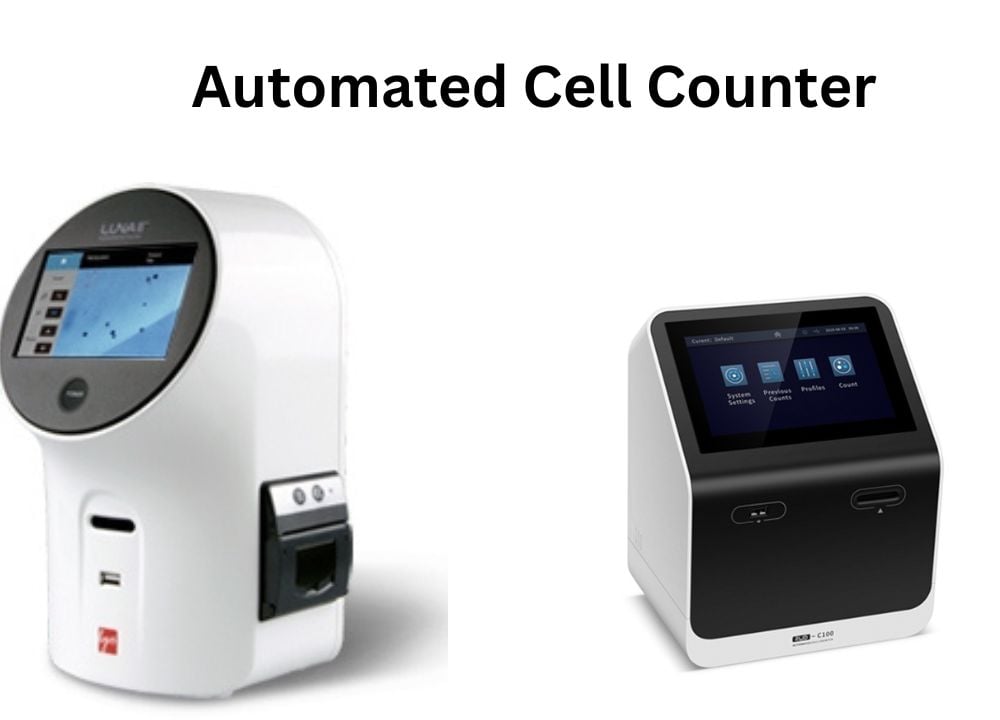 Figure: Automated Cell Counter
Principle of Automated Cell Counter
In the market, different types of automated cell counters are available. This instrument works based on either of two distinct principles. These include; the electrical impedance method or optical method.
Electrical impedance cell counting principle
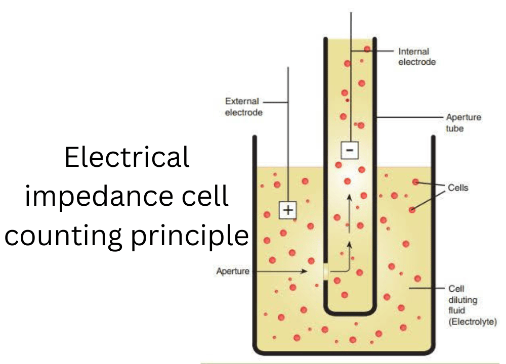 Figure: Illustration of electrical impedance cell counting principle
Also called the Coulter principle, after Wallace Coulter who patented it in 1953.
Cells are suspended in a conductive electrolyte solution and drawn through a narrow aperture with an electrode on each side. A steady current flows through the aperture. The aperture is deliberately made larger than the cells, typically several times their diameter, so that cells pass through one at a time rather than blocking it. If the aperture matched the cell size, nothing would flow.
Here is the mechanism that matters. A cell is a poor conductor compared with the electrolyte around it. As each cell passes through the aperture, it displaces its own volume of conductive fluid, so the electrical resistance across the aperture briefly rises. That rise is registered as a voltage pulse.
Two pieces of information come out of this:
- The number of pulses gives the number of cells.
- The height of each pulse is proportional to the volume of fluid displaced, which is the volume of the cell. This is why a Coulter counter reports cell size distribution as well as count.
The limitation follows directly from the mechanism. The instrument detects displaced volume, not biology. It cannot distinguish a live cell from a dead one, nor a cell from a similarly sized particle of debris, because both displace electrolyte identically. Coulter counters therefore give count and size, but not viability.
Optical cell counting principle
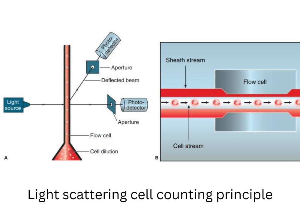 Figure: Illustration of light-scattering cell counting principle
Also called the light-scattering principle.
A stream of diluted cell suspension passes through a focused beam of light. Each cell that crosses the beam scatters light, and a detector registers the interruption as a single event. Counting the events counts the cells.
The scattering pattern carries information beyond the count. Light scattered in the forward direction, at a small angle, corresponds broadly to cell size. Light scattered at right angles corresponds to internal complexity, such as granularity and nuclear structure. This is the basis on which flow cytometers distinguish cell populations, and it is why a differential white cell count can be produced optically.
Hydrodynamic focusing makes this possible. The sample stream is injected into the center of a faster-moving sheath fluid, which squeezes it into a narrow core so cells travel single file through the beam. Without it, cells would cross the beam in clumps and at varying positions, and neither the count nor the scatter measurement would be reliable.
Image analysis principle
Rather than detecting cells as they flow past a sensor, an image-based counter photographs a static sample in a counting chamber and analyzes the image with software.
The instrument captures a bright-field or fluorescence image of the loaded slide, and an algorithm identifies objects by size, shape, and contrast, separating cells from debris and attempting to resolve clumps into individual cells. Because the image is retained, the operator can view what was counted and confirm the instrument has not miscounted debris or missed clustered cells.
This is essentially an automated version of the hemocytometer, with the software replacing the human eye at the microscope. It is the principle used by most benchtop counters in cell culture laboratories.
Types of Automated Cell Counters
Based on the above-explained principles, automated cell counters are of three types;
Coulter counter
- These are particle counters based on the electrical impedance cell counting principle.
- It provides the number and size of cells per particle within the sample.
- They are unable to provide cell viability information.
- It has applications in particle characterization, hematology, and counting cells such as fat cells, plant cell aggregates, bacteria, etc.
Flow Cytometer
- Measures individual cells in a hydrodynamically focused stream, recording forward scatter, side scatter, and fluorescence at several wavelengths simultaneously.
- Counting is only one of its functions and arguably the least of them. Its real purpose is characterization: identifying and quantifying cell subpopulations by surface markers, measuring DNA content, and assessing viability and apoptosis.
- Substantially more expensive and more complex to operate than a dedicated cell counter, and requires fluorochrome-conjugated antibodies for most of its applications.
In short: every flow cytometer can count cells, but using one purely to count cells is like using a mass spectrometer to weigh something.
Image-based cell counter
- Uses a bright-field or fluorescence optical path with a digital camera, and analyzes the captured image in software.
- Bright-field instruments assess viability with a colorimetric dye, usually trypan blue. Fluorescence instruments use fluorescent dyes such as acridine orange with propidium iodide, which improves discrimination in samples containing debris.
- Retains the image, so the operator can verify what was counted. This is a real practical advantage over flow-based instruments, where a spurious count cannot be inspected after the fact.
- The most common format in cell culture laboratories, using disposable counting slides.
Procedure for the operation of Automated Cell Counter
Automated cell counter instrument works differently. So, it’s highly recommended to go through the user manual that comes with the commercial instrument.
How viability is measured: the dye exclusion principle
Trypan blue is a dye that a living cell keeps out. An intact plasma membrane actively excludes it, so live cells stay unstained and appear bright. When a cell dies, its membrane loses integrity, the dye enters freely, and the cell appears blue.
This is why the method is called dye exclusion: what is being measured is not death itself but membrane integrity, which serves as its proxy. The distinction matters, because a cell in early apoptosis may still exclude trypan blue and be counted as viable.
Sample preparation:
- Without viability assessment: pipette 10 µL of the cell suspension directly.
- With viability assessment: mix 10 µL of suspension with 10 µL of 0.4% trypan blue, giving a 1:2 dilution. Take 10 µL of that mixture for loading. The instrument applies the dilution factor automatically, but confirm this in the settings rather than assuming it.
Timing matters. Read the sample within about 3 to 5 minutes of adding trypan blue. The dye is itself toxic, and cells that were viable at mixing will begin taking it up if left standing, so viability falls the longer the sample waits.
The general operating procedure of the automated cell counter is given below;
- Switch on the instrument by pressing the power switch.
- Handle the counting slide using the edges and avoid touching the optical surface of a slide.
- Pipette 10 μl of the cell suspension into the outer opening of either chamber of the counting slide.
- Insert the counting slide in a slide slot. The cell counter will automatically initiate the cell count.
- The count results appear on the Current Count screen as the total cell count per ml. Suppose the number of cells is above or below the specified range. Value out of range is displayed on the screen. Select the View Image key from the Current Count screen to see the image.
- The dilution calculator calculates the volume adjustments needed to achieve the cell concentration required for the experiments.
- Once the instrument completes the cell count, remove the slide.
- Lastly, switch off the instrument after use.
Applications of Automated Cell Counter
Automated cell counter has wide range of applications in various fields from research to diagnostic purposes. The applications of automated cell counter are as follows:
- It is used to determine cell count to check the viability of cell culture line used for research purposes.
- Automated cell counter is used in blood analysis. Determination of the concentration of various types of blood cells helps to determine the health condition of individuals.
- It is also applicable in urine analysis to determine the number and types of cells in the urine sample.
- Automated cell counter is used for measuring cell viability, i.e., measurement of fraction of viable and dead cells.
- In cell therapy, it is used to control the dose of cells administered to patients.
- Studies that examine the growth rate of microorganisms require cell counting.
Advantages of Automated Cell Counter
The automated cell counter is a great substitute for laboratory opting for automation. It has following advantages:
- It provides accurate and reliable results in a fraction of the time.
- It significantly reduces the requirement of the workforce and cell count variance.
- It is more efficient and cost-effective than the manual count method.
- Unlike the manual method, it does not require many replicate counts at low cell concentrations.
Limitations of Automated Cell Counter
Although automated cell counter is highly advantageous, it has some limitations which are as follows:
- Debris is counted as cells. Impedance instruments cannot distinguish a cell from a similarly sized particle. Image-based instruments do better but still misread debris in dirty samples.
- Clumped cells are undercounted. Two adherent cells passing together register as one large cell. Adequate dissuspension before loading is essential.
- Concentration limits are real. Above the upper limit, coincidence (two cells passing simultaneously) causes undercounting. Below the lower limit, too few cells are sampled for a reliable statistic.
- Dye exclusion measures membrane integrity, not viability as such. Cells in early apoptosis may still exclude trypan blue and be reported as live.
- Cost. Both the instrument and the consumable counting slides carry ongoing cost, which is a significant consideration in resource-limited settings where a hemocytometer and a microscope already exist.
- In automated hematology analyzers specifically, atypical cells such as immature neutrophils are poorly classified, and platelet clumps may be misread as a single leucocyte or erythrocyte. These limitations apply to clinical blood analyzers rather than to benchtop cell counters used in culture work.
Cell Counter, Colony Counter, and Hemocytometer: What Each One Counts
| Instrument | Counts | Sample | Output | Counts dead cells? |
|---|---|---|---|---|
| Automated cell counter | Individual cells in suspension | Liquid cell suspension | Cells/mL, size distribution, viability | Yes, and can distinguish them with dye |
| Colony counter | Colonies growing on agar | Inoculated and incubated plate | CFU per plate, converted to CFU/mL | No, only organisms that grew |
| Hemocytometer (manual) | Individual cells in suspension | Liquid cell suspension | Cells/mL | Yes, with trypan blue |
The distinction students most often miss. A cell counter counts cells that are present. A colony counter counts colonies that grew, and each colony arose from one viable organism (or one clump of them), which is why the unit is the colony forming unit rather than the cell. A suspension containing ten million dead bacteria gives a high cell count and zero colonies.
This is why the two instruments answer different questions. Use a cell counter to ask "how many cells are in this suspension?" Use a colony counter to ask "how many viable organisms could grow?"
For colony enumeration on plates, CFU calculation, and the countable range, see the Colony Counter article.
Against the hemocytometer. The manual counting chamber remains accurate in skilled hands and costs almost nothing to run, which is why it is still standard in many laboratories. What the automated counter buys is speed and freedom from operator-to-operator variation, at the cost of the instrument and its consumable slides. It is a workflow decision, not an accuracy one.
Precautions
Some of the precautionary measure to apply while using the automated cell counter as follows:
1. Mix immediately before every aliquot. Cells settle within a minute. Pipette or vortex the suspension and draw the sample straight away. Discordant repeat counts almost always trace back to this rather than to the instrument.
2. Dilute samples above the instrument's range. Overloaded samples undercount, because two cells passing the sensor together register as one.
3. Handle counting slides by the edges. Fingerprints on the optical surface interfere with image-based counting.
4. Do not overfill the chamber. Overflow can contaminate the instrument's optics or fluidics.
5. Read trypan blue samples within 3 to 5 minutes. The dye is toxic and apparent viability falls with standing time.
6. Dispose of slides as biohazardous waste according to local regulations.
7. Run a control suspension periodically. Beads of known concentration, or a well-characterized cell line, confirm the instrument is still counting accurately.
How to remember
Coulter counts holes in the current. A cell is a poor conductor. As it squeezes through the aperture it pushes aside conductive fluid, resistance jumps, and that jump is one pulse. Count the pulses, count the cells. Measure how big each pulse is, and you have measured the cell.
The aperture must be bigger than the cell. This is the point students get backwards. If the hole matched the cell, nothing would pass. It is deliberately several times wider so cells travel through one at a time.
Pulse height equals cell volume. The bigger the cell, the more fluid it displaces, the taller the pulse. That single relationship is why a Coulter counter can report a size distribution and not just a number.
Live cells keep the dye out. Trypan blue exclusion measures membrane integrity. An intact membrane refuses the dye, so live cells stay bright and dead cells turn blue. The cell is not staining itself; it is failing to keep the stain out.
Cells versus colonies. A cell counter counts what is there. A colony counter counts what grew. Ten million dead bacteria give a large cell count and no colonies at all.
Key exam facts in one table
| Question a student actually gets asked | The answer, with the reasoning that makes it stick |
|---|---|
| What is the Coulter principle? | Cells suspended in electrolyte pass through an aperture between two electrodes. Each cell displaces conductive fluid, raising resistance briefly and producing a voltage pulse. Pulse count gives cell number; pulse height gives cell volume. |
| Is the aperture the same size as the cell? | No. It is deliberately larger, several times the cell diameter, so cells pass one at a time. A matched aperture would block flow entirely. |
| Why can a Coulter counter not assess viability? | It detects displaced volume, not biology. A live cell, a dead cell, and a similarly sized debris particle all displace electrolyte identically. |
| What is the optical counting principle? | Cells crossing a focused light beam scatter light, and each interruption is counted. Forward scatter reflects size; side scatter reflects internal complexity. |
| What is hydrodynamic focusing? | A faster sheath fluid squeezes the sample stream into a narrow core so cells pass single file through the detection point. Without it, counts and scatter measurements are unreliable. |
| How does trypan blue indicate viability? | By dye exclusion. An intact membrane keeps the dye out, so live cells remain unstained; dead cells with compromised membranes take it up and appear blue. |
| Standard trypan blue preparation? | Equal volumes of cell suspension and 0.4% trypan blue, giving a 1:2 dilution. Read within 3 to 5 minutes, since the dye is toxic. |
| Three types of automated cell counter? | Coulter (impedance), flow cytometer (optical, multiparameter), and image-based (camera plus software analysis). |
| Typical working concentration range? | Broadly 10⁴ to 10⁷ cells/mL, though instrument-specific. Dilute above the range; counts below it are statistically unreliable. |
| Cell counter versus colony counter? | Cell counter counts cells in suspension and reports cells/mL. Colony counter counts colonies on a plate and reports CFU, which only reflects organisms that were viable enough to grow. |
| Why do clumped cells cause undercounting? | Two adherent cells passing the sensor together register as a single larger cell. Adequate dissuspension before loading is essential. |
| Commonest cause of discordant repeat counts? | Failure to remix the suspension. Cells settle within a minute, so successive aliquots come from different parts of the tube. |
Where students get confused
"The aperture is the same size as the cell." It is not, and this is the most common misstatement of the Coulter principle. The aperture is deliberately several times wider than the cells so they pass through singly. An aperture matched to cell size would simply block.
"Higher resistance means the cell is conducting." The opposite. The cell is a poorer conductor than the electrolyte it displaces, so the fluid available to carry current is reduced and resistance rises. The pulse is the absence of electrolyte, not the presence of a cell conducting anything.
"An automated counter counts only live cells." By default it counts everything of the right size, including dead cells and debris. Viability requires a dye, and even then trypan blue reports membrane integrity rather than true viability.
"Cell count and colony count are the same measurement." They answer different questions. A cell count includes dead cells; a colony count includes only organisms that grew. A heat-killed suspension gives a high cell count and zero CFU.
"Trypan blue stains live cells." It stains dead ones. Live cells actively exclude it. The name of the method, dye exclusion, states what is being measured: the cell's ability to keep the dye out.
"The instrument is more accurate than a hemocytometer." It is more consistent, which is not the same thing. A skilled operator with a hemocytometer is accurate; the automated counter removes operator variability and saves time. Where budget is limited, the manual chamber remains entirely respectable.
"A flow cytometer is just an expensive cell counter." Counting is incidental to what it does. Its purpose is characterizing populations by surface markers, DNA content, and fluorescence. Buying one to count cells wastes most of the instrument.
References
1. Green R, Wachsmann-Hogiu S. Development, history, and future of automated cell counters. Clin Lab Med. 2015;35(1):1-10. https://doi.org/10.1016/j.cll.2014.11.003
2. Crocker J, Burnett D. The Science of Laboratory Diagnosis. 2nd ed. Chichester: John Wiley & Sons; 2005.
3. Strober W. Trypan blue exclusion test of cell viability. Curr Protoc Immunol. 2015;111:A3.B.1-A3.B.3. https://doi.org/10.1002/0471142735.ima03bs111
4. Graham MD. The Coulter principle: foundation of an industry. J Assoc Lab Autom. 2003;8(6):72-81. https://doi.org/10.1016/S1535-5535(03)00023-6
5. Adan A, Alizada G, Kiraz Y, Baran Y, Nalbant A. Flow cytometry: basic principles and applications. Crit Rev Biotechnol. 2017;37(2):163-176. https://doi.org/10.3109/07388551.2015.1128876
Frequently Asked Questions
What is the principle of an automated cell counter?
Why must the aperture be larger than the cell in a Coulter counter?
Why does resistance increase when a cell passes through the aperture?
Can an automated cell counter tell live cells from dead ones?
How does trypan blue work?
How should trypan blue be prepared and how quickly should it be read?
What is the difference between a cell counter and a colony counter?
Is an automated cell counter more accurate than a hemocytometer?
Why do repeat counts of the same sample disagree?
What causes an automated counter to undercount?

Tankeshwar Acharya, MSc (Medical Microbiology)
Tankeshwar Acharya is an Assistant Professor in the Department of Microbiology at Patan Academy of Health Sciences (PAHS), Nepal, where he has been teaching and practicing clinical microbiology for over 14 years. He is the founder of Microbe Online, one of the leading free microbiology education resources on the web, covering bacteriology, mycology, parasitology, immunology, and clinical laboratory diagnostics written from direct experience in both the classroom and the diagnostic laboratory.
Comments
No comments yet. Be the first to share your thoughts.
Leave a comment
All comments are reviewed before they appear.