Hanging Drop Method: Principle, Procedure & How to Read True Motility
Step-by-step hanging drop technique — how to tell true motility from Brownian movement and passive drift, plus its real use in flagging cholera and ruling out Bacillus anthracis.
The hanging drop method is a wet-mount technique used to observe living, unstained bacteria moving freely in a suspended drop of broth culture — the gold-standard way to distinguish genuinely motile organisms from those that only appear to move.
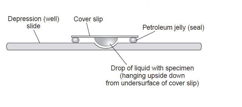 Figure: Hanging Drop Technique
Why It Matters
When a patient presents with sudden, profuse rice-watery stool during a suspected cholera outbreak, a hanging drop prep can give a presumptive answer in minutes — long before culture results are back. Vibrio cholerae shows a distinctive darting motility: fast, erratic, shooting-star-like movement that looks visibly different from the slower, purposeful motility of typical Enterobacteriaceae. In an outbreak setting, that visual alone is often enough to start oral rehydration and alert public health authorities while confirmatory culture is still pending.
The same basic technique also plays a role in a very different, high-stakes scenario: distinguishing Bacillus anthracis from its close, harmless relatives in the B. cereus group. True B. anthracis is classically nonmotile, while B. cereus and most other Bacillus species are motile — so a quick motility check is one of the first practical steps in ruling anthrax in or out when a concerning Bacillus isolate turns up, well before more specialized confirmatory testing is run.
And on the obstetric/neonatal side: Listeria monocytogenes shows a characteristic tumbling motility in hanging drop preps from overnight broth culture — a useful clue in suspected listerial meningitis or perinatal infection workups.
Principle
In a true hanging drop, the culture drop hangs freely from the coverslip with no pressure or compression from above — unlike a standard wet mount, where the coverslip presses directly onto the slide. That free-hanging environment is what allows organisms to move naturally and lets you reliably tell apart three different things that can all look like motion under the microscope:
| Movement type | What it looks like | What it means |
|---|---|---|
| True motility | Organisms actively change position relative to each other — purposeful, directional | Genuinely motile organism |
| Brownian movement | Organisms jiggle or vibrate in place but stay in the same position relative to one another | Not motility — molecular bombardment by water molecules, seen in motile and nonmotile organisms alike |
| Passive/convectional drift | The entire field drifts uniformly in one direction together | Not motility — usually evaporation or temperature currents in the fluid, not the organism's own movement |
This table is the single most important thing to internalize before reading any result — every other interpretation in this test depends on telling these three apart correctly.
Materials Required
- Glass slides with a concave depression (or a regular slide with a paraffin/adhesive-tape ring to create one)
- Petroleum jelly (vaseline) or another sticky sealant
- Inoculating loop
- Coverslip
- Microscope — ideally with oil immersion capability for closer inspection once organisms are located
- Bunsen burner
- A young, actively growing broth culture (4–6 hours for fast growers, or as fresh as practical for the organism) — see the culture-age note below
Quality Control
| Organism | Expected result |
|---|---|
| Proteus mirabilis or E. coli | Motile (positive control) |
| Klebsiella pneumoniae or Shigella sonnei | Nonmotile (negative control) |
Technologist competency should also be periodically validated using known motile organisms such as enterococci or Listeria in a broth assay — motility in these groups can be subtle, and they're a good test of whether a technologist is reading the slide correctly, not just whether the slide was prepared correctly.
Slide Preparation
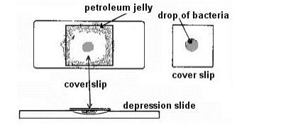 Figure: Slide Preparation for Hanging Drop Method
- If using a flat slide, apply a paraffin or adhesive-tape ring to create a circular concavity. (Skip this step if using a true depression slide.)
- Hold a clean coverslip by its edges and dab vaseline on its corners with a toothpick.
- Place a light loopful of fresh broth culture in the center of the coverslip — it should not look visibly turbid. A heavy inoculum overcrowds the field, making it impossible to track individual cells and increasing the chance of convection currents that can mimic motility.
- Invert the concave slide over the drop so the vaseline seals the coverslip to the slide around the concavity.
- Turn the slide right-side up so the coverslip is on top, and let organisms settle for about a minute.
Microscopic Observation
- Position the slide so an edge of the drop sits under the low-power objective.
- Lower the objective to its lowest position and close the diaphragm. This isn't an arbitrary step — unstained bacteria are nearly transparent and have very little contrast against the surrounding fluid. Closing the diaphragm reduces stray light and dramatically improves your ability to actually see them.
- Raise the objective slowly until the edge of the drop appears as an irregular line crossing the field, then center it.
- Swing in the high-dry objective without changing the focus height, and fine-adjust until the edge appears as a thick, dark line.
- Look just inside that line for small, dark or faintly greenish rods or spheres — these are the bacteria. (Remember: high-dry magnifies a little less than half of oil immersion.)
- Adjust the diaphragm as needed to maximize visibility.
- Observe cell morphology and grouping, then assess motility using the true-motility-vs-Brownian-vs-drift distinction above.
- If you need a closer look once organisms are located, oil immersion can be applied directly on top of the coverslip — most labs read motility reliably at high-dry, but oil immersion can help confirm subtler patterns like Listeria tumbling.
- Dispose of the slide and coverslip per your institution's biohazard protocol (see Safety note below) — don't reuse a depression slide without proper decontamination.
Where people actually get confused (students and bench staff alike)
- Brownian movement will be visible on every slide, motile or not. Its presence doesn't confirm motility, and its absence doesn't rule it out — what matters is whether organisms change position relative to each other over time.
- Convection drift at the edge of the drop can fool you. Evaporation at the drop's margin creates currents that sweep the whole field in one direction together. If everything in view is moving the same way at the same speed, that's drift, not motility — read closer to the center of the drop, not right at the edge.
- Culture age matters more than people expect. Many organisms lose motility as a culture ages past log phase — flagella can be shed or expression downregulated in stationary phase. A nonmotile-looking result from an old culture may be a false negative, not a true one. Always use the youngest practical culture.
- An incomplete vaseline seal lets the drop dry out before you finish reading it — if the drop is visibly shrinking or the edges look ragged partway through observation, the seal likely failed; remake the prep rather than trying to interpret a drying sample.
- Vibrio cholerae and Campylobacter darting motility can look almost too fast to be real — tiny dots flickering in and out of the field. That's the expected appearance, not a preparation error.
Safety Note
This test is frequently performed on organisms that may include high-risk pathogens — V. cholerae in outbreak settings, or Bacillus species being screened in a possible anthrax-exclusion context. Handle suspect isolates under your institution's biosafety protocol (appropriate containment level, no unnecessary aerosol generation), and dispose of slides and coverslips in a biohazard sharps/waste stream — soaking used depression slides in a disinfectant (e.g., a lysol-based solution) before reuse, or discarding flat slides outright, rather than simply washing and reusing without decontamination.
Result Interpretation Summary
| Observation | Interpretation |
|---|---|
| Organisms change position relative to each other, directionally | True motility — positive |
| Organisms jiggle in place, stay in the same relative position | Brownian movement only — negative |
| Entire field drifts uniformly together | Passive/convectional drift — not a valid reading, re-examine |
| Fast, erratic, "darting" movement | Consistent with Vibrio cholerae / Campylobacter pattern |
| Tumbling motion, especially from overnight culture | Consistent with Listeria monocytogenes |
| No movement at all, even Brownian artifact ruled out | Nonmotile |
For any organism that reads negative on initial wet mount, repeat after further incubation in broth, or confirm by tube method:
- Nonfermenting Gram-negative rods: incubate at 30°C for 24 hours before re-testing
- Enterococci and Listeria: incubate at 30°C for 2 hours before re-testing
- Most other organisms: incubate at their usual optimal growth temperature, typically 35°C
References
- Jordan, E. O., Caldwell, M. E., & Reiter, D. (1934). Bacterial Motility. Journal of bacteriology, 27(2), 165–174. https://doi.org/10.1128/jb.27.2.165-174.1934
- Luna, V. A., Peak, K. K., Veguilla, W. O., Reeves, F., Heberlein-Larson, L., Cannons, A. C., Amuso, P., & Cattani, J. (2005). Use of two selective media and a broth motility test can aid in identification or exclusion of Bacillus anthracis. Journal of clinical microbiology, 43(9), 4336–4341. https://doi.org/10.1128/JCM.43.9.4336-4341.2005
Frequently Asked Questions
How do I tell true motility from Brownian movement?
My drop dried out before I finished reading it — what went wrong?
Can hanging drop be used to help rule out anthrax?

Tankeshwar Acharya, MSc (Medical Microbiology)
Tankeshwar Acharya is an Assistant Professor in the Department of Microbiology at Patan Academy of Health Sciences (PAHS), Nepal, where he has been teaching and practicing clinical microbiology for over 14 years. He is the founder of Microbe Online, one of the leading free microbiology education resources on the web, covering bacteriology, mycology, parasitology, immunology, and clinical laboratory diagnostics written from direct experience in both the classroom and the diagnostic laboratory.