
Listeria monocytogenes: Properties, Virulence Factors, Pathogenesis, and Lab Diagnosis
Listeria monocytogenes is a psychrotolerant, facultative intracellular food-borne pathogen. Learn its cold enrichment, differential motility, virulence factors (InlA/B, listeriolysin O, ActA actin tails), intracellular lifecycle, three-barrier crossing, listeriosis clinical syndromes, lab diagnosis (CAMP test, culture), and ampicillin treatment.

On this page
In 2011, a listeriosis outbreak linked to whole cantaloupes from a Colorado farm killed 33 people and hospitalized 147 across 28 US states (the deadliest foodborne illness outbreak in the United States in nearly 90 years). The contamination occurred during post-harvest handling and packing; the cantaloupes were not cooked before consumption. Many victims were elderly, immunocompromised, or pregnant.
Listeria monocytogenes is responsible for one of the most clinically severe foodborne diseases despite relatively low incidence. While Salmonella and Campylobacter infect millions annually, listeriosis is far rarer but its case fatality rate is approximately 20–30%, compared to less than 1% for most other foodborne pathogens. This lethality reflects two biological features that make L. monocytogenes uniquely dangerous:
- It grows in the refrigerator at 4°C (the temperature most people assume eliminates bacterial risk in stored food).
- It lives inside host cells, protected from antibodies, complement, and many antibiotics making it disproportionately dangerous in people with impaired cell-mediated immunity.
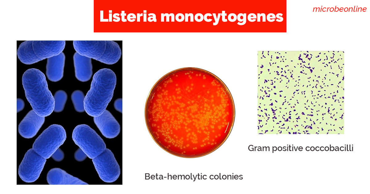 Figure: Listeria monocytogenes in Gram staining and Culture
General Properties
Listeria monocytogenes is a facultative intracellular, Gram-positive coccobacillus with several distinctive properties that set it apart from other food-borne pathogens:
- It shows tumbling type of motility at 25°C but non-motile at 37°C (called differential motility, which is due to the temperature-dependent flagella expression).
- Unlike most other harmful bacteria, L. monocytogenes grow slowly on foods stored in a refrigerator (4°C). It is commonly found in stored foods especially aged soft cheeses, packaged meats, milk, and cold salads.
- Growth improves if the specimen is refrigerated at 4°C and cultured at frequent intervals, this procedure is called cold enrichment.
- It shows a positive CAMP test when streaked at a right angle to beta-hemolytic Staphylococcus aureus (enhanced arrowhead hemolysis).
- Beta-hemolytic (narrow zone of hemolysis) on blood agar; this is the key visual identification feature that distinguishes L. monocytogenes from non-pathogenic Listeria species and from similar-appearing organisms like Enterococcus
- Catalase-positive, oxidase-negative distinguishes from streptococci (catalase-negative) and Gram-positive rods
- Temperature range: 1–45°C; grows at refrigerator temperatures (psychrotolerant); optimal 30–37°C
- Salt tolerance: Grows in up to 10% NaCl; relevant to growth in processed meats and cheeses
High-risk foods for Listeria contamination:
| Food category | Examples | Risk note |
|---|---|---|
| Soft cheeses | Brie, Camembert, feta, queso fresco | Unpasteurised or post-pasteurisation contamination; not cooked before consumption |
| Ready-to-eat deli meats | Sliced ham, salami, hot dogs | Processing equipment contamination; may not be reheated |
| Smoked fish | Smoked salmon, gravlax | Cold-smoked; not fully cooked |
| Raw sprouts | Alfalfa, bean sprouts | Soil contamination; not cooked |
| Refrigerated paté | Liver paté, meat spreads | Long refrigerator shelf life allows multiplication |
Epidemiology and High-Risk Groups
L. monocytogenes is ubiquitous in the environment. It is found in soil, water, vegetation, animal feces, and silage. Human infection occurs almost exclusively through ingestion of contaminated food.
Unlike most foodborne pathogens:
- Incubation period is long: 1–70 days (median ~21 days); patients may not connect illness to a specific meal
- Infectious dose is uncertain: immunocompromised individuals may be infected by much lower doses than healthy adults
- Small proportion of infections become invasive — most healthy adults clear the organism at the gut level; invasive disease requiring hospitalization occurs almost exclusively in the high-risk groups below
High-risk groups:
| Group | Risk and mechanism | Clinical manifestation |
|---|---|---|
| Pregnant women | T-cell immunity naturally suppressed during pregnancy | Usually mild flu-like illness in mother; severe fetal disease: premature labor, stillbirth, neonatal sepsis, neonatal meningitis |
| Neonates | Immature immune system; maternal–fetal transmission | Early-onset (≤7 days): septicemia, granulomatosis infantiseptica; late-onset (>7 days): meningitis |
| Elderly (>65 years) | Declining cell-mediated immunity | Bacteremia, meningitis, meningoencephalitis |
| Immunocompromised | HIV/AIDS, transplant, chemotherapy, corticosteroids | Bacteremia; CNS disease; mortality up to 30% |
| Healthy adults | Rarely invasive; high-dose exposure | Febrile gastroenteritis (self-limiting); no invasive disease |
The immune system clue: The pattern of who gets invasive listeriosis (pregnant, neonates, elderly, immunosuppressed) points directly to the pathogenesis: L. monocytogenes depends on cell-mediated immunity for clearance. Anyone whose cell-mediated immunity is compromised is vulnerable. This is why listeriosis is considered an AIDS-defining illness and why transplant recipients are warned about soft cheeses.
Transmission
Listeria monocytogenes can be found in a variety of dairy products, vegetables, fish, and meat products. Unlike most other harmful bacteria, it can grow on foods stored in a refrigerator. Consumption of food contaminated with Listeria monocytogenes is the primary mode of transmission of listeriosis. Listeriosis can also be transmitted from mother to fetus if infection occurs during pregnancy.
As L. monocytogenes is able to cross the placenta, listeriosis is a major cause of perinatal and neonatal disease, typically resulting in abortion.
Pathogenesis: The Intracellular Lifecycle
The pathogenesis of listeriosis is defined by the organism's extraordinary ability to invade non-phagocytic cells, escape the phagosome, replicate in the cytoplasm, and spread directly to adjacent cells. All of these steps happen without ever being exposed to the extracellular immune environment.
The lifecycle in six steps:
1. INTERNALISATION (InlA/InlB)
   L. monocytogenes attaches to E-cadherin (via InlA) and Met receptor (via InlB)
   on intestinal epithelial cells → bacteria are engulfed into a membrane vacuole
                    ↓
2. PHAGOSOME ESCAPE (Listeriolysin O + Phospholipases)
   LLO creates pores in the vacuolar membrane
   PlcA and PlcB digest the membrane → bacteria escape into the cytoplasm
   (before lysosomal fusion, bacterium avoids intracellular killing)
                    ↓
3. INTRACELLULAR REPLICATION
   Bacteria multiply freely in the nutrient-rich cytoplasm
   PrfA (master regulator) coordinates virulence gene expression
                    ↓
4. ACTIN TAIL FORMATION (ActA)
   ActA on bacterial surface nucleates host actin filaments
   Rocket-like F-actin tails form → bacteria propelled at 11 μm/minute
                    ↓
5. CELL-TO-CELL SPREAD
   Bacteria propelled to cell surface → protrusion into adjacent cell
   Engulfed in double-membrane vacuole in new cell
                    ↓
6. DOUBLE VACUOLE ESCAPE (LLO + PlcB again)
   Same mechanism as before: LLO and PlcB lyse the double membrane, and a new cycle begins
Why this lifecycle is clinically unique: At no point in steps 3–6 does L. monocytogenes re-enter the extracellular environment. It spreads from cell to cell entirely within host cells protected from:
- Antibodies (cannot reach intracellular bacteria)
- Complement (same)
- Many antibiotics (poor intracellular penetration)
This is why cell-mediated immunity (CD8+ T cells and NK cells) is the primary defense against Listeria, not antibody-mediated immunity. Immunosuppression of cell-mediated immunity (pregnancy, steroids, AIDS, transplant) = vulnerability.
Virulence Factors
PrfA (The Master Regulator)
All L. monocytogenes virulence genes are co-regulated by PrfA (Positive Regulatory Factor A), a transcriptional activator encoded within LIPI-1 (Listeria Pathogenicity Island 1):
- Activates expression of hly (LLO), actA, plcA, plcB, inlA, inlB, and other virulence genes
- Expression is temperature-regulated, upregulated at 37°C (host temperature), low at environmental temperatures
- Mutations in prfA abolish virulence, strains without functional PrfA cannot cause disease
- PrfA is the single most important target for understanding Listeria virulence gene regulation
 Figure: Listeria cell cycle (Image source: Pasteur Institute)
Internalin A and B (InlA and InlB): Invasion Proteins
Internalin A (InlA):
- Surface protein; binds E-cadherin on intestinal epithelial cells
- Mediates entry across the intestinal epithelium, the first barrier crossing
- Also mediates placental invasion, E-cadherin is expressed on syncytiotrophoblasts; explains maternal-fetal transmission
- Species specificity: InlA binds human and guinea pig E-cadherin but NOT murine E-cadherin. This explains why standard mouse models underestimate listeriosis risk.
Internalin B (InlB):
- Binds Met receptor (hepatocyte growth factor receptor) and complement component C1q receptor
- Mediates entry into hepatocytes and other cell types not expressing E-cadherin
- Contributes to hepatic invasion and hematogenous spread
Listeriolysin O (LLO): The Phagosome Escape Factor
LLO is a cholesterol-dependent cytolysin (CDC): the same family as streptolysin O (S. pyogenes), pneumolysin (S. pneumoniae), and perfringolysin (C. perfringens):
- Creates pores in the phagosomal membrane at low pH → bacteria escape to cytoplasm before lysosomal fusion
- pH-dependent activation (active at pH 5–6 of phagosome; less active at neutral cytoplasmic pH). It prevents bacterial self-destruction in cytoplasm
- Beta-hemolysis on blood agar is the laboratory manifestation of LLO activity, the narrow hemolysis zone around colonies is diagnostic
- LLO is also active in the double-membrane vacuole during cell-to-cell spread (with PlcB)
Actin Assembly-Inducing Protein (ActA): The Motility Engine
ActA is the uniquely Listeria-specific virulence factor that enables intracellular motility:
- Surface protein expressed on the bacterial pole
- Nucleates host actin monomers via the Arp2/3 complex, same mechanism as cell migration
- Produces rocket-like F-actin tails propelling bacteria at up to 11 µm/minute
- Enables bacteria to be propelled through the cytoplasm and into adjacent cells which enables direct cell-to-cell spread with no extracellular exposure
 Figure: Actin filament and intracellular spread of Listeria monocytogenes.
Phospholipases C (PlcA and PlcB)
Two distinct phospholipases act sequentially in phagosome escape:
- PlcA (PI-PLC): Phosphatidylinositol-specific; cleaves GPI-anchored proteins on phagosomal membrane; acts early in single-membrane phagosome escape
- PlcB (PC-PLC): Broad-range; essential for escaping the double-membrane vacuole during cell-to-cell spread; acts with LLO in the second escape step
Virulence Factor Summary
| Factor | Function | Barrier/Step |
|---|---|---|
| PrfA | Master regulator of all virulence genes | Controls everything |
| InlA | Binds E-cadherin → intestinal epithelial entry; placental crossing | Intestinal + placental barriers |
| InlB | Binds Met/C1q → hepatocyte and diverse cell entry | Hepatic invasion |
| LLO | Pore formation → single-membrane phagosome escape; double-membrane escape | Step 2 + Step 6 |
| ActA | Actin tail formation → intracellular motility; cell-to-cell spread | Steps 4–5 |
| PlcA | PI-specific membrane cleavage; early phagosome disruption | Step 2 |
| PlcB | Broad-range; double-membrane vacuole disruption | Step 6 |
Three Barriers: Why Listeria Reaches the Brain and Placenta
L. monocytogenes can cross three host tissue barriers that most bacteria cannot:
| Barrier | Mechanism | Clinical consequence |
|---|---|---|
| Intestinal epithelium | InlA binds E-cadherin on enterocytes; transcytosis | Systemic bacteremia |
| Blood-brain barrier | InlA + InlB enable crossing of brain capillary endothelium; "Trojan horse" in monocytes | Meningitis, meningoencephalitis, brain abscess |
| Placenta (fetoplacental barrier) | InlA binds E-cadherin on syncytiotrophoblasts | Fetal infection: abortion, stillbirth, neonatal sepsis |
The "Trojan horse" mechanism: L. monocytogenes also uses infected circulating monocytes and macrophages as vehicles to cross the blood-brain and placental barriers; the bacteria are transported inside the immune cells that were supposed to destroy them.
Diseases
Listeriosis is one of the most serious foodborne diseases. Clinical manifestation of listeriosis depends on the age of the patient and other underlying conditions such as pregnancy, immunosuppression, etc.
Depending on the immune status of the person and the part of the body affected, listeriosis can cause variety of symptoms ranging from milder flu-like illness to convulsion and death.
- Pregnant women: It affects both the mother and the fetus. Mother mostly experiences mild illness (flu-like symptoms) but it causes severe disease in the fetus or newborn baby (such as abortion, preterm delivery, newborn death, and neonatal meningitis).
- People other than pregnant women: Fever, muscle aches, headache, stiff neck, confusion, loss of balance, and convulsions.
Most people with invasive listeriosis require hospital care, and about one in five people with the infection die.
Laboratory Diagnosis
Sample: CSF, blood, feces or placental tissue or any other tissue specimens depending on the suspected disease and/or clinical presentation.

Direct wet mount: Listeria monocytogenes exhibits characteristic end-over-end tumbling motility when incubated in nutrient broth at room temperature for 1 to 2 hours. Presumptive identification of L. monocytogenes can be done by observation of such motility by direct wet mount.
Alternatively, suspected colonies of L. monocytogenes can be stabbed into a tube of semisolid agar and incubated at 25°C for 18 hours. Characteristics umbrella-shaped motility pattern is seen after overnight incubation.
Gram staining: Regular, short, gram-positive rods or coccobacilli occurring in pairs (resembles streptococci).
Cold enrichment
Cold enrichment is done for the isolation of Listeria monocytogenes from the placenta, tissues as well as specimens from heavily contaminated sources such as feces. The specimen is inoculated into a nutrient broth or trypticase soy broth and incubated at 4°C for one month and sub cultured weekly.
 Figure: Beta-hemolytic colonies of L. monocytogenes
Culture: Listeria monocytogenes can be cultured on conventional media incorporating sheep, horse or rabbit blood. Listeria monocytogenes produces round, smooth, translucent colonies with a narrow zone of beta-hemolysis. Colonies are small and give a blue-green sheen under obliquely transmitted (Henry's) illumination.
Other characteristics that helps in the identification of this bacteria are:
- Catalase positive
- Voges-Proskauer-positive
- Esculin positive: L. monocytogenes is able to hydrolyze esculin in the presence of 40% bile. Esculin hydrolysis is indicated by the blackening of the medium.
- CAMP test positive
- L. monocytogenes shows an arrowhead (flame-shaped) enhancement of beta-hemolysis when streaked perpendicular to a beta-hemolytic Staphylococcus aureus streak. The zone of enhanced hemolysis widens toward the S. aureus streak. This is the positive CAMP test for Listeria.
- Streptococcus agalactiae (GBS) gives the same arrowhead enhancement in the forward CAMP test, so the shape does not separate the two organisms. They are distinguished instead by catalase (L. monocytogenes positive, GBS negative) and Gram stain (Listeria is a Gram-positive rod, GBS a Gram-positive coccus in chains). The bow-tie pattern belongs to the reverse CAMP test (Clostridium perfringens), not to GBS. For full procedure and controls, see the CAMP test article.
Molecular diagnosis
Detection of Listeria monocytogenes DNA in CSF and tissue (fresh or paraffin blocks)
Commercial methods
- AccuPROBE Listeria monocytogenes Culture Identification kit: Rapid DNA probe test, which utilizes the technique of nucleic acid hybridization for the identification of Listeria monocytogenes isolated from culture.
- Matrix-assisted laser desorption/ionization-time of flight (MALDI-TOF) method
- GeneQuence for Listeria monocytogenes
Treatment
Treatment decisions belong to the treating clinician; what follows is the microbiological basis for them: the susceptibility and intrinsic-resistance pattern of L. monocytogenes that guides therapy.
The single most important microbiological fact is that L. monocytogenes is intrinsically resistant to all cephalosporins. This matters because cephalosporins are a mainstay of empirical therapy for bacterial meningitis before the organism is known, and they do not cover Listeria.
L. monocytogenes is consistently susceptible to ampicillin (and amoxicillin), which is why ampicillin is regarded as the agent of choice in published guidance. In severe invasive disease such as meningitis or bacteremia, ampicillin is often combined with gentamicin, because the combination is synergistic in vitro and gentamicin contributes activity against the intracellular organism. In patients who cannot receive penicillins, the organism's susceptibility to trimethoprim-sulfamethoxazole (co-trimoxazole) makes it the usual reported alternative for invasive listeriosis. The choice of agent, dose, and duration is a clinical decision made by the treating physician.
No vaccine is available. Prevention relies on food safety: avoiding high-risk foods in high-risk groups, adequate cooking, and proper refrigerator hygiene.
Key exam facts
| Feature | Listeria monocytogenes |
|---|---|
| Gram reaction / shape | Gram-positive short rod or coccobacillus (can resemble streptococci) |
| Catalase / oxidase | Catalase-positive, oxidase-negative |
| Motility | Tumbling motility at 25°C, non-motile at 37°C (differential motility) |
| Stab-culture pattern | Umbrella (sombrero) growth at 25°C |
| Hemolysis | Beta-hemolytic (narrow zone) on blood agar |
| CAMP test | Positive (arrowhead), like Streptococcus agalactiae; separated by catalase and Gram stain |
| Esculin / VP | Esculin-positive (bile esculin), Voges-Proskauer-positive |
| Growth temperature | 1 to 45°C; grows at 4°C (psychrotolerant); optimum 30 to 37°C |
| Salt tolerance | Grows in up to 10% NaCl |
| Habitat / source | Ubiquitous; soft cheese, deli meats, smoked fish, refrigerated ready-to-eat foods |
| Lifestyle | Facultative intracellular |
| Key virulence factors | InlA/InlB (entry), listeriolysin O (phagosome escape), ActA (actin tails), PlcA/PlcB |
| Master regulator | PrfA (temperature-regulated) |
| Main defense against it | Cell-mediated immunity (not antibody) |
| High-risk groups | Pregnant women, neonates, elderly, immunocompromised |
| Case fatality (invasive) | About 20 to 30% |
| Drug of choice | Ampicillin (with gentamicin in severe disease); intrinsically resistant to cephalosporins |
Where students get confused
Run the motility test at 25°C, not 37°C. Listeria expresses flagella and tumbles at room temperature but is non-motile at body temperature. Testing at 37°C (the default incubator setting) gives a false non-motile result and misses the classic clue. This temperature dependence is called differential motility.
Cephalosporins do not cover Listeria, which matters in meningitis. Empirical meningitis therapy often uses a third-generation cephalosporin, but L. monocytogenes is intrinsically resistant to all cephalosporins. This is why ampicillin is added when Listeria is a possibility, such as in neonates, the elderly, and the immunocompromised.
The CAMP arrowhead does not by itself mean Streptococcus agalactiae. Both L. monocytogenes and group B Streptococcus give a positive arrowhead CAMP test. They are separated by catalase (Listeria positive, GBS negative) and Gram stain (Listeria is a Gram-positive rod, GBS a coccus in chains), not by the CAMP shape.
It grows in the refrigerator, so cold storage is not protective. Unlike most foodborne pathogens, L. monocytogenes multiplies at 4°C. This is why long-refrigerated ready-to-eat foods (deli meats, soft cheese, smoked fish) are the classic sources, and why "it was refrigerated" does not make a food safe.
Cell-mediated immunity, not antibody, is the defense. Because Listeria lives and spreads inside host cells, antibodies and complement cannot reach it. This is why the people who get invasive disease are those with impaired cell-mediated immunity (pregnancy, steroids, transplant, HIV), and why it is an AIDS-defining illness.
A Gram stain can mislead toward streptococci. Listeria is a short Gram-positive rod that can appear in pairs and look coccoid, resembling Streptococcus. Catalase separates them quickly: Listeria is catalase-positive, streptococci are catalase-negative.
How to Remember
Listeria is defined by two unique properties:
- Grows at 4°C (the refrigerator temperature) → infects refrigerated ready-to-eat food
- Intracellular lifecycle → cell-mediated immunity is the only defense → dangerous in pregnancy, immunosuppression, elderly
The intracellular lifecycle as a logical chain: InlA/B (get in) → LLO + PlcA (escape phagosome) → ActA (make actin rocket) → spread to next cell → LLO + PlcB (escape double membrane) → repeat
Every step has a named virulence factor. Each step explains the next.
The differential motility trick: 25°C: motile (tumbling), flagella expressed but at 37°C: non-motile as flagella suppressed. The motility test is run at 25°C (room temperature), not 37°C (incubator temperature). Testing at the wrong temperature gives a false non-motile result.
Three barriers, three outcomes: Intestinal barrier → bacteremia Blood-brain barrier → meningitis/encephalitis Placental barrier → fetal death / neonatal sepsis
Frequently Asked Questions
Is Listeria monocytogenes Gram-positive or Gram-negative?
Is Listeria monocytogenes Gram-positive or Gram-negative?
Gram-positive. It is a short Gram-positive rod or coccobacillus that can occur in pairs and resemble streptococci. It is also catalase-positive, which distinguishes it from the catalase-negative streptococci it can look like.
What is the tumbling motility of Listeria?
What is the tumbling motility of Listeria?
Listeria shows a characteristic end-over-end "tumbling" motility when grown in nutrient broth at room temperature (about 25°C) for 1 to 2 hours, seen on a wet mount. In a semisolid agar stab incubated at 25°C, it produces an umbrella-shaped (sombrero) growth pattern. It is motile at 25°C but non-motile at 37°C, because the flagella are only expressed at the lower temperature. This is why the motility test must be done at 25°C.
Why is Listeria dangerous in pregnancy?
Why is Listeria dangerous in pregnancy?
Cell-mediated immunity is naturally suppressed during pregnancy, and cell-mediated immunity is the main defense against this intracellular organism. The mother usually has only a mild flu-like illness, but Listeria can cross the placenta (using InlA to bind E-cadherin on the placenta) and cause miscarriage, stillbirth, preterm labor, or neonatal sepsis and meningitis.
Why doesn't refrigeration prevent Listeria?
Why doesn't refrigeration prevent Listeria?
Because L. monocytogenes is psychrotolerant: it grows at refrigerator temperatures (4°C), unlike most foodborne bacteria. Long-refrigerated ready-to-eat foods such as deli meats, soft cheeses, and smoked fish are therefore classic sources, and cold storage actually allows the organism to multiply over time.
How is Listeria meningitis treated, and why not a cephalosporin?
How is Listeria meningitis treated, and why not a cephalosporin?
The drug of choice is ampicillin, often combined with gentamicin in severe disease. Listeria is intrinsically resistant to all cephalosporins, so the cephalosporin used empirically for bacterial meningitis does not cover it. This is why ampicillin is added when Listeria is a possible cause, particularly in neonates, the elderly, and immunocompromised patients. Specific doses are clinical decisions.
What is differential motility?
What is differential motility?
It is the property of being motile at one temperature but not another. Listeria is motile (tumbling) at 25°C, where it expresses flagella, and non-motile at 37°C, where flagella expression is suppressed. This temperature-dependent motility is a key identifying feature and the reason the motility test is performed at 25°C.
References
- Procop, G. W., & Koneman, E. W. (2017). Koneman's Color Atlas and Textbook of Diagnostic Microbiology (7th ed.). Wolters Kluwer.
- Tille, P. (2022). Bailey & Scott's Diagnostic Microbiology (15th ed.). Elsevier.
- Listeria (Listeriosis)| Listeria | CDC. https://www.cdc.gov/listeria/
- Smith GA et al. (1995). The two distinct phospholipases C of L. monocytogenes have overlapping roles in escape from a vacuole and cell-to-cell spread. Infection and Immunity, 63(11), 4231.
- Wei Z et al. (2005). L. monocytogenes phosphatidylinositol-specific phospholipase C has evolved for virulence. PNAS, 102(36), 12927. doi:10.1073/pnas.0501725102
- Swaminathan B & Gerner-Smidt P. (2007). The epidemiology of human listeriosis. Microbes and Infection, 9(10), 1236–1243. https://doi.org/10.1016/j.micinf.2007.05.011
- Cossart P. (2011). Illuminating the landscape of host-pathogen interactions with the bacterium Listeria monocytogenes. PNAS, 108(49), 19484–19491. https://doi.org/10.1073/pnas.1112371108

Tankeshwar Acharya, MSc (Medical Microbiology)
Tankeshwar Acharya is an Assistant Professor in the Department of Microbiology at Patan Academy of Health Sciences (PAHS), Nepal, where he has been teaching and practicing clinical microbiology for over 14 years. He is the founder of Microbe Online, one of the leading free microbiology education resources on the web, covering bacteriology, mycology, parasitology, immunology, and clinical laboratory diagnostics written from direct experience in both the classroom and the diagnostic laboratory.
Comments
No comments yet. Be the first to share your thoughts.
Leave a comment
All comments are reviewed before they appear.