Peptidoglycan: Structure and Why It Is the Target of Penicillin
Peptidoglycan (murein) — structure, NAG-NAM backbone, cross-linking, transpeptidase mechanism, gram-positive vs gram-negative differences, and why it is the single most important antibiotic target in medicine. With mnemonics and clinical stories.
Why peptidoglycan is the single most important molecule in antibacterial medicine
Before diving into the chemistry, understand this: peptidoglycan is the reason penicillin works.
Alexander Fleming discovered penicillin in 1928, but it took decades to understand exactly why it killed bacteria without harming human cells. The answer is peptidoglycan; a molecule that exists in virtually every bacterium but in no human cell whatsoever. This single fact is the foundation of an entire category of medicine: beta-lactam antibiotics, which today include penicillin, cephalosporin, carbapenems, and monobactams: collectively the most widely prescribed class of antibiotics in the world.
The core principle that makes this clinically powerful: any drug that disrupts peptidoglycan synthesis will kill bacteria while leaving human cells completely unharmed because human cells have no peptidoglycan to disrupt. This is called selective toxicity, and it is the holy grail of pharmacology. Few other antibiotic targets achieve this degree of safety margin.
Understanding peptidoglycan structure, therefore, is not abstract biochemistry — it is understanding the molecular basis of how a huge proportion of all antibiotics work, and why bacteria that modify or hide their peptidoglycan become resistant.
The term peptidoglycan was derived from the peptides and the sugars (glycan) that make a molecule; it is also called ‘murein’ or ‘mucopeptide.’ This complex interwoven network of sugar polymer and amino acids surrounds the entire bacterial cell. It provides structural rigidity and gives the characteristic shape for that bacterium.
Peptidoglycan is found only in bacterial cell walls but not in human cells. Peptidoglycan is also absent from the cell walls of Archaea, so it is regarded as a key biomarker of bacteria. Peptidoglycan is a good target for antibacterial drugs such as penicillin, cephalosporin, and vancomycin, which inhibit the synthesis of peptidoglycan by inhibiting transpeptidase reactions.
Penicillin and cephalosporin are not effective for Mycoplasma pneumoniae as it lacks cell wall (peptidoglycan).
Structure of Peptidoglycan
Peptidoglycan consists of a carbohydrate backbone (glycan chain) composed of alternating units of N-acetylglucosamine (NAG) and N-acetylmuramic acid (NAM) molecules attached through β-1,4-glycosidic bonds. The covalent bonds between NAG-NAM form a sheet-like structure around the bacterium.
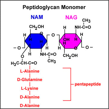 Figure: Peptidoglycan monomer
Adjacent glycan chains are held together by cross-links between the short peptide stems that project from each NAM. This cross-linking, not the sugar backbone, is what gives the wall its strength in the second dimension, and the density of cross-linking varies between species. Staphylococcus aureus is very highly cross-linked; E. coli much less so
A short peptide stem hangs from each NAM. As it is first built, this stem is a pentapeptide: L-alanine, D-glutamic acid, then either L-lysine (most gram-positives) or diaminopimelic acid (DAP) (most gram-negatives and Bacillus, Clostridium), followed by two terminal D-alanine (D-alanyl-D-alanine).
That terminal D-Ala-D-Ala pair is the single most important detail in the whole molecule for medicine: it is the handle transpeptidase grabs to make a cross-link, the structure beta-lactams imitate, and the site vancomycin binds. During cross-linking the outermost D-alanine is cleaved off, so the mature, cross-linked stem is a tetrapeptide. This is why textbook diagrams sometimes show five residues and sometimes four; they are showing the stem before and after cross-linking.
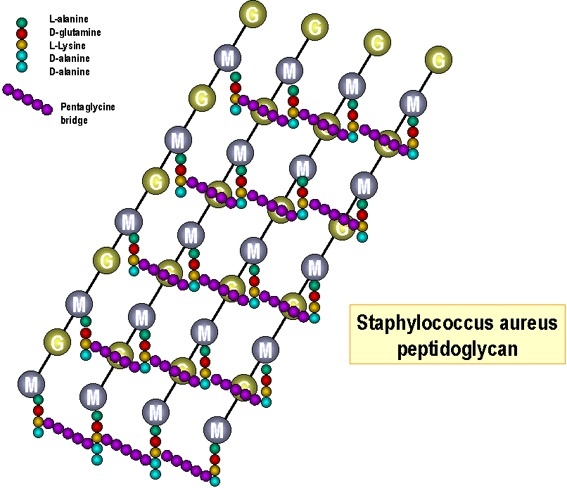 Figure: Peptidoglycan structure of Staphylococcus aureus
Peptidoglycan is unusual in containing D-amino acids: D-glutamic acid and D-alanine (two D-alanines in the newly made stem). Proteins, by contrast, are built almost entirely from L-amino acids. These D-forms resist most host proteases, which cannot cleave them, and this is part of why the wall is durable.
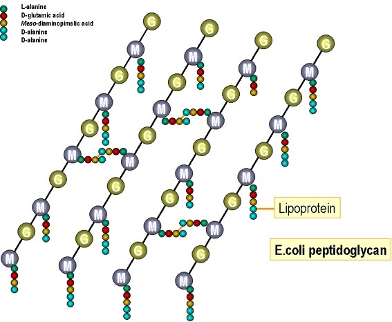 Figure: Peptidoglycan structure of E. coli
In gram-negative bacteria such as Escherichia coli, peptidoglycan cross-linkage occurs by a peptide bond formed between DAP and the terminal D-alanine of another glycan chain.
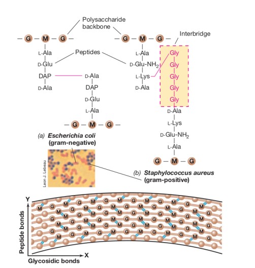 In gram-positive bacteria, cross-linkage may occur through a short peptide inter-bridge (e.g., glycine inter-bridge in Staphylococcus aureus) between L-lysine of one glycan chain and the D-alanine on the adjacent glycan chain.
The analogy that makes peptidoglycan structure unforgettable
Think of peptidoglycan as a chain-link fence wrapped around a balloon.
- The glycan backbone (alternating NAG-NAM units) is like the long horizontal wires of the fence, strong in one direction but not rigid on their own
- The peptide cross-links are like the vertical connectors welding each horizontal wire to its neighbors. These create the actual mesh structure that gives the fence its strength
- The balloon inside is the bacterial plasma membrane, under enormous internal pressure. The cytoplasm is far more concentrated than the watery environments most bacteria live in, so water constantly tries to rush in by osmosis.
- Without the fence, the balloon would simply burst from internal pressure. This is exactly what happens to a bacterium when its peptidoglycan is destroyed
This is why every mechanism that disrupts peptidoglycan (whether an antibiotic, lysozyme, or an immune attack) has the same ultimate effect: osmotic lysis. The cell, no longer able to contain its own internal pressure, bursts.
The pressure is real and enormous: the internal osmotic pressure of a typical gram-positive bacterium is comparable to the pressure inside a car tire (15–25 atmospheres). The peptidoglycan sacculus must withstand this continuously, every second of the bacterium's life.
Diversity of Peptidoglycan
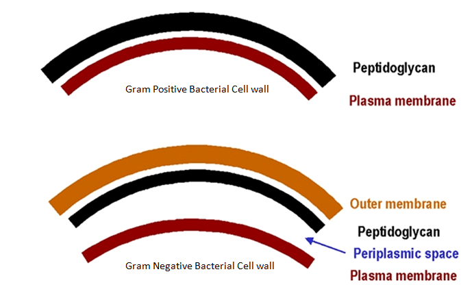 Figure: Difference between Gram-positive and Gram-negative bacterial cell wall
Peptidoglycan is the outermost cell wall layer of gram-positive bacteria. In gram-negative bacteria, additional layers are present outside this rigid layer, called lipopolysaccharide. The peptidoglycan layer is much thicker in gram-positive than in gram-negative bacteria. In gram-positive bacteria, as much as 90% of the cell wall is peptidoglycan, whereas, in gram-negative bacteria, it is only about 10%.
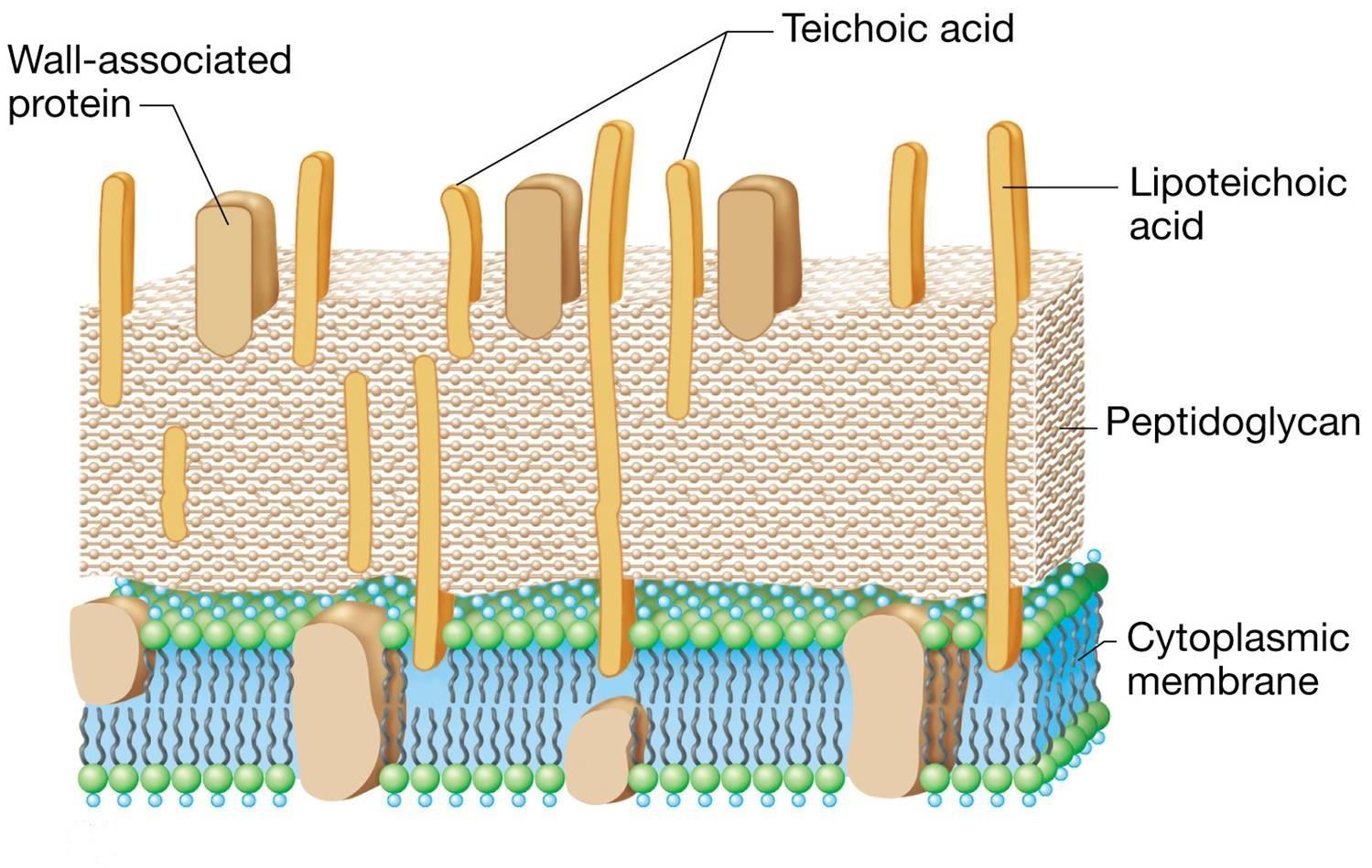 Figure: Gram-positive Cell wall with Teichoic acid
Many Gram-positive bacteria also have teichoic acid and lipoteichoic acid, which are either glycerol phosphate or ribitol phosphate polymers. Lipoteichoic acid penetrates the peptidoglycan layer and is covalently linked to the lipid in the cytoplasmic membrane, whereas teichoic acids mostly anchor to the muramic acid of the peptidoglycan.
Peptidoglycan Across Different Bacterial Groups
| Group | Peptidoglycan present? | Thickness | Clinical implication |
|---|---|---|---|
| Gram-positive bacteria | Yes, abundant | Thick (20–80 nm; up to 90% of cell wall dry weight) | More susceptible to lysozyme and beta-lactams (less barrier to penetrate) |
| Gram-negative bacteria | Yes, present but thin | Thin (2–7 nm; ~10% of cell wall dry weight) | Outer membrane provides additional barrier; some beta-lactams need porins to enter |
| Acid-fast bacteria (Mycobacterium) | Yes, present, modified | Variable; covered by thick mycolic acid layer | Mycolic acid barrier makes most antibiotics ineffective; requires specialized drugs (rifampicin, isoniazid) |
| Mycoplasma | No. Completely absent | None | Intrinsically and completely resistant to ALL beta-lactam antibiotics; no target exists |
| Archaea | No. Different cell wall (pseudopeptidoglycan or other polymers) | — | Peptidoglycan absence is a key taxonomic distinction between Bacteria and Archaea domains |
| Human cells | No | — | The basis of selective toxicity — beta-lactams are safe for humans |
A classic exam fact: Mycoplasma pneumoniae causes "atypical" or "walking" pneumonia, and because it has no cell wall (no peptidoglycan), penicillins and cephalosporins are completely ineffective against it. Treatment requires macrolides (azithromycin) or tetracyclines (doxycycline), which target protein synthesis instead.
How Beta-Lactam Antibiotics Actually Work
The peptide cross-links between adjacent glycan strands are formed by an enzyme called transpeptidase (also called penicillin-binding protein, or PBP). This enzyme catalyzes the final cross-linking step, i.e., joining the D-alanine of one peptide stem to the diaminopimelic acid (or L-lysine) of an adjacent peptide stem, releasing a terminal D-alanine in the process.
Why beta-lactam antibiotics work: the "molecular mimicry" story
Beta-lactam antibiotics (penicillin, amoxicillin, ceftriaxone, meropenem) share the beta-lactam ring which closely mimics the D-alanine-D-alanine terminus of the peptidoglycan peptide stem, which is the natural substrate of transpeptidase.
The transpeptidase enzyme is fooled. It binds to the antibiotic instead of its true substrate and the beta-lactam ring then covalently and irreversibly binds to the active site of the enzyme, permanently inactivating it. With transpeptidase disabled:
- New peptidoglycan cannot be cross-linked
- The cell wall becomes progressively weaker as the bacterium continues trying to grow and divide
- The cell's own autolysins, which normally nick the wall open so new material can be inserted during growth, keep cutting, but no properly cross-linked replacement is laid down
- The weakened cell wall cannot withstand internal osmotic pressure
- The cell undergoes osmotic lysis and dies
This explains a critical clinical fact: beta-lactam antibiotics only kill actively growing and dividing bacteria. A dormant or non-dividing bacterium has no urgent need for new peptidoglycan synthesis, and beta-lactams have minimal effect on it. This is why beta-lactams are described as bactericidal against actively dividing cells but largely ineffective against dormant persister cells or endospores.
→ Beta-Lactam Antibiotics: Mechanism of Action and Resistance
Vancomycin: a different attack on the same target
Beta-lactams disable the transpeptidase enzyme. Vancomycin attacks the same cross-linking step from the opposite side: instead of blocking the enzyme, it binds directly onto the D-Ala-D-Ala terminus of the peptide stem, the enzyme's substrate. With the substrate capped, transpeptidase physically cannot reach it, and cross-linking fails.
Two consequences follow, both heavily examined. First, vancomycin is a large molecule that cannot pass through the porins of the gram-negative outer membrane, so it works only against gram-positive bacteria. Second, resistance has a beautifully logical mechanism: vancomycin-resistant enterococci (VRE) remodel the stem terminus from D-Ala-D-Ala to D-Ala-D-Lactate, a single change that drops vancomycin binding roughly a thousandfold while still allowing cross-linking to proceed. The drug is left grasping for a terminus that is no longer there.
The contrast is the point worth carrying: same target molecule, two enzymes' worth of chemistry, two entirely different drug strategies. Beta-lactams inhibit the worker; vancomycin hides the bricks.
Unique amino acids and sugars found in peptidoglycan layer
- N-acetylmuramic acid and diaminopimelic acid are unique to bacterial cells and have never been found in Archaea’s cell walls or Eukarya.
- Amino acids of the D stereoisomer: D-alanine, and D-glutamic acid are not found in animal proteins.
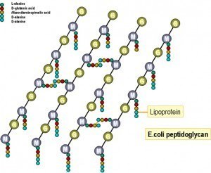 Figure: Peptidoglycan layer of Gram-negative (left) and Gram-positive bacteria (right)
Functions
- Peptidoglycan provides rigid support to bacterial cells and maintains the characteristic shape of the cell.
- Allows bacterial cells to withstand media of low osmotic pressure, such as water.
- Bacteria are divided into two major groups, called gram-positive and gram-negative based on Gram-stain reaction. The difference in the cell-wall structure (thickness of the peptidoglycan layer) plays a major role in the differential staining reactions of bacteria.
Medical Significance
- Peptidoglycan is a good target for antibacterial drugs. Drugs like penicillins, cephalosporins, etc. inhibit the transpeptidase reaction that cross-links adjacent peptide stems involved in peptidoglycan synthesis.
- Lysozyme enzymes in human tears, mucus, and saliva cleave the peptidoglycan backbone, breaking the glycosyl bonds of peptidoglycan, thus providing a major line of defense against bacterial infection.
- Gram-positive bacteria are generally resistant to complement-mediated lysis because the thick peptidoglycan layer in their cell wall prevents the insertion of the membrane attack complex (MAC) into the inner membrane.
How to Learn and Remember Peptidoglycan
The "NAG-NAM" memory trick
NAG and NAM alternate along the backbone, never repeating consecutively, like a checkerboard: NAG-NAM-NAG-NAM. Remember the pattern by thinking "Never Alone, Go together; Never Alone, Meet repeatedly." And to keep straight which is which, read the last letter: NAG ends in G for Glucosamine, NAM ends in M for Muramic acid.
One sentence that captures the entire clinical relevance
"Peptidoglycan is the wall; transpeptidase is the bricklayer; beta-lactams fire the bricklayer; and the cell wall collapses without him."
D-Ala-D-Ala is the target on the target. Peptidoglycan is the target molecule, and within it the two terminal D-alanines are the exact spot every cross-linking drug aims at. Transpeptidase grabs it, beta-lactams imitate it, vancomycin caps it, and VRE bacteria survive by changing the second D-Ala to D-lactate. If you carry one three-letter pair out of bacteriology, make it D-Ala-D-Ala.
Three clinical stories that make peptidoglycan unforgettable
Story 1: Fleming's accidental discovery In 1928, Alexander Fleming returned from holiday to find his Staphylococcus aureus culture plates contaminated with mold and a clear zone around the mold where bacteria had stopped growing. He had discovered penicillin, though he did not yet know it worked by attacking peptidoglycan cross-linking. It took until the 1960s for the precise transpeptidase mechanism to be elucidated. Today, every medical student understands in minutes what took the scientific world more than 30 years to discover.
Story 2: The newborn with "walking pneumonia" that won't respond to amoxicillin A pediatrician treats a child with mild pneumonia symptoms using standard amoxicillin, which is the first-line choice for typical bacterial pneumonia. After 48 hours, there is no improvement. The diagnosis is reconsidered: Mycoplasma pneumoniae; "atypical" pneumonia. Because Mycoplasma has no peptidoglycan at all, amoxicillin had zero chance of working from the start, no matter how high the dose. Switching to azithromycin (targeting ribosomes, not peptidoglycan) resolves the infection within days.
Story 3: Tears as a weapon Human tears, saliva, and nasal secretions contain lysozyme. It is an enzyme that cleaves the β-1,4 glycosidic bond between NAG and NAM, directly destroying the peptidoglycan backbone. This is part of the body's innate immune defense. Every time you cry or your nose runs, you are deploying a peptidoglycan-destroying weapon against invading gram-positive bacteria. Gram-negative bacteria are relatively protected from lysozyme by their outer membrane, which is one reason lysozyme alone is insufficient against gram-negative infections.
Key exam facts in one table
| Question | Answer |
|---|---|
| What are the two sugars in the glycan backbone? | N-acetylglucosamine (NAG) and N-acetylmuramic acid (NAM) |
| What bond links NAG and NAM? | β-1,4 glycosidic bond |
| What enzyme cross-links peptide stems? | Transpeptidase (penicillin-binding protein) |
| What enzyme destroys the glycan backbone? | Lysozyme |
| What is the unique amino acid in gram-negative peptidoglycan? | DAP (in most gram-negatives and in Bacillus/Clostridium); most other gram-positives use L-lysine instead. |
| Which group has thicker peptidoglycan; gram-positive or negative? | Gram-positive (up to 90% of dry weight vs ~10%) |
| Which organism completely lacks peptidoglycan? | Mycoplasma |
| Does Archaea have peptidoglycan? | No. This is a key distinguishing feature from Bacteria |
| Why are beta-lactams useless against Mycoplasma? | No peptidoglycan = no target |
| What structural feature of beta-lactams mimics D-Ala-D-Ala? | The beta-lactam ring |
References and further readings
- Madigan, M. T., Bender, K. S., Buckley, D. H., Sattley, W. M., & Stahl, D. A. (2021). Brock Biology of Microorganisms (16th ed.). Pearson.
- Tille, P. M. (2022). Bailey & Scott's Diagnostic Microbiology (15th ed.). Elsevier.
- Vollmer, W., Blanot, D., & de Pedro, M. A. (2008). Peptidoglycan structure and architecture. FEMS Microbiology Reviews, 32(2), 149–167. https://doi.org/10.1111/j.1574-6976.2007.00094.x
- Silhavy, T. J., Kahne, D., & Walker, S. (2010). The bacterial cell envelope. Cold Spring Harbor Perspectives in Biology, 2(5), a000414. https://doi.org/10.1101/cshperspect.a000414
Frequently Asked Questions
Why are beta-lactam antibiotics safe for human cells?
Why is Mycoplasma pneumoniae resistant to all penicillins and cephalosporins?
What is the difference between peptidoglycan in gram-positive and gram-negative bacteria?
How does lysozyme destroy peptidoglycan?
Why does Archaea lack peptidoglycan?
What is diaminopimelic acid and why is it significant?
How do vancomycin and beta-lactams differ in targeting peptidoglycan?
Why can beta-lactam antibiotics only kill actively dividing bacteria?

Tankeshwar Acharya, MSc (Medical Microbiology)
Tankeshwar Acharya is an Assistant Professor in the Department of Microbiology at Patan Academy of Health Sciences (PAHS), Nepal, where he has been teaching and practicing clinical microbiology for over 14 years. He is the founder of Microbe Online, one of the leading free microbiology education resources on the web, covering bacteriology, mycology, parasitology, immunology, and clinical laboratory diagnostics written from direct experience in both the classroom and the diagnostic laboratory.