
Lipopolysaccharide (LPS): Structure, Endotoxin, and Functions
Lipopolysaccharide (LPS) is the outer-membrane molecule of gram-negative bacteria. Learn its three parts (lipid A, core, O-antigen), what each one does, why lipid A is the toxic endotoxin, and how LPS compares with gram-positive LTA.

On this page
Lipopolysaccharide (LPS) is the large molecule that makes up most of the outer leaflet of the outer membrane, the outermost layer found only in gram-negative bacteria. The outer membrane is a second lipid bilayer, sitting outside the cytoplasmic membrane, and Braun's lipoprotein covalently anchors it to the underlying peptidoglycan layer. So "LPS" names the molecule, and "outer membrane" names the layer that LPS is built into. Keep the two ideas separate.
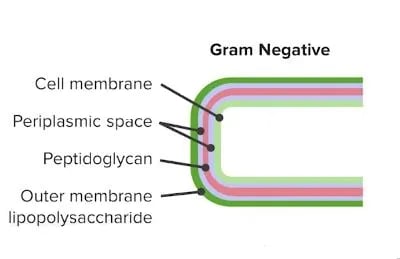 Figure: Cell wall of Gram-negative bacteria
Unlike the cytoplasmic membrane, which is made of phospholipids only, the outer membrane's outer leaflet is built largely from LPS, which carries polysaccharides and proteins.
As in peptidoglycan biosynthesis, LPS molecules are assembled at the plasma or inner membrane.
LPS is a defining feature of gram-negative bacteria and is absent from gram-positive bacteria, which have lipoteichoic acid in that functional role instead.
Why LPS matters
LPS matters for three reasons, and it is worth knowing them before the details so you can see where the topic is going:
- It is the reason gram-negative bacteria cause fever and shock. The toxic part of LPS, called lipid A, is also known as endotoxin. When gram-negative bacteria are killed, lipid A is released and the immune system reacts strongly to it. This is covered later on this page under clinical significance.
- Pharmaceutical quality control: every injectable drug and implantable medical device must be tested for LPS contamination before it can be administered to patients. A contaminated intravenous solution can cause an immediate pyrogenic reaction (fever, rigors, hypotension) from the LPS alone, even with no live bacteria present. How this contamination is detected is covered in our articles on the LAL (bacterial endotoxin) test and on pyrogen and bacterial endotoxin testing methods.
- Vaccine and serotyping tool: the O-antigen component of LPS is so variable between strains that it serves as the basis for identifying and typing many gram-negative pathogens (e.g. E. coli O157:H7, Salmonella Typhimurium (antigenic formula 4,5,12:i:1,2). This same O-antigen is a target for some gram-negative vaccines.
Structure and Composition
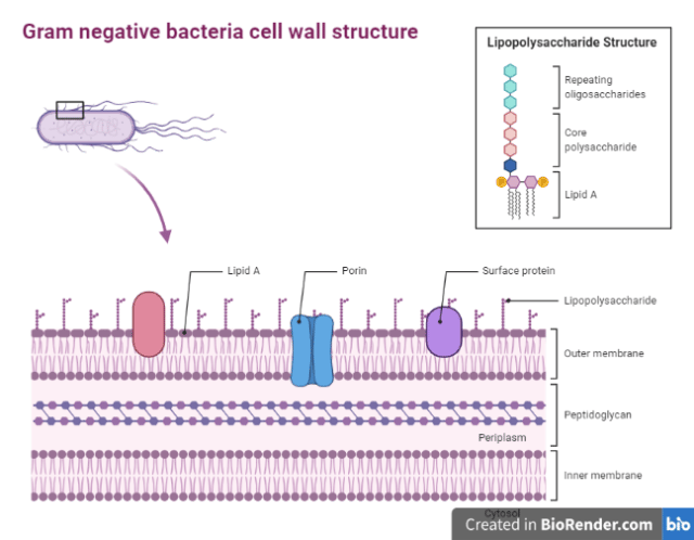 Figure: Cell wall structure of Gram-negative bacteria
The LPS is composed of three distinct units;
- A glucosamine-based glycolipid called Lipid A embeds in the outer leaflet of the outer membrane. Also known as endotoxin, Lipid A is responsible for the toxic effects (fever and shock). Most LPS is released when the cell is killed and the outer membrane disintegrates, though live gram-negatives also shed LPS continuously in outer membrane vesicles. Neisseria meningitidis is notable for shedding large amounts of LPS-bearing outer membrane blebs even while alive, which contributes to the rapid, fulminant course of meningococcal sepsis.
- A core oligosaccharide, a relatively conserved short sugar chain, linked to Lipid A through the characteristic sugar KDO (ketodeoxyoctonate). KDO is a reliable chemical marker of LPS.
- O antigen: An outer polysaccharide consisting of up to 25 repeating units of 3-5 sugars. These are hydrophilic in nature. O antigen is highly varied among species. Example: E. coli O157:H7, which causes food poisoning and hemolytic uremic syndrome. O antigens are used to identify certain organisms in microbiology laboratories.
The O-antigen contributes to virulence not by being toxic (the toxicity of LPS resides entirely in Lipid A) but by helping the bacterium evade the immune system: a long O-antigen shields the cell surface from complement, and its variability lets the organism stay ahead of antibody responses.
Note: unlike protein exotoxins, endotoxin (Lipid A) is heat stable and cannot be converted into a toxoid, so there is no classic endotoxin toxoid vaccine. The O-antigen, by contrast, is immunogenic and is used in some gram-negative vaccines.
Reading the three parts from the membrane outward
The three units are not just stacked. Their order from the membrane outward maps directly onto their jobs, and this is the single most useful thing to hold in your head.
Lipid A (buried in the membrane, points inward). It is a glucosamine backbone carrying six or seven fatty acid chains. Those chains are what anchor the whole molecule in the outer leaflet, and their exact number and length are what the host receptor reads. This is why lipid A is the toxic part: TLR4/MD-2 recognizes this fatty-acid pattern specifically. A bacterium that trims a fatty acid from its lipid A (some do) becomes less inflammatory and harder for the immune system to detect. So the "toxic end" and the "membrane anchor" are the same end, by design.
Core oligosaccharide (the short bridge). A conserved sugar chain joined to lipid A by KDO (ketodeoxyoctonate). Because the core is nearly the same across gram-negative species, KDO is used in the lab as a reliable chemical marker that LPS is present. Think of the core as the fixed connector: not variable, not the target of typing, just the bridge.
O-antigen (the long outward chain, unique to each strain). Up to about 25 repeating units of 3 to 5 sugars, hydrophilic, waving out into the environment. Two consequences follow directly from its position and variability. First, it is the part antibodies and the lab see, so it is the basis of O-serotyping. Second, a long O-antigen physically shields the cell surface from complement, and changing its sugar pattern lets the organism stay one step ahead of antibody responses. The O-antigen is not toxic. It is camouflage and armor.
Put the geometry together: the toxic anchor sits hidden in the membrane, the identifying flag sticks out where it can be typed, and a conserved bridge connects them. When the bacterium is killed and the membrane falls apart, the toxic anchor is what floods out.
The analogy that makes LPS structure unforgettable
Think of LPS as a molecular anchor, a three-part flagpole embedded in the outer membrane.
Lipid A is the anchor: its fatty acid chains hold the whole structure in the membrane. This is also the toxic end.
Core oligosaccharide is the flagpole base: the short sugar chain connecting anchor to flag.
O-antigen is the flag: the long, outward-facing chain, unique to each strain, that lets the laboratory identify the organism.
The critical clinical point: when the flagpole falls (when gram-negative bacteria are killed), the flag and anchor are released together into the bloodstream, and it is the anchor (Lipid A) that activates the immune system with devastating consequences.
The one-line version to carry into the exam: the part you can see (O-antigen) is for hiding; the part you cannot see (lipid A) is for killing.
Functions of Lipopolysaccharides (LPS) Layer
- Outer membrane serves as an impermeable barrier to prevent the escape of important enzymes, such as those involved in cell wall growth, from the periplasmic space. It also serves as a barrier to various external chemicals and enzymes that could damage the cell.
- Outer membrane allows transport of smaller molecules, such as nucleotides, oligosaccharides, monosaccharides, peptides, and amino acids, to pass across via porin channels.
- Lipopolysaccharide is a pyrogenic (fever-causing) substance and can also cause endotoxic shock. It does this by activating macrophages, which release inflammatory signals that produce fever, a drop in blood pressure, and in severe cases widespread clotting (DIC). The full step-by-step mechanism is in the clinical significance section below.
Clinical significance of LPS (endotoxin)
This is the part that matters at the bedside. It is more advanced than the structure above, so if you are learning LPS for the first time, know that the key idea is simple: lipid A is what triggers the body's overreaction, and that overreaction, not the bacterium itself, is what makes the patient dangerously ill.
How LPS triggers shock
When gram-negative bacteria are killed, lipid A is released into the blood. The body has a receptor built specifically to detect it: TLR4, working with a partner protein called MD-2, on the surface of macrophages. (Two helper proteins, LBP and CD14, hand the LPS to this receptor.) Once TLR4 detects lipid A, the macrophage pours out inflammatory signals: TNF-α, IL-1β, IL-6, and IL-8. These cause the fever, and they drive the vessel wall to release nitric oxide, which makes blood vessels widen and blood pressure fall. This is why gram-negative septic shock is a "distributive" shock: the problem is not lost blood, it is blood vessels that will no longer tighten. In severe cases the same signals set off widespread clotting (DIC) and multi-organ failure. Gram-negative septic shock carries a mortality of 20 to 50 percent even with intensive care.
The one comparison to remember: the gram-positive equivalent molecule, lipoteichoic acid, does the same job through a different receptor (TLR2), and LPS is about 1,000 times more potent.
The antibiotic paradox (a favorite exam point)
Antibiotics kill bacteria, but they do not neutralize LPS. In fact, killing a large number of bacteria at once releases a burst of LPS, which can make a septic patient briefly worse in the first hours of treatment. This is not treatment failure. It is a sign the drug is working, and the patient is supported through it with fluids and vasopressors rather than by stopping the antibiotic. (This is the same idea as the Jarisch-Herxheimer reaction seen when treating some other infections.)
Why injectable drugs are tested for LPS
Because lipid A is heat stable, autoclaving a solution kills the bacteria but leaves the LPS behind, still able to cause fever when injected. "Sterile" and "endotoxin-free" are two different claims. This is why every injectable drug, vaccine, and implantable device is screened for LPS before use, using the LAL (Limulus amebocyte lysate) test and related methods. Those methods, including the recombinant Factor C alternative, are covered in detail in our articles on the LAL (bacterial endotoxin) test and pyrogen and bacterial endotoxin testing methods.
LPS vs LTA : How Gram-Negative and Gram-Positive Bacteria Cause Sepsis
| Feature | Lipopolysaccharide (LPS) | Lipoteichoic acid (LTA) |
|---|---|---|
| Found in | Gram-negative bacteria only | Gram-positive bacteria only |
| Location | Outer leaflet of outer membrane | Anchored to plasma membrane |
| Toxic component | Lipid A | Lipid-anchored polymer backbone |
| Pattern recognition receptor | TLR4/MD-2 | TLR2 |
| Cytokines stimulated | TNF-α, IL-1β, IL-6, IL-8 | TNF-α, IL-1β, IL-6, IL-8 (same) |
| Clinical syndrome caused | Gram-negative septic shock | Gram-positive septic shock |
| Relative potency | High. ~1,000× more potent than LTA | Lower. Requires higher concentrations |
| Heat stability | Heat stable | Heat labile |
| Detection test | LAL test (Limulus Amebocyte Lysate) | No equivalent routine clinical test |
| Vaccine target | O-antigen (some vaccines) | Less developed |
| Serotyping use | Extensively used. E. coli O-types, Salmonella O:H typing, Widal test O-antigen | Not used for serotyping |
How to Learn and Remember LPS
LPS is abstract chemistry until you connect it to what it does in a patient. One sentence and one case make it concrete.
One sentence that captures the entire clinical relevance
"LPS is the bacterial molecule that kills the patient; not directly, but by tricking the immune system into destroying the patient's own organs in an attempt to fight the infection."
The story that makes LPS stick
A hospital pharmacy prepares an IV nutrition bag. The water used was contaminated with gram-negative bacteria. The bacteria are later filtered out, but the LPS they released stays dissolved in the solution, small enough to slip through the sterilizing filter that removed the bacteria. The patient receives the bag and within 30 minutes develops rigors, a fever of 40°C, and a falling blood pressure. Blood cultures are drawn and come back negative. There are no bacteria in the patient or in the bag. The culprit is LPS alone.
This one case captures the whole topic: LPS is toxic even with no living bacteria present, it is heat stable so sterilizing does not remove it, and lipid A is what the body reacts to. It is also why injectable fluids are screened for endotoxin. That screening test, the LAL test, was built from the blood of the horseshoe crab (Limulus polyphemus), whose own immune system clots on contact with LPS.
Key exam facts
| Question | Answer |
|---|---|
| What are the three components of LPS? | Lipid A + Core oligosaccharide + O-antigen |
| Which component is the toxic endotoxin? | Lipid A |
| Why is lipid A both the anchor and the toxin? | Its fatty acid chains anchor LPS in the membrane, and that same fatty-acid pattern is what TLR4/MD-2 recognizes |
| What directly causes the hypotension in gram-negative septic shock? | Nitric oxide, driven by cytokines, causing distributive (vasodilatory) shock |
| Which TLR does LPS activate? | TLR4/MD-2 complex |
| Which TLR does LTA activate? | TLR2 |
| What is the function of the O-antigen? | Serotyping; immune evasion (camouflage); protective barrier |
| Is LPS heat stable or labile? | Heat stable. Autoclaving kills bacteria but does not inactivate LPS |
| What test detects LPS in pharmaceuticals? | Limulus Amebocyte Lysate (LAL) test |
| What is the gram-positive equivalent of LPS? | Lipoteichoic acid (LTA) |
| Why is treating gram-negative sepsis paradoxically dangerous? | Antibiotics lyse bacteria releasing LPS, triggering cytokine storm |
| What does E. coli O157:H7 mean? | O157 = specific O-antigen serotype; H7 = specific flagellar (H) antigen serotype |
Where students get confused
"Is LPS the same as endotoxin?" Almost, but be precise. LPS is the whole three-part molecule. Endotoxin is specifically lipid A, the toxic part. In everyday clinical speech people use the two words interchangeably, and that is usually fine, but on an exam the toxic activity is lipid A, not the O-antigen and not the core.
"If antibiotics release LPS and make the patient worse, should we avoid antibiotics in gram-negative sepsis?" No. The infection kills faster than the LPS release does. The point of understanding the paradox is not to withhold antibiotics; it is to expect the early worsening, support the patient through it with fluids and vasopressors, and not mistake it for treatment failure and switch drugs.
"Does autoclaving make a solution safe to inject?" No, and this trips up a lot of students. Autoclaving kills the bacteria, but LPS is heat stable. A sterile solution can still be pyrogenic because the lipid A survives the autoclave intact. Sterile and endotoxin-free are two different claims. This is the entire reason a separate endotoxin test exists on top of sterility testing.
"Do gram-positive bacteria have LPS?" No. LPS is exclusive to gram-negative bacteria. Gram-positive bacteria use lipoteichoic acid (LTA) in the analogous inflammatory role, signaling through TLR2 instead of TLR4. Both can cause septic shock, but LPS is roughly 1,000 times more potent.
"O157:H7, what do the numbers mean?" O157 is the O-antigen (the LPS O-side-chain) serotype. H7 is the flagellar (H) antigen serotype. The O number comes straight from the molecule this article is about. The H number is a separate structure (the flagellum) and has nothing to do with LPS.
Frequently Asked Questions
What is the difference between LPS, Lipid A, and endotoxin?
How does LPS cause septic shock?
Why is LPS heat stable and why does this matter clinically?
LPS is a glycolipid that doesn't denature at sterilization temperatures. Autoclaving kills bacteria but does not inactivate LPS, pharmaceutical depyrogenation requires dry heat at 250°C or specific removal methods, not just sterilization.
What is the Limulus Amebocyte Lysate (LAL) test?
What is the O-antigen and why is it used for serotyping?
The outermost, highly variable polysaccharide component of LPS. Variation between strains allows precise serotyping by agglutination (e.g. E. coli O157, Salmonella O:H typing, Widal test O-antigen detection).
Why is treating gram-negative sepsis sometimes paradoxically dangerous?
Antibiotics killing bacteria release LPS simultaneously, triggering a massive cytokine storm that can acutely worsen hemodynamic status in the hours after treatment starts. This is not treatment failure, it requires intensified supportive care (vasopressors, fluids) alongside continued antibiotics.
Can LPS be removed from pharmaceutical solutions?
What is the difference between smooth and rough strain LPS?
Smooth (S) strains have complete LPS with full O-antigen so they are more resistant to complement/phagocytosis. Rough (R) strains lack O-antigen (truncated LPS) and are generally less virulent but their exposed Lipid A is often a more potent TLR4 stimulant.
References
- Madigan, M. T., Bender, K. S., Buckley, D. H., Sattley, W. M., & Stahl, D. A. (2021). Brock Biology of Microorganisms (16th ed.). Pearson.
- Tille, P. M. (2022). Bailey & Scott's Diagnostic Microbiology (15th ed.). Elsevier.
- Whitfield, C., & Trent, M. S. (2014). Biosynthesis and export of bacterial lipopolysaccharides. Annual Review of Biochemistry, 83, 99–128. https://doi.org/10.1146/annurev-biochem-060713-035600
- VanOtterloo, L. M., & Trent, M. S. (2024). Lipopolysaccharide: a remarkable component of the gram-negative bacterial surface. Microbiology, 170(3). https://doi.org/10.1099/mic.0.001439
- Rietschel, E. T., Kirikae, T., Schade, F. U., et al. (1994). Bacterial endotoxin: molecular relationships of structure to activity and function. FASEB Journal, 8(2), 217–225. https://doi.org/10.1096/fasebj.8.2.8119492

Tankeshwar Acharya, MSc (Medical Microbiology)
Tankeshwar Acharya is an Assistant Professor in the Department of Microbiology at Patan Academy of Health Sciences (PAHS), Nepal, where he has been teaching and practicing clinical microbiology for over 14 years. He is the founder of Microbe Online, one of the leading free microbiology education resources on the web, covering bacteriology, mycology, parasitology, immunology, and clinical laboratory diagnostics written from direct experience in both the classroom and the diagnostic laboratory.
Comments
No comments yet. Be the first to share your thoughts.
Leave a comment
All comments are reviewed before they appear.