
Sporothrix schenckii (Rose Gardener's Disease): Pathogenesis, and Lab Diagnosis
Sporothrix schenckii causes sporotrichosis (rose gardener's disease), appearing as cigar-shaped yeast in tissue and rosette conidia in the mold form. Learn its morphology, lymphocutaneous spread, cat-transmitted disease, and treatment.

On this page
A gardener pricks a finger on a rose thorn, and weeks later a painless nodule appears at the site. Then more nodules march up the arm, one after another, following the line of a lymphatic vessel. This is sporotrichosis, and that ascending chain of nodules is its signature. In tissue the culprit is a small cigar-shaped yeast, and recognizing it, along with the story of how it got in, is what names the disease.
Sporothrix schenckii is a medically important thermally dimorphic fungus. It is the classic cause of sporotrichosis, commonly known as rose gardener's disease (also rose handler's disease or rose thorn disease), because it is typically acquired from plants such as rose thorns and sphagnum moss. Other species that cause human infection include Sporothrix brasiliensis, Sporothrix globosa, and Sporothrix luriei. Sporotrichosis is the most common subcutaneous mycosis in many parts of Latin America.
Sporothrix Overview
| Natural Habitat | Soil, plants |
|---|---|
| Infectious form | Conidia/hyphae |
| Mode of Transmission | Trauma, rarely inhalation |
| Common site of infection | Skin and lymphatics, lungs, and meninges |
| Form in tissue | Cigar-shaped yeast |
| Yeast size | 3 to 5 μm (micrometers) |
Morphology, the cigar-shaped yeast
Sporothrix looks completely different in its two forms, and each has a signature feature.
In tissue (yeast form, 37°C): the cigar-shaped yeast
In infected tissue and at body temperature, Sporothrix appears as a small yeast, about 3 to 5 μm, that is round, oval, or characteristically elongated into a cigar shape. These cigar-shaped budding yeast cells are the identifying feature of Sporothrix and the single most searched fact about it. A practical caution: the yeast cells are often sparse and hard to find in clinical specimens, so a negative direct smear does not exclude the diagnosis, and culture is more reliable.
Asteroid bodies
Sometimes the tissue yeast is surrounded by a star-shaped, radiating deposit of eosinophilic host material, called an asteroid body (the Splendore-Hoeppli phenomenon). When present, it is a helpful clue, though it is not unique to sporotrichosis.
In the environment and culture (mold form, 25 to 30°C): rosette conidia
At environmental temperature the fungus grows as a mold with delicate, thin, septate hyphae. Small oval conidia are borne at the tips of conidiophores in a cluster that radiates outward, described as a rosette, daisy, or bouquet arrangement. Conidiophores rise at right angles from the hyphae. This flowerette pattern of conidia is the identifying feature of the mold form. Colonies grow within a few days and darken from white to brown or black with a leathery, wrinkled surface as they age.
The switch between these forms is temperature-dependent (mold in the cold environment, yeast in the warm body), which is what makes Sporothrix thermally dimorphic.
Pathogenesis
Transmission and who is affected
Sporothrix schenckii lives worldwide in soil and on living or dead vegetation. Humans usually acquire it by traumatic implantation, when a thorn, splinter, or plant material carrying the fungus is pushed into the skin, typically of the hand or arm. Because of this, sporotrichosis is an occupational disease of gardeners, nursery and florist workers, farmers, and forestry workers, hence "rose gardener's disease." Inhalation causing lung infection is rare.
Cat-transmitted (zoonotic) sporotrichosis
An important and growing form is zoonotic sporotrichosis, spread from animals, especially cats, to people. This is caused mainly by Sporothrix brasiliensis and has driven a large ongoing epidemic in Brazil and other parts of South America. It spreads through scratches, bites, or contact with infected cats, so it affects cat owners, veterinarians, and animal handlers rather than only gardeners. Infections with S. brasiliensis tend to be more severe and more likely to disseminate than the classic plant-acquired disease. This makes sporotrichosis both a sapronosis (acquired from the environment) and a zoonosis (acquired from animals).
Virulence and pathogenesis
Sporothrix causes disease through a few linked features.
Thermal dimorphism. What: the switch from environmental mold to tissue yeast at body temperature. Why: converting to the yeast form is required to establish infection in tissue.
Cell wall components (peptidorhamnomannan and melanin). What: molecules in the fungal cell wall. Why: they help the yeast resist the host immune response and killing, and melanin protects against oxidative attack.
Adherence and tissue tropism. What: the ability to attach to skin and subcutaneous tissue after implantation. Why: it lets the fungus establish a local lesion and then spread along lymphatics.
Putting it together
The fungus is implanted into the skin by trauma (attach and establish). At body temperature it converts to the yeast form and resists the local immune response using its cell wall components (resist defenses). It multiplies at the site, forming a nodule that ulcerates (damage). It then commonly spreads up the lymphatic vessels, producing a chain of nodules along the lymphatic channel (spread). This ascending lymphocutaneous pattern, sometimes called sporotrichoid spread, is the classic and most common presentation.
Clinical forms
- Lymphocutaneous (most common): a primary nodule at the site of injury, often on a finger or the back of the hand, that ulcerates and does not heal, followed by a chain of nodules ascending along the lymphatics.
- Fixed cutaneous: a single lesion that stays localized at the inoculation site without spreading along lymphatics. This is more common where the disease is very common and there is partial immunity.
- Extracutaneous and disseminated (uncommon): in people with weak immunity or with the more severe S. brasiliensis, the fungus can involve the lungs, bones and joints, sinuses, eyes, and rarely the meninges.
Lab Diagnosis of Sporotrichosis
Clinical specimen: Exudate aspirated from unopened subcutaneous nodules or from open draining lesions. Sporothrix can be recovered from respiratory secretions, skin, subcutaneous tissue, maxillary sinuses, synovial fluid, bone marrow, bone, CST, ear, and conjunctiva.
Direct microscopy
Culture is the gold standard for diagnosis. Direct microscopy has limited value, because the cigar-shaped yeast cells are often sparse and difficult to find in clinical specimens. When seen, Sporothrix schenckii appears as small (3 to 5 μm), round to oval to cigar-shaped budding yeast cells. Special stains such as periodic acid-schiff (PAS) and Gomori methenamine silver help show the yeast in tissue sections, and asteroid bodies may be seen. Because the yeast is hard to find directly, the sample is cultured, and a mold that grows and then converts to the cigar-shaped yeast form at 37°C confirms the identification.
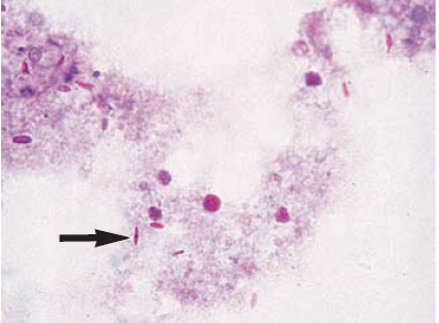 Figure: PAS exudate staining shows the cigar to oval-shaped yeast cells (arrow) of Sporothrix spp.
Culture
Colonies of Sporothrix schenckii grow rapidly (3-5 days) and maybe mistaken with yeast colonies. On further incubation, these colonies become membranous, wrinkled, dark brown, or black in color. The consistency of the colony may be leathery. Sporothrix schenckii hyphae are delicate, septate, and branching.
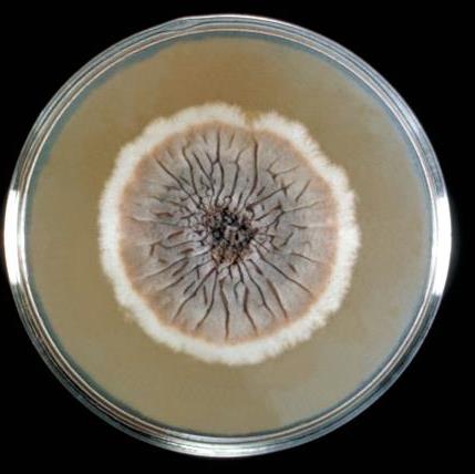 Figure: Sporothrix schenckii growing on Sabouraud agar (Image source: CDC)
If the culture of Sporothrix schenckii is incubated at 37°C colony transforms into a soft, cream-colored white yeast-like colony (dimorphism). Conversion from mold form to yeast form takes place within 1-5 days. Flowerette arrangement of conidia (single-celled conidia borne in clusters from the tip of single conidiophore) is seen.
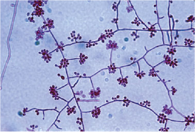 Figure: Mold phase of S. schenckii mounted on Parker-Quink stain showing thin septated hyphae with right-angled conidiophores and ovoid-shaped conidia in a “rosette”-like arrangement. Reproduced with permission from A/Prof. David Ellis, University of Adelaide.
If the yeast-like colony is taken from a plate cultured at 37°C singly or multiply budding oval or elongate, cigar-shaped yeast cells are seen.
Treatment
Treatment depends on the form and severity, and this article gives drug-of-choice and class-level guidance only.
Lymphocutaneous and cutaneous disease (the usual forms): an azole, itraconazole, is the treatment of choice, given for several months until the lesions resolve.
Potassium iodide (a saturated solution, SSKI) taken by mouth is an older but effective and inexpensive treatment for lymphocutaneous disease, still used in low-resource settings. How it works is not fully understood.
Severe, disseminated, or pulmonary disease, and infection in immunocompromised patients: a polyene, amphotericin B, is used first, often followed by itraconazole.
Actual regimens, doses, and durations are decided by the treating clinician.
How to Remember
Cigar-shaped yeast means Sporothrix. In tissue, the small elongated cigar-shaped budding yeast is the signature. Cigar equals Sporothrix.
Rosette conidia in the mold. In culture, conidia cluster in a daisy or rosette at the tip of the conidiophore. Flowers on the mold, cigars in the body.
Rose thorns and sphagnum moss. The classic entry is traumatic implantation from plants, hence rose gardener's disease.
Nodules climb the arm. Lymphocutaneous spread is a chain of nodules ascending along the lymphatics from the injury.
Cats and Brazil mean brasiliensis. Cat-transmitted, more severe, epidemic sporotrichosis is S. brasiliensis.
Itraconazole first, iodide the old classic. Itraconazole is first-line; potassium iodide is the traditional low-cost option.
Key exam facts
| Item | Fact |
|---|---|
| Organism | Sporothrix schenckii (and related species) |
| Disease | Sporotrichosis (rose gardener's disease) |
| Tissue form | Cigar-shaped budding yeast, 3 to 5 μm |
| Mold form | Rosette/daisy arrangement of conidia on delicate hyphae |
| Classic tissue clue | Asteroid body (Splendore-Hoeppli phenomenon) |
| Route | Traumatic implantation from plants (thorns, moss) |
| Signature presentation | Lymphocutaneous, ascending nodules along lymphatics |
| Zoonotic form | Sporothrix brasiliensis, cat-transmitted, more severe (Brazil) |
| At-risk groups | Gardeners, florists, farmers; and cat owners/vets for zoonotic form |
| Diagnosis gold standard | Culture (direct microscopy low-yield) |
| Treatment | Itraconazole (first-line); potassium iodide (classic); amphotericin B (severe) |
Where Students Get Confused
"You can diagnose it easily by seeing the yeast on a smear." No. The cigar-shaped yeast is often sparse and hard to find in specimens. Culture is the gold standard, and a negative direct smear does not exclude sporotrichosis.
"Sporotrichosis only comes from plants." Not anymore. Classic disease comes from plant trauma, but a large and growing form, caused by Sporothrix brasiliensis, is transmitted from cats and is more severe.
"The cigar-shaped yeast is seen in the environment." No. The cigar-shaped yeast is the tissue form at 37°C. In the environment the fungus is a mold with rosette conidia. It is thermally dimorphic.
"Asteroid bodies confirm sporotrichosis." They are a helpful clue when present, but they are not unique to sporotrichosis and are not always seen.
"All sporotrichosis needs amphotericin B." No. The usual lymphocutaneous disease is treated with itraconazole (or potassium iodide). Amphotericin B is reserved for severe, disseminated, or pulmonary disease.
References and further readings
- Tille, P. M. (2022). Bailey and Scott's Diagnostic Microbiology (15th ed.). Elsevier.
- Larone, D. H. (2018). Larone's Medically Important Fungi: A Guide to Identification (6th ed.). ASM Press.
- Centers for Disease Control and Prevention. Sporotrichosis. https://www.cdc.gov/sporotrichosis/
Frequently Asked Questions
What does Sporothrix look like under the microscope?
What does Sporothrix look like under the microscope?
In tissue at body temperature it is a small (3 to 5 μm) cigar-shaped budding yeast. In the environment it is a mold with delicate hyphae and conidia arranged in a rosette or daisy pattern.
What is cigar-shaped yeast?
What is cigar-shaped yeast?
It describes the elongated, cigar-like shape of the Sporothrix yeast cells seen in infected tissue. It is the identifying feature of Sporothrix schenckii.
Why is it called rose gardener's disease?
Why is it called rose gardener's disease?
Because it is classically acquired when the fungus is implanted into the skin by a thorn or plant material, such as from roses or sphagnum moss, so gardeners and florists are commonly affected.
How does sporotrichosis spread in the body?
How does sporotrichosis spread in the body?
Most often as a lymphocutaneous infection: a nodule forms at the site of injury, and further nodules appear in a chain ascending along the lymphatic vessels.
What is cat-transmitted sporotrichosis?
What is cat-transmitted sporotrichosis?
A zoonotic form caused mainly by Sporothrix brasiliensis, spread from infected cats through scratches and bites. It is common in Brazil, tends to be more severe, and affects cat owners and veterinarians.
How is sporotrichosis diagnosed?
How is sporotrichosis diagnosed?
Culture is the gold standard, because the yeast is hard to see directly. The mold that grows is confirmed by converting it to the cigar-shaped yeast form at 37°C. PAS and silver stains can show the yeast in tissue.

Tankeshwar Acharya, MSc (Medical Microbiology)
Tankeshwar Acharya is an Assistant Professor in the Department of Microbiology at Patan Academy of Health Sciences (PAHS), Nepal, where he has been teaching and practicing clinical microbiology for over 14 years. He is the founder of Microbe Online, one of the leading free microbiology education resources on the web, covering bacteriology, mycology, parasitology, immunology, and clinical laboratory diagnostics written from direct experience in both the classroom and the diagnostic laboratory.
Comments
No comments yet. Be the first to share your thoughts.
Leave a comment
All comments are reviewed before they appear.