
Quality Control of Culture Media: Why a Plate Can Look Perfect and Still Mislead

On this page
The plate that looked fine and still got it wrong
A batch of MacConkey agar plates, prepared weeks earlier and stored a little longer than ideal, looks completely normal on visual inspection: correct color, no cracks, no contamination. A stool sample from a suspected Shigella case is plated onto it. The colonies that grow are pale and non-lactose-fermenting, exactly the pattern expected for a genuine enteric pathogen.
Except the organism recovered turns out, on confirmatory biochemical testing, to be an ordinary E. coli, one that should have fermented lactose and turned distinctly pink. The plate hadn't failed visually. It had failed functionally: its selective and differential ingredients had degraded just enough, invisible to the eye, to distort the very reaction the medium exists to reveal.
This is exactly the gap between a visual check and genuine quality control. A plate can pass every physical inspection and still mislead a workup, which is exactly why performance testing with known QC strains, not just a look at the plate, is the step that actually catches this kind of failure before a real patient sample ever depends on it.
Culture media are pivotal in any microbiology laboratory for isolating, identifying, and sensitivity testing different pathogens. Ensuring media quality requires checking growth-supporting characteristics, physical characteristics, and sterility, three genuinely different kinds of checks, before use.
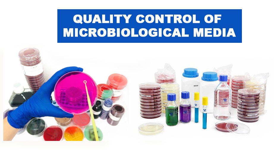 Figure: Quality control of Culture Media
Media quality control is one part of the laboratory's wider quality system. For how it fits alongside strain, reagent, equipment, and reporting checks, see Quality Control in the Microbiology Laboratory.
Preparation of Media
Ready-to-use media purchased from vendors offer little opportunity to control preparation beyond trusting the supplier, though transport and storage conditions can still degrade quality after manufacture. In-house prepared media require attention to several additional factors during preparation itself.
Testing of Physical and Chemical Parameters
Checking of physical parameters such as the color of the prepared medium, thickness, formation of excessive bubbles, uneven surface, shrinking of the media, cracks or crystallization in the media, and chemical parameters such as the pH of the medium is necessary.
Perform visual checks to rule out the following anomalies;
- Agar detached from the plates
- Cracked or damaged plates
- Frozen or melted agar
- Changes in the expected color of the media
- Obvious contamination
- Unequal filling of the plates or insufficient agar depth (<3 mm) (Note: For Mueller-Hinton agar, the agar depth should be 4± 0.5 mm)
- Hemolysis of blood-containing media
- Excessive bubbles or rough surfaces
- Excessive moisture or dehydration
- Presence of precipitates
- The integrity of the packaging
- Presence of broken or cracked Petri plates or tubes
- Presence of leakage from the Petri plates or tubes
- Accuracy of the labeling
Testing the pH of Medium
The simplest way of testing the pH of a culture medium is to use narrow-range pH paper or a calibrated pH meter, tested during preparation before or after autoclaving; broth is tested by dipping paper directly, agar by pouring a sample onto a plate, letting it solidify, and laying paper on the surface. Adjustments use dilute NaOH for acidic media or dilute HCl for alkaline media, with 1N NaOH specifically for alkaline peptone water.
Measuring pH of Broth
- Dip a narrow-range pH paper into a sample of the test medium at room temperature.
- Compare the color of the paper against the pH color chart.
Measuring pH of Agar
- To test the pH of an agar medium, pour a sample of the molten medium into a Petri plate and let it solidify.
- Lay a narrow range pH paper on its surface.
- Compare the color of the paper against the pH color chart.
Adjustment of pH
In most cases, adjustment of the pH won’t be needed. Minor pH adjustments can be made by using
- 0.1 mol/L sodium hydroxide when the medium is too acidic, and
- 0.1 mol/L hydrochloric acid when the medium is too alkaline.
- Use 1 mol/L (1N) sodium hydroxide to adjust the pH of alkaline peptone water.
Sterility Testing
Every media prepared, following stringent quality control parameters, is sterile. Incubate in-house prepared culture media for 48 hours at 35-37°C (varying from 24 h to 5 days, depending on the reference). For the batch with <100 units, a 2% sample must be subjected to sterility testing, but for the batch with >100 units, ten random units are sufficient to ascertain no growth or contamination.
If growth is seen after incubation (i.e., contamination), reject or discard the whole batch and prepare a new one. Do not re-use the plates used for sterility check. Note that an additional visual check should always be done before use.
Why Performance Testing Is the Step Visual Checks Can't Replace
This is exactly the gap illustrated at the beginning of this article: physical and chemical checks confirm a plate looks and measures correctly. Only performance testing, running actual QC strains through the medium and confirming the expected reaction, confirms the medium still does the job it's supposed to do. A medium can pass every visual and pH check while its selective agents or differential indicators have quietly degraded past the point of giving a reliable result. This is why performance testing isn't a redundant extra step after physical inspection, it's checking something visual inspection cannot see at all.
Performance Testing
After sterility testing, test the media for supporting growth and giving desired reactions. Use a standardized suspension of QC strains to inoculate the culture media.
Check and record growth (colony size and morphology), selectivity, and differentiation:
- Support of target organism: Use at least one organism, and record growth and biochemical reaction. e.g., If a strain of Escherichia coli ATCC 25922 is inoculated into a sterile MacConkey agar plate, after proper incubation, it should show lactose fermenting pink colonies.
- Selective media: Use at least one organism that is expected to grow and at least one organism that is not expected to grow
- Differential media: Use organisms that will display the intended growth and reactions (e.g., MacConkey agar: Shigella flexneri, Escherichia coli, and Staphylococcus aureus)
- Biochemical media (e.g., urease-test): Use at least one organism that will produce a negative reaction and one that will produce a positive reaction.
The results should be examined both qualitatively and quantitatively, and while testing new lots, both previous and new batches should be included. Perform disc diffusion to check Mueller-Hinton agar plates.
Always Remember
"looks fine isn't the same as works fine." A plate can pass every physical and chemical check and still fail the one job that actually matters, producing the correct reaction. Performance testing with known QC strains is the only check that catches that specific failure.
Use of QC Strains
Media are performance-tested with well-characterized reference strains, most commonly ATCC strains, whose expected reaction on each medium is already known. For what reference strains are, what ATCC means, and how the designations work, see Quality Control Strains and ATCC in Microbiology.
Choose the QC organism from the standards or the manufacturer's instructions; previously characterized clinical or EQA strains with documented, stable phenotypes are also acceptable. Store and maintain QC organisms per the governing CLSI guidance (M22).
| S.no | Quality control strain | Media | Expected output |
|---|---|---|---|
| 1. | Escherichia coli (ATCC® 25922™) | MacConkey agar | Lactose fermenting pink colonies |
| 2. | Acinetobacter baumannii (ATCC® 19606™) | MacConkey agar | Non- Lactose fermenting pale colonies |
| 3. | Staphylococcus aureus (ATCC® 25923™) | Mannitol Salt agar | Golden Yellow colonies |
| 4 . | Streptococcus pyogenes (ATCC® 19615™) | Blood agar | Beta hemolytic, grey colonies |
| 5. | Streptococcus pneumoniae | Blood agar | Alpha hemolytic, small greenish colonies |
| 6. | Shigella sonnei (ATCC® 25931™) | XLD agar | Red pink colonies |
| 7. | Salmonella Typhimurium (ATCC® 14028™) | XLD agar | Red pink colonies with black center |
| 8. | Pseudomonas aeruginosa (ATCC® 27853™) | Mueller hinton Agar | Flat serrated greenish colonies |
| 9. | Neisseria gonorrhoeae (ATCC® 49226™) | Thayer martin agar | Water droplet-like colonies |
| 10. | Enterococcus faecalis (ATCC® 29212™) | Bile esculin agar | Small transparent colonies with brown-black halos. |
| 11. | Escherichia coli (ATCC® 25922™) | EMB agar | Blue-black bull’s eye-like colonies with a green metallic sheen |
A list of quality control (QC) strains to be tested against routinely used culture media is as follows. However, one strain can be inoculated on various other media. Escherichia coli ATCC 25922, Staphylococcus aureus ATCC 25923, and Pseudomonas aeruginosa ATCC 27853 are used as cumulative control for testing all media.
The Cumulative Control Strains
For media QC, Escherichia coli ATCC 25922, Staphylococcus aureus ATCC 25923, and Pseudomonas aeruginosa ATCC 27853 act as cumulative controls: one of these three can validate performance across many general-purpose and selective media, so a unique strain is not needed for every medium.
Specialized media (XLD, Thayer-Martin, Bile Esculin, and the like) still need their own specific strain in addition, because they test for reactions the cumulative three cannot demonstrate.
These same three strains recur across laboratory quality control; see Quality Control Strains and ATCC in Microbiology for the full picture.
NOTE: "three strains do double duty." E. coli 25922, S. aureus 25923, and P. aeruginosa 27853 cover routine QC across most general-purpose and selective media. Specialized media still need their own specific strain in addition, since the cumulative three can't demonstrate every specialized reaction.
Where Students Get Confused
- Treating a visual/physical check as sufficient quality control. As mentioned in the beginning of this article, a plate can look completely normal while its selective or differential components have degraded enough to distort results.
- Reversing the sterility testing sample-size rule. Batches under 100 units need a 2% sample; batches over 100 units need 10 random units, not the other way around.
- Assuming every medium needs its own unique QC strain. The three cumulative strains (E. coli 25922, S. aureus 25923, P. aeruginosa 27853) cover routine testing across many media; only specialized media need an additional, specific strain.
- Assuming ready-to-use commercial media need no ongoing QC. Manufacturing quality is the supplier's responsibility, but transport and storage conditions after purchase can still degrade the medium before use.
References and further readings
- Basu S, Pal A, Desai PK (2005) Quality control of culture media in a microbiology laboratory, Indian journal of medical microbiology,23:3;159-163
- Quality Control of Microbiological Culture Media by Scott Sutton, Ph.D.
- Orekan J, Barbé B, Oeng S, Ronat JB, Letchford J, Jacobs J, Affolabi D, Hardy L. Culture media for clinical bacteriology in low- and middle-income countries: challenges, best practices for preparation and recommendations for improved access. Clin Microbiol Infect. 2021 Oct;27(10):1400-1408. doi: 10.1016/j.cmi.2021.05.016. Epub 2021 May 18. PMID: 34015533.
Frequently Asked Questions
What are the three components of quality control for culture media?
Quality control of culture media has three components: (1) Physical/visual inspection — checking appearance, color, clarity, pH, and agar depth before use; (2) Sterility testing — incubating 5-10% of each new batch at 35°C for 48-72 hours without inoculation to confirm no contamination occurred during preparation; (3) Performance testing: inoculating with known ATCC reference strains to confirm the medium supports expected growth, selectivity, and differential reactions. All three must pass before a batch is released for clinical use. A batch that fails any component must be quarantined and investigated.
Which ATCC strains are used for quality control of MacConkey agar?
MacConkey agar QC requires testing with both a target organism and a selectivity control: Escherichia coli ATCC 25922 should produce good growth with pink lactose-fermenting colonies (positive performance); Staphylococcus aureus ATCC 25923 should be inhibited or show no growth (selectivity check, confirming gram-positive organisms are suppressed). Both results must be as expected before the batch is used for clinical specimens. Using only a positive control without a selectivity control can miss medium batches where the selective agents have degraded, allowing gram-positive contamination to go undetected.
What should happen to clinical results when a batch of culture media fails quality control?
When a batch of culture media fails QC (whether sterility testing, performance testing, or visual inspection) the entire batch must be quarantined and not used for clinical specimens. If clinical specimens were already processed on a failed batch before the failure was detected, all results from those specimens must be flagged for clinical review and the requesting clinicians notified. Repeat testing of available specimens should be offered. The root cause of the failure must be investigated (autoclave records, pH records, preparation logbook) and documented before the next batch is prepared. QC failures must be recorded in the laboratory QC logbook regardless of outcome.

Tankeshwar Acharya, MSc (Medical Microbiology)
Tankeshwar Acharya is an Assistant Professor in the Department of Microbiology at Patan Academy of Health Sciences (PAHS), Nepal, where he has been teaching and practicing clinical microbiology for over 14 years. He is the founder of Microbe Online, one of the leading free microbiology education resources on the web, covering bacteriology, mycology, parasitology, immunology, and clinical laboratory diagnostics written from direct experience in both the classroom and the diagnostic laboratory.
Comments
No comments yet. Be the first to share your thoughts.
Leave a comment
All comments are reviewed before they appear.