
XLD Agar: Composition, Principle, Colony Characteristics, and Uses
XLD (Xylose Lysine Deoxycholate) agar is the best single medium for isolating Salmonella and Shigella from stool. Learn its three-step differentiation mechanism, why Salmonella produces red colonies with black centers, and how XLD compares to DCA and SS agar.

On this page
A family of four presents to the emergency department with 48 hours of bloody diarrhea after attending a wedding feast. Stool specimens are collected. In the laboratory, the technician has a specific challenge: distinguishing the causative pathogen (likely Salmonella or Shigella) from the millions of normal E. coli and other coliforms in each specimen. MacConkey agar will grow every gram negative enteric bacilli; blood agar will grow everything and tell you nothing about enteric pathogens specifically.
XLD agar was designed precisely for this moment. It uses a three-step biochemical sequence; xylose fermentation, lysine decarboxylation, and hydrogen sulfide production, to produce colony colors that are diagnostic on their own, before a single biochemical test is run.
Principle
Xylose lysine deoxycholate (XLD) agar is a selective and differential medium for the isolation, cultivation, and differentiation of gram-negative enteric microorganisms. This media primarily isolates and differentiates Salmonella and Shigella from clinical and non-clinical specimens.
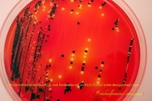 Figure: Mixed culture of E coli and Salmonella in XLD agar
XLD agar selectively promotes the growth of Salmonella and Shigella by inhibiting other enteric pathogens and differentiates Gram-negative enteric bacteria based on xylose fermentation, lysine decarboxylation, and the production of hydrogen sulfide from the sodium thiosulphate.
XLD medium was formulated by Taylor for the isolation and differentiation of enteric pathogens.
Composition of XLD Agar
The key ingredients of XLD agar are three sugars (xylose, lactose, and sucrose), lysine, and ferric ammonium citrate.
Ingredients in Per liter formulations:
| Ingredient | Amount (g/L) | Function |
|---|---|---|
| Lactose | 7.5 | Fermentable carbohydrate: most coliforms ferment this, producing yellow colonies |
| Sucrose | 7.5 | Fermentable carbohydrate: Proteus and some coliforms ferment sucrose; adds to yellow colony production |
| Sodium thiosulfate | 6.8 | Sulphur source; H₂S indicator (reacts with ferric ammonium citrate) |
| L-Lysine | 5.0 | Substrate for lysine decarboxylation: Salmonella decarboxylates lysine to produce alkaline cadaverine; restores red color |
| Sodium chloride | 5.0 | Osmotic balance |
| Xylose | 3.75 | Fermentable carbohydrate: primary first-step sugar; fermented rapidly by Salmonella and most enteric GN but not Shigella |
| Yeast extract | 3.0 | Nitrogen, carbon, vitamins |
| Sodium deoxycholate | 2.5 | Selective agent that inhibits Gram-positive organisms |
| Ferric ammonium citrate | 0.8 | H₂S indicator: reacts with H₂S to form black iron sulfide precipitate |
| Phenol red | 0.08 | pH indicator: red in alkaline/neutral, yellow in acid |
| Agar | 15.0 | solidifying agent |
Final pH: 7.4 ± 0.2, medium appears bright red at baseline
A note on reading XLD agar: Unlike most enteric media (which start pale or colorless), XLD agar is bright red to red-orange when freshly prepared, due to the phenol red indicator at its baseline alkaline pH (7.4). Organisms that produce acid turn colonies and surrounding medium yellow. Organisms that produce alkaline reactions or no fermentation maintain or restore the red color. This means yellow colonies on XLD indicate fermentation, and red colonies indicate alkalinity.
Principle of XLD Agar
Xylose is rapidly fermented by most Gram-negative enteric bacteria, including Salmonella. This causes acidification of the medium, turning the phenol red indicator yellow. Since Shigella spp doesn’t utilize xylose, acidification does not occur, and red colonies are produced. This property aids in the differentiation of Shigella spp.
After the xylose supply is exhausted, Salmonella spp decarboxylates lysine, increasing the pH to alkaline condition, and also produces red colonies like Shigella spp. However, Salmonellae also metabolize thiosulfate to produce hydrogen sulfide, which leads to the formation of colonies with black centers and allows them to be differentiated from the similarly colored Shigella colonies.
Organisms that ferment lactose, sucrose, and xylose but are lysine decarboxylase negative cause an acid pH and produce yellow colonies.
The three-step sequence can be summarized as a decision tree:
| Step | Reaction | pH change | Colony color |
|---|---|---|---|
| Step 1: Xylose fermentation | Most GN enteric organisms ferment xylose first. Shigella cannot. | Acid → yellow | Salmonella goes yellow initially; Shigella stays red |
| Step 2: Lysine decarboxylation (after xylose exhausted) | Salmonella decarboxylates lysine → cadaverine (alkaline). Coliforms and Shigella cannot. | Alkaline → red restored | Salmonella reverts to red; coliforms stay yellow; Shigella stays red |
| Step 3: H2S production | Salmonella metabolizes thiosulfate → H2S → reacts with ferric citrate → black precipitate | No pH change | Black center appears within red colony |
The result: Salmonella ends up as red colonies with black centers because all three steps occur. Shigella ends up as red colonies without black centers because it skips step 1 but cannot do step 3. Coliforms (E. coli, Klebsiella) stay yellow because they ferment all three sugars but cannot reverse the acid pH.
Why this three-step system is better than single-sugar media: MacConkey and EMB use only lactose , both Salmonella and Shigella appear colorless and are indistinguishable. XLD adds two more reactions (lysine decarboxylation and H2S) that specifically differentiate Salmonella from Shigella with a single overnight incubation.
Preparation of Media
- Suspend 55 gm of the medium in one liter of purified water.
- Heat with frequent agitation until the medium reaches the boiling point.
- Avoid overheating. Do not autoclave.
- Transfer immediately to a water bath at 50°C.
- After cooling, pour into sterile Petri plates.
It is advisable not to prepare large volumes that will require prolonged heating, thereby producing precipitate.
Colony Characteristics on XLD Agar
After incubation of the plates with test organisms, various colored colonies develop, and differentiation is based on it.
| Organism | Colony color | Black center | Mechanism | Notes |
|---|---|---|---|---|
| Salmonella typhi | Red/pink | Yes (variable; may be small or faint) | Xylose+ → lysine decarboxylase+ → H₂S+ | Small black center; can be missed. Always pick red colonies regardless of center size |
| Salmonella typhimurium (and most non-typhi) | Red/pink | Yes (prominent) | Xylose+ → lysine decarboxylase+ → H₂S+ | Large black center; classic XLD appearance |
| Salmonella paratyphi A | Red/pink | No black center | Xylose+ → lysine decarboxylase+; H₂S negative | Mimics Shigella, cannot differentiate on XLD alone; serology required |
| Salmonella choleraesuis, S. pullorum, S. gallinarum | Red/pink | No or faint black center | Variable H₂S production | See Limitations: these species produce red colonies without black centers |
| Shigella sonnei | Red/pink | No black center | Xylose negative → no acid; lysine decarboxylase negative | Cannot be distinguished from S. paratyphi A on colony appearance alone |
| Shigella flexneri | Red/pink | No black center | Same as S. sonnei | TSI + serology for confirmation |
| Shigella dysenteriae | Red/pink (may be small) | No | Most inhibited Shigella — medium is not highly inhibitory to it (unlike SS agar) | Best Shigella recovery of all enteric selective media |
| Escherichia coli | Yellow | No | Xylose+ lactose+ sucrose+; lysine variable; no H₂S | Partially inhibited on XLD; yellow colonies are conspicuous against red background |
| Klebsiella/Enterobacter | Yellow, mucoid | No | Lactose+, sucrose+; lysine decarboxylase+ (Enterobacter) but yellow due to excess sugar fermentation | Mucoid appearance; typically larger than E. coli |
| Proteus mirabilis | Yellow (variable) | Small black center possible | Sucrose+ in some; H₂S+ | Proteus mimicry warning: Small black-centered colonies; distinguished from Salmonella by urease test (Proteus strongly urease positive) |
| Proteus vulgaris | Yellow | Variable black center | Similar to P. mirabilis | |
| Pseudomonas aeruginosa | Red/colorless | No | Non-fermenter | Grows on XLD; red or colorless colonies; may produce pigment |
The two critical mimicry problems on XLD:
- S. paratyphi A produces red colonies without black centers, indistinguishable from Shigella on XLD alone. Always confirm with TSI and serology.
- Proteus spp. can produce small black-centered colonies resembling Salmonella. Urease test immediately differentiates them: Salmonella is urease-negative; Proteus is strongly urease-positive.
Quality Control
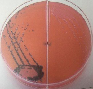 Figure: XLD Cultures: Left: Red-pink black centered colonies of Salmonella typhimurium Right: Red-pink Shigella colonies
| Organism | Growth and colony morphology |
|---|---|
| S. Typhimurium ATCC 14028 | Good growth, Red colonies with a black center |
| S. flexneri ATCC 12022 | Luxuriant growth, red colonies |
| Escherichia coli ATCC 25922 | Partially inhibited, Large, flat, yellow colonies |
| Enterococcus faecalis ATCC 29212 | Partial to complete inhibition, clear pinpoint colonies |
The commercially available dehydrated media should be homogeneous, free-flowing, and light pink-beige in color. After preparation, the medium is bright red to reddish-orange, trace to slightly with neutral pH (7.20-7.60).
Quality testing of the prepared media plates should be done by performing sterility and performance testing.
- Sterility testing: Incubate un-inoculated plates of XLD agar for 48 hours at 35-37°C and observe any growth. After 48 hours, the sterility test plate should remain clear. Discard the whole lot if any colonies are seen.
- Performance testing: Inoculate known standard strains on XLD agar plates, incubate for 18- 24 hours at 35-37°C, and observe for growth and colony characteristics.
Uses of XLD Agar
- For isolation and differentiation of Salmonella and Shigella spp from other enteric pathogens.
- Isolation and screening of samples containing mixed flora suspected of harboring enteric pathogens, e.g., medical specimens or food products.
- Detection of Salmonella in non-sterile pharmaceutical products (in accordance with EP, USP) and food, water dairy products, etc.
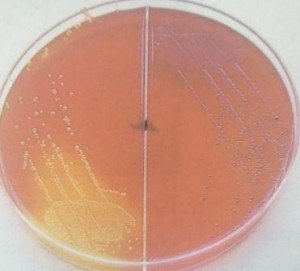 Figure: Xylose Lysine deoxycholate (XLD) Cultures. Left: Yellow Escherichia coli colonies Right: Red-pink Salmonella colonies (Some Proteus species look identical)
How to Remember
XLD = three ingredients doing three jobs:
- Xylose: first sugar fermented; separates Shigella (can't ferment) from everything else (can)
- Lysine: decarboxylated by Salmonella after xylose runs out; restores red color; distinguishes Salmonella from coliforms
- Deoxycholate: inhibits Gram-positives
The three-step story as a clinical narrative:
Imagine Salmonella as a marathon runner passing three checkpoints: Checkpoint 1 (xylose): Salmonella ferments xylose → turns yellow → passes checkpoint 1 Checkpoint 2 (lysine): When xylose runs out, Salmonella decarboxylates lysine → turns alkaline → reverts to red → passes checkpoint 2 Checkpoint 3 (H2S): Salmonella reduces thiosulfate → H2S → black precipitate → passes checkpoint 3
Shigella can't pass Checkpoint 1 (no xylose fermentation) thus stays red throughout, no black center. Coliforms pass Checkpoint 1 (ferment everything) so stay yellow, never revert.
The medium starts red: Yellow colonies on XLD = fermenters = coliforms. Red colonies on XLD = pathogens or non-fermenters. This is counterintuitive because on most media, color usually signals the presence of something (a positive result). On XLD, the red is the baseline, the absence of fermentation.
XLD in the enteric media trio:
- XLD: Best for Shigella recovery (especially S. dysenteriae); clearest Salmonella signature (red + black)
- DCA: Moderate selectivity; good for Salmonella; poorer for S. dysenteriae
- SS agar: Highest selectivity; best for suppressing coliforms; poorest for Shigella
If only one enteric medium: choose XLD. If two: XLD + MacConkey.
Limitations
- A single medium may not be enough to recover all pathogens, so a less selective medium such as MacConkey agar should also be used.
- Non-enteric pathogens such as Pseudomonas may grow in the medium producing red colonies like Shigella; therefore, further, identification is necessary.
- Proteus mirabilis may produce small black-centered colonies similar to Salmonella but can be distinguished from Salmonella colonies, the latter being large, with a big black center.
- Prolonged incubation (more than 48 hours) may lead to false-positive results.
- Salmonella Paratyphi, S. Choleraesius, S. Pullorum and S. Gallinarum produce red colonies without a black center, hence may appear like Shigella.
- Since it is a selective medium, further subculture onto a basal medium is essential before performing biochemical tests, serotyping, etc.
References
- Tille, P. M. (2017). Bailey and Scott's Diagnostic Microbiology (14th ed.). Elsevier.
- Taylor, W. I. (1965). Isolation of Shigellae. I. Xylose lysine agars; new media for isolation of enteric pathogens. American Journal of Clinical Pathology, 44(4), 471–475.
- Cheesbrough, M. (2006). District Laboratory Practice in Tropical Countries, Part 2 (2nd ed.). Cambridge University Press.

Tankeshwar Acharya, MSc (Medical Microbiology)
Tankeshwar Acharya is an Assistant Professor in the Department of Microbiology at Patan Academy of Health Sciences (PAHS), Nepal, where he has been teaching and practicing clinical microbiology for over 14 years. He is the founder of Microbe Online, one of the leading free microbiology education resources on the web, covering bacteriology, mycology, parasitology, immunology, and clinical laboratory diagnostics written from direct experience in both the classroom and the diagnostic laboratory.
Comments
No comments yet. Be the first to share your thoughts.
Leave a comment
All comments are reviewed before they appear.