
Mannitol Salt Agar (MSA): Principle, Composition, Uses, and Colony Characteristics
Mannitol Salt Agar (MSA) is a selective and differential medium for isolating Staphylococcus aureus. Learn its principle, composition, colony colors of S. aureus vs CONS, and why Enterococcus and Micrococcus can give false-positive results.

On this page
An infection control nurse in a hospital ward is conducting an MRSA screening program. Nasal swabs are collected from all newly admitted patients in the ICU. In the laboratory, these swabs are plated onto a specialized medium and incubated overnight. The next morning, yellow colonies on a red-pink background immediately flag a sample as potentially positive for Staphylococcus aureus, before any further testing is done.
That immediate visual signal: yellow colonies on a pink medium is the differential principle of Mannitol Salt Agar, and it is the reason MSA became the standard screening medium for S. aureus isolation from specimens with mixed flora.
Mannitol salt agar (MSA) is a selective, differential, and indicator medium used to isolate and identify Staphylococcus aureus from the clinical specimen. As its name suggests, mannitol salt agar (MSA) contains 1% mannitol (sugar), 7.5% salt, and agar as a solidifying agent. Members of the genus Staphylococcus can tolerate high salt concentration (7.5%) and grow on mannitol salt agar.
Principle
Mannitol salt agar (MSA) is a selective and differential medium.
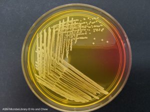 Figure: Yellow colonies of S. aureus in Mannitol Salt Agar (MSA). Image source: ASM
Selective medium
Incorporating 7.5% sodium chloride in the medium helps select only those bacteria that can tolerate high salt concentrations. Staphylococci species can tolerate this salt concentration, but other pathogenic bacteria may not. This concentration inhibits the growth of most other gram-positive and gram-negative bacteria. Thus MSA selectively isolates Staphylococcus spp.
Differential Medium
Pathogenic staphylococci, i.e. Staphylococcus aureus is able to ferment mannitol, but coagulase-negative Staphylococcus (CONS) are not. So, if that particular specimen contains S. aureus, it ferments mannitol and changes the pH of the medium to acidic.
As MSA contains phenol red as a pH indicator, at pH below 6.9 the medium turns yellow. If CONS grow (which cannot ferment mannitol), the medium around the colony does not turn yellow. It stays red (the uninoculated color), not pink.
So, MSA is also a differential medium.
Remember that in the neutral pH (6.9 to 8.4) the color of phenol red is red; while above pH 8.4, the color of phenol red is pink. Other commonly used media that contain Phenol red as pH indicator are; TSI Agar, urea base agar, and XLD agar.
Uses of Mannitol Salt Agar
1. Primary isolation of Staphylococcus aureus from clinical specimens MSA is used as a selective plating medium for specimens where S. aureus is suspected but where mixed flora is expected to obscure its growth on non-selective media. Common specimens include:
- Skin and wound swabs (particularly from skin infections, abscesses, and burn wounds)
- Nasal and throat swabs
- Food samples in public health investigations
2. MRSA screening MSA supplemented with oxacillin (6 µg/mL) or cefoxitin is used in some laboratories as a screen plate for methicillin-resistant S. aureus (MRSA) from nasal, axillary, groin, and perianal surveillance swabs in hospital settings. Chromogenic MRSA agars have largely replaced this in high-resource laboratories, but oxacillin-MSA remains in use in resource-limited settings.
3. Environmental and food microbiology MSA is used to enumerate staphylococci in food samples (particularly dairy products and processed meats) and environmental surfaces during hygiene audits, where the high salt tolerance of staphylococci is the key selection principle.
4. Teaching and demonstration MSA is one of the clearest visual teaching media in microbiology. The yellow vs. pink colony distinction gives immediate, unambiguous results that demonstrate selective and differential media principles simultaneously.
Composition of Mannitol Salt Agar
| Ingredient | Amount (g/L) | Function |
|---|---|---|
| Enzymatic digest of casein | 5.0 | Nitrogen, carbon, amino acids |
| Enzymatic digest of animal tissue | 5.0 | Nitrogen, vitamins, growth factors |
| Beef extract | 1.0 | Additional nitrogen, vitamins, minerals |
| D-Mannitol | 10.0 | Sole carbohydrate: fermented by S. aureus, not by most CONS |
| Sodium chloride (NaCl) | 75.0 | Selective agent: inhibits most organisms except halotolerant staphylococci |
| Phenol red | 0.025 | pH indicator: yellow below pH 6.9; red/pink at neutral to alkaline pH |
| Agar | 15.0 | Solidifying agent |
Final pH: 7.4 ± 0.2 at 25°C
Note on NaCl concentration: MSA contains 7.5% NaCl, i.e., approximately 15 times the salt concentration of normal saline (0.9%). This extreme osmotic environment is lethal or severely inhibitory to most bacteria, including Gram-negative rods and most streptococci. Staphylococci survive because they possess active osmoregulation mechanisms, including accumulation of compatible solutes (such as glycine betaine) that counterbalance external osmotic pressure.
Preparation of Mannitol Salt Agar
You can purchase prepared mannitol salt agar from commercial suppliers, get the powder, and prepare the media in your laboratory. Mannitol salt agar is best prepared from ready-to-use dehydrated powder, available from most suppliers of culture media. The medium is usually used at 11.1 g in every 100 mL of distilled water (concentration may vary depending on the manufacturer).
- Prepare the medium as instructed by the manufacturer. Sterilize by autoclaving at 121oC for 15 minutes.
- When the medium has cooled to 50-55oC, mix well, and dispense it aseptically in sterile Petri dishes. Date the medium and give it a batch number.
- Store the plates at 2-8oC in plastic bags to prevent moisture loss.
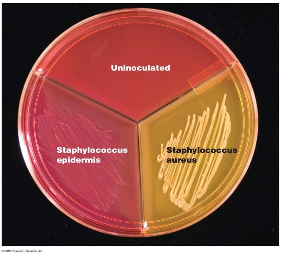 Figure: Growth of S. aureus and CONS on MSA (Image source: Pearson Education)
Shelf life: Several weeks, providing there is no change in the appearance of the medium to suggest contamination, deterioration, or alteration of pH.
The medium's pH should be 7.4 ± 0.2 at room temperature.
Staphylococcus saprophyticus (coagulase-negative Staphylococcus) may ferment mannitol, producing yellow halo around colonies in MSA thus resembling S. aureus.
Colony Characteristics in Mannitol Salt Agar
| Organism | Colony color | Halo | Mannitol fermentation | Notes |
|---|---|---|---|---|
| Staphylococcus aureus | Yellow | Yellow | Yes | Acid production turns phenol red yellow; typical target organism |
| S. epidermidis | Pink to colorless | None | No | Most common CONS; does not ferment mannitol |
| S. saprophyticus | Yellow (variable) | Yellow (variable) | Yes (variable) | Important: may produce yellow colonies resembling S. aureus; confirm with coagulase test and novobiocin susceptibility |
| Enterococcus faecalis | Yellow | Yellow | Yes | False positive: salt-tolerant; ferments mannitol; differentiate from Staphylococcus by catalase test (Enterococcus is catalase-negative) |
| Micrococcus luteus | Yellow | Variable | Variable | False positive: some Micrococcus spp. produce yellow colonies; differentiate from Staphylococcus using modified oxidase test (microdase) |
| M. roseus | Pink to red | None | No | Pink colony; does not mimic S. aureus |
| Escherichia coli | No growth | — | — | Inhibited by 7.5% NaCl |
| Streptococcus pyogenes | No growth | — | — | Inhibited by 7.5% NaCl |
Critical note on coagulase testing from MSA: Do not perform the coagulase test directly from colonies isolated on mannitol salt agar. The high salt concentration of the medium can cause false-positive results in the tube coagulase test. Subculture the suspect colony onto blood agar or TSA and perform the coagulase test from fresh growth (18–24 hours) on the subcultured plate.
Troubleshooting
- When grown on mannitol salt agar, some Micrococcus (Micrococcus is a normal flora of human skin, mucosa, and oropharynx), such as M. luteus (yellow) can produce yellow colonies. M. roseus (red) produces pink colonies on MSA. Find out the difference between Micrococcus and Staphylococcus here
- Enterococcus faecalis and Enterococcus faecium(the most common enterococcal species that have been isolated from human infections) are salt-tolerant bacteria. They can ferment mannitol and produce lactic acid, producing yellow-colored colonies on MSA. Catalase test can help to differentiate between Enterococcus (-ve) and Staphylococcus (+ve).
How to Remember
The name is the mechanism:
- Mannitol: the only sugar in the medium; S. aureus ferments it, most CONS do not
- Salt: 7.5% NaCl selects for salt-tolerant staphylococci; almost everything else is inhibited
- Agar: solidifying agent
The color logic: two questions, two answers
| Question | Positive answer | Negative answer |
|---|---|---|
| Did anything grow? | Yes. Organism is salt-tolerant (Staphylococcus or occasional Enterococcus/Micrococcus) | No growth, inhibited by salt |
| Are the colonies yellow? | Yes. Mannitol fermenter; likely S. aureus (confirm with coagulase) | No. Mannitol non-fermenter; likely CONS |
The false-positive trap: three organisms to remember:
- S. saprophyticus: a CONS that can ferment mannitol; yellow colonies; UTI pathogen in young women
- Enterococcus faecalis: salt-tolerant; ferments mannitol; yellow colonies; ruled out by catalase test
- Micrococcus: skin commensal; some species produce yellow colonies; ruled out by microdase (modified oxidase) test
References
- Kampf, G., Lecke, C., Cimbal, A. K., Weist, K., & Rüden, H. (1998). Evaluation of mannitol salt agar for detection of oxacillin resistance in Staphylococcus aureus by disk diffusion and agar screening. Journal of Clinical Microbiology, 36(8), 2254–2257.
- Han, Z., Lautenbach, E., Fishman, N., & Nachamkin, I. (2007). Evaluation of mannitol salt agar, CHROMagar Staph aureus and CHROMagar MRSA for detection of methicillin-resistant Staphylococcus aureus from nasal swab specimens. Journal of Medical Microbiology, 56(1), 43–46.
- Tille, P. M. (2022). Bailey and Scott's Diagnostic Microbiology (15th ed.). Elsevier.
- Clinical and Laboratory Standards Institute (CLSI). (2023). M22: Quality Control for Commercially Prepared Microbiological Culture Media (5th ed.). CLSI.

Tankeshwar Acharya, MSc (Medical Microbiology)
Tankeshwar Acharya is an Assistant Professor in the Department of Microbiology at Patan Academy of Health Sciences (PAHS), Nepal, where he has been teaching and practicing clinical microbiology for over 14 years. He is the founder of Microbe Online, one of the leading free microbiology education resources on the web, covering bacteriology, mycology, parasitology, immunology, and clinical laboratory diagnostics written from direct experience in both the classroom and the diagnostic laboratory.
Comments
No comments yet. Be the first to share your thoughts.
Leave a comment
All comments are reviewed before they appear.