Staphylococcus saprophyticus: Properties, Pathogenesis, and Lab Diagnosis
Staphylococcus saprophyticus morphology, urease and adhesin virulence factors, and the novobiocin test used to confirm this cause of UTI in young women
Staphylococcus saprophyticus is one of the pathogenic species of staphylococci, the other two are S. aureus and S. epidermidis. Staphylococcus epidermidis and S. saprophyticus are often referred to as coagulase-negative staphylococci (CONS).
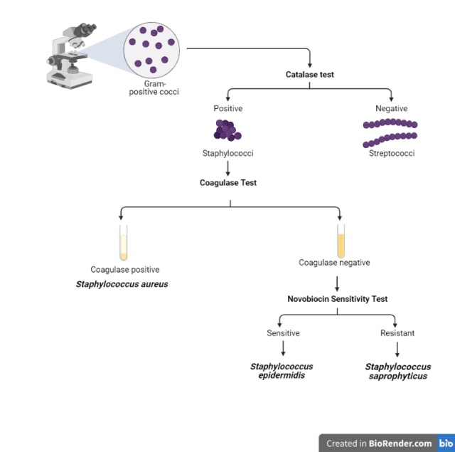 Figure: Flow chart for the identification of Staphylococcus saprophyticus
S. saprophyticus causes urinary tract infections (UTI), especially cystitis in sexually active young women, second only to E. coli as a cause of community-acquired UTI in this group. The infection often follows sexual intercourse closely enough that it has a clinical nickname worth knowing: honeymoon cystitis.
General Features
- In Gram staining, Staphylococcus saprophyticus appears as Gram-positive cocci in clusters.
- Catalase test: Positive
- Coagulase production: No
- Typical hemolysis: None
- Resistant to novobiocin in contrast to S. epidermidis which is sensitive.
- Causes community-acquired UTI in young, sexually active women
Transmission
Staphylococcus saprophyticus is a normal flora of the perineum, rectum, urethra, cervix, and gastrointestinal tract. It is found primarily on the mucosa of the genital tract in young women and from that site can ascend into the urinary bladder to cause urinary tract infections. Most women with this infection have had sexual intercourse within the previous 24 hours. Patients usually present with dysuria, pyuria, and hematuria.
Why This Organism Causes Disease Where It Does
Three things explain why this particular CoNS, out of dozens of staphylococcal species living harmlessly on skin, is the one that consistently causes UTIs:
- Adhesins (including a surface protein called Aas) let it stick tightly to uroepithelial cells lining the bladder, resisting the mechanical washout that urination would otherwise provide.
- Urease, present in essentially all strains, hydrolyzes urea into ammonia, which alkalinizes the urine. Clinical link: this is the same mechanism behind Proteus-associated struvite stones. S. saprophyticus is a less common but real cause of infection-associated struvite (magnesium ammonium phosphate) stones, worth remembering as an exception to "Proteus is always the urease answer."
- Biofilm formation lets it persist on the bladder lining and on indwelling catheters, contributing to why recurrence is common.
Sexual intercourse mechanically displaces the organism from its normal home, the perineum, rectum, and periurethral area, into the short female urethra, which is the direct explanation for honeymoon cystitis and why symptoms typically appear within 24 hours of intercourse.
How to Remember
Pair it directly against its CoNS sibling, since that contrast is the entire reason the novobiocin test exists: Saprophyticus Stands its ground, Epidermidis Eases up. Saprophyticus resists novobiocin, epidermidis is sensitive to it. The alliteration (S-S, E-E) matches the organism's own initial to its behavior, so you're not memorizing an arbitrary pairing.
For the clinical picture, "honeymoon cystitis" already does the work, a young, sexually active woman with dysuria within a day of intercourse should put this organism, not just E. coli, on your list.
Laboratory Diagnosis
Sample: Clean-catch mid-stream urine sample.
Find detailed information about the urine sample collection procedure here.
Colony Morphology in 5% sheep blood agar
S. saprophyticus usually gives white colonies in blood agar, but colonies can be yellow to orange. Colonies are large; entire, very glossy, smooth, opaque, butyrous, and convex.
Urine culture: Though a positive urine culture is indicated by the presence of 100,000 colony-forming units per mL, S. saprophyticus is usually present in quantities less than or equal to 100,000 cfu/mL, but will be detected in sequential specimens.
If significant bacteriuria of gram-positive cocci is seen, the organism is identified by performing catalase and coagulase test. Suppose the organism is catalase-positive and coagulase-negative, novobiocin test should be performed to differentiate S. epidermidis from S. saprophyticus. If the organism is sensitive to novobiocin, it is S. epidermidis, most likely to be the contaminant (normal flora).
If the organism is novobiocin-resistant, it is identified as S. saprophyticus. It is a pathogen for women of the reproductive age group, so antimicrobial testing should be performed, and the antibiogram result should be reported to the physician.
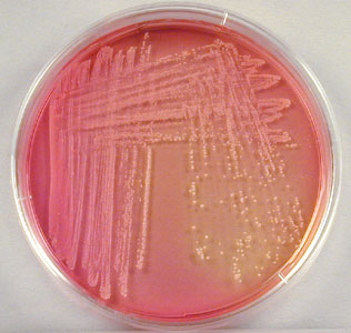 Figure: Staphylococcus saprophyticus growing on Mannitol Salt Agar (Image source: Gary E. Kaiser)
Where students actually get confused
- A colony count below the usual 100,000 CFU/mL threshold doesn't rule this organism out. S. saprophyticus UTIs are a recognized exception to the standard significant-bacteriuria cutoff, lower counts can still be clinically significant, especially in sequential specimens. Treating this organism with the same threshold logic as E. coli will cause you to dismiss real infections.
- Catalase-positive, coagulase-negative is not the end of the workup. Both S. epidermidis and S. saprophyticus fit that description. Novobiocin is the test that actually separates them, not catalase or coagulase.
- "CoNS in urine" used to mean contaminant by default. Historically true for most CoNS, but S. saprophyticus specifically is a real uropathogen in young women, not a skin contaminant, context (young, sexually active, symptomatic) matters as much as the organism ID.
Key exam facts in one table
| Feature | S. saprophyticus | Memory hook |
|---|---|---|
| Catalase | Positive | All Staph are catalase-positive, this is genus-defining |
| Coagulase | Negative | CoNS, like S. epidermidis |
| Novobiocin | Resistant | "Saprophyticus Stands its ground" |
| Hemolysis | None | Unlike S. aureus |
| Urease | Positive | Same mechanism as Proteus struvite stones |
| Typical patient | Young, sexually active woman | Honeymoon cystitis |
| Rank as UTI cause | 2nd after E. coli | In this specific demographic |
| Significant colony count | Can be <100,000 CFU/mL | Exception to the usual cutoff |
Treatment
Staphylococcus saprophyticus urinary tract infections can be treated with nitrofurantoin, trimethoprim-sulfamethoxazole, or a quinolone such as ciprofloxacin, with the specific choice depending on local resistance patterns.
Self-check questions
- Why is a colony count of 50,000 CFU/mL not automatically dismissed as insignificant when S. saprophyticus is suspected?
- A urine isolate is catalase-positive, coagulase-negative, and novobiocin-sensitive. Is this S. saprophyticus? What is it instead?
- By what mechanism can S. saprophyticus contribute to kidney stone formation?
- Why does honeymoon cystitis happen specifically in women rather than men with comparable sexual activity?
References
- Kuroda, M., Yamashita, A., Hirakawa, H., Kumano, M., Morikawa, K., Higashide, M., Maruyama, A., Inose, Y., Matoba, K., Toh, H., Kuhara, S., Hattori, M., & Ohta, T. (2005). Whole genome sequence of Staphylococcus saprophyticus reveals the pathogenesis of uncomplicated urinary tract infection. Proceedings of the National Academy of Sciences, 102(37), 13272-13277. https://doi.org/10.1073/pnas.0502950102
- Gatermann, S., John, J., & Marre, R. (1989). Staphylococcus saprophyticus urease: characterization and contribution to uropathogenicity in unobstructed urinary tract infection of rats. Infection and Immunity, 57(1), 110-116. https://doi.org/10.1128/IAI.57.1.110-116.1989
- Raz, R., Colodner, R., & Kunin, C. M. (2005). Who are you, Staphylococcus saprophyticus? Clinical Infectious Diseases, 40(6), 896-898. https://doi.org/10.1086/428353
- Gupta, K., Hooton, T. M., Naber, K. G., Wullt, B., Colgan, R., Miller, L. G., Moran, G. J., Nicolle, L. E., Raz, R., Schaeffer, A. J., & Soper, D. E. (2011). International clinical practice guidelines for the treatment of acute uncomplicated cystitis and pyelonephritis in women: A 2010 update by the Infectious Diseases Society of America and the European Society for Microbiology and Infectious Diseases. Clinical Infectious Diseases, 52(5), e103-e120. https://doi.org/10.1093/cid/ciq257
Frequently Asked Questions
Why is Staphylococcus saprophyticus often missed if labs use the standard 100,000 CFU/mL cutoff for UTI?
What's the difference between Staphylococcus saprophyticus and Staphylococcus epidermidis?

Tankeshwar Acharya, MSc (Medical Microbiology)
Tankeshwar Acharya is an Assistant Professor in the Department of Microbiology at Patan Academy of Health Sciences (PAHS), Nepal, where he has been teaching and practicing clinical microbiology for over 14 years. He is the founder of Microbe Online, one of the leading free microbiology education resources on the web, covering bacteriology, mycology, parasitology, immunology, and clinical laboratory diagnostics written from direct experience in both the classroom and the diagnostic laboratory.