
Staphylococcus vs. Micrococcus: Key Differences and Tests
How to tell Staphylococcus and Micrococcus apart: morphology, catalase, bacitracin, furazolidone, and microdase test results compared.

On this page
Staphylococcus and Micrococcus both are Gram-positive cocci (GPC). Both of these cocci are non-motile, non-sporing, and catalase-positive.
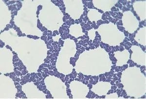 Figure: Staphylococcus in Gram Stain
Both of these organisms are normal flora of the skin and mucous membranes of man and animals. Member of the genus Staphylococcus is associated with clinical infections whereas that of Micrococcus is rarely involved in infection.
Why this distinction matters in practice: both organisms are catalase-positive Gram-positive cocci, so a catalase test alone won't separate them, and Micrococcus is usually dismissed as a contaminant. Getting this wrong in either direction has consequences, calling a true coagulase-negative Staphylococcus infection "probably Micrococcus, ignore it" can mean missing a real infection, especially in immunocompromised patients or line-associated bacteremia where Micrococcus itself can occasionally turn pathogenic.
Some of the notable differences between Micrococcus and Staphylococcus are
| Characteristics /Tests | Micrococcus | Staphylococcus |
|---|---|---|
| Morphology | Large Gram-positive cocci, usually arranged in tetrads or in pairs. | Gram-positive cocci in clusters, sometimes in pairs and short chains. |
| Clinical Significance | Usually considered contaminants of clinical specimen; rarely implicated as cause of infections. May cause opportunistic infection in an immunocompromised host. | Bacteria of this genus are of primary clinical significance. S. aureus is a notorious pathogen. S. saprophyticus causes honeymoon cystitis. |
| Lysostaphin Sensitivity Test | Not lysed with lysostaphin (resistant) | Lysed with lysostaphin (sensitive) |
| Furazolidone Sensitivity Test | Resistant to antibiotic furazolidone | Susceptible to antibiotic furazolidone |
| Bacitracin sensitivity test | Susceptible to 0.04 U of bacitracin | Resistant to 0.04 U of bacitracin |
| Microdase Test | Microdase positive | Microdase negative |
| Oxygen requirements | They only grow aerobically (obligate aerobe) | Facultative anaerobic (can use either aerobic respiration and/or fermentation depending on the availability of oxygen, does not solely depend on aerobic respiration for growth). |
| Carbohydrate utilization | Utilize sugars oxidatively or not at all in the O/F test. | Fermentative |
How to remember these tests?
Every test in this panel is a clean flip between the two genera, there's no exception to memorize around: Micrococcus is bacitracin-sensitive where Staph is resistant, furazolidone-resistant where Staph is sensitive, lysostaphin-resistant where Staph is sensitive, and microdase-positive where Staph is negative. If you know Staph's result on any one of these four tests, Micrococcus is simply the opposite, every time.
A. Bacitracin Sensitivity test is used for the rapid identification of group A streptococci. It can be used to distinguish staphylococci (resistant) from micrococci (sensitive).
B. Lysostaphin Sensitivity test distinguishes staphylococci (sensitive) from micrococci (resistant).
Procedure and Result:
- Emulsify the test isolate in 2 ml phosphate-buffered saline at pH 7.3 to a concentration equivalent to McFarland’s opacity standard no. 1.
- Divide the suspension into two portions. To one add a lysostaphin disc and shake vigorously. The other is the control.
- Incubate both portions in a water bath at 37°C for 2 hours.
- Clearing the test suspension relative to the control indicates sensitivity to lysostaphin.
Where students actually get confused?
- Both are catalase-positive, so catalase alone rules out Streptococcus, not Micrococcus. Bacitracin, furazolidone, lysostaphin, or microdase are what actually separate Staph from Micrococcus.
- Micrococcus being "rarely pathogenic" doesn't mean never, it can cause real opportunistic infection in immunocompromised or catheterized patients, treat it as a low-probability default, not an automatic dismissal.
Key exam facts
| Test | Staphylococcus | Micrococcus |
|---|---|---|
| Catalase | Positive | Positive (doesn't separate them) |
| Bacitracin | Resistant | Sensitive |
| Furazolidone | Sensitive | Resistant |
| Lysostaphin | Sensitive | Resistant |
| Microdase | Negative | Positive |
| Oxygen requirement | Facultative anaerobe | Obligate aerobe |
References
- Falk, D., & Guering, S. J. (1983). Differentiation of Staphylococcus and Micrococcus spp. with the Taxo A bacitracin disk. Journal of Clinical Microbiology, 18(3), 719-721. https://doi.org/10.1128/jcm.18.3.719-721.1983
- Forbes, S., Sahm, D. F., & Weissfeld, A. S. (2002). Bailey & Scott's Diagnostic Microbiology. Mosby. https://amzn.to/2WZxPHL
- Baker, J. S. (1984). Comparison of various methods for differentiation of staphylococci and micrococci. Journal of Clinical Microbiology, 19(6), 875-879. https://doi.org/10.1128/jcm.19.6.875-879.1984
- Faller, A., & Schleifer, K. H. (1981). Modified oxidase and benzidine tests for separation of staphylococci from micrococci. Journal of Clinical Microbiology, 13(6), 1031-1035. https://doi.org/10.1128/jcm.13.6.1031-1035.1981
Frequently Asked Questions
How can you tell Staphylococcus and Micrococcus apart if both are catalase-positive?
How can you tell Staphylococcus and Micrococcus apart if both are catalase-positive?
Catalase doesn't separate them since both genera are catalase-positive. Differentiation relies on other tests: bacitracin (Staph resistant, Micrococcus sensitive), furazolidone (Staph sensitive, Micrococcus resistant), lysostaphin (Staph sensitive, Micrococcus resistant), and the microdase test (Staph negative, Micrococcus positive).
Is Micrococcus ever a real pathogen, or always a contaminant?

Tankeshwar Acharya, MSc (Medical Microbiology)
Tankeshwar Acharya is an Assistant Professor in the Department of Microbiology at Patan Academy of Health Sciences (PAHS), Nepal, where he has been teaching and practicing clinical microbiology for over 14 years. He is the founder of Microbe Online, one of the leading free microbiology education resources on the web, covering bacteriology, mycology, parasitology, immunology, and clinical laboratory diagnostics written from direct experience in both the classroom and the diagnostic laboratory.
Comments
No comments yet. Be the first to share your thoughts.
Leave a comment
All comments are reviewed before they appear.