
Type III Hypersensitivity: When Antibody and Antigen Clump in the Blood and Deposit Where They Should Not
Type III hypersensitivity explained by mechanism: why the antigen-antibody ratio decides everything, why small complexes at antigen excess are the dangerous ones, and how immune complexes pick the kidney, joints, and vessels. Arthus reaction, serum sickness, SLE, and PSGN, plus the exam points students miss.

On this page
In World War I and again in World War II, soldiers were given large doses of horse serum to protect them against tetanus and diphtheria. It worked. But about a week to ten days later, many of them developed fever, joint pain, rashes, and swollen lymph nodes. The horse serum was not infected and was not toxic. The delay was the clue. It took that long for the soldier's own antibodies to build up, meet the still-circulating horse protein in the bloodstream, and form clumps that lodged in joints, skin, and kidneys.
This delayed, clump-driven illness was named serum sickness, and it is the classic picture of Type III hypersensitivity: the damage is done not by the antigen, and not by the antibody, but by the two of them stuck together in the wrong place.
Type III hypersensitivity reaction is an abnormal immune response mediated by the formation of antigen-antibody complexes or immune complexes. In general, hypersensitivity means an inappropriate or overreaction against an antigen (which is otherwise harmless); symptoms are seen in those individuals who have had at least one previous exposure to such antigen.
Type III is different from the other antibody-mediated types in one specific way. The antigen-antibody complexes are formed in the circulation first, while both antigen and antibody are still soluble, and only later do these complexes deposit in tissues and cause damage.
Type III hypersensitivity reactions can be associated with medications, autoimmune, or infectious conditions. The response may be systemic or localized, depending on the site involved.
Usually, an immune complex is formed to clear antigens by phagocytic cells and red blood cells. And such an immune complex is removed by phagocytes of the spleen and lymph nodes. But, when the excess amount of immune complexes form and persist, these cause tissue damage by complement and immune cell activation.
The one idea that explains everything below
Type III is the "floating complex" reaction, and one number controls the whole disease: the ratio of antigen to antibody.
Start with the contrast to Type II. In Type II the antigen is fixed on a cell or tissue, and the antibody travels to it. In Type III the antigen is soluble and free in the blood. Antibody meets it there, in the circulation, and the two clump together into immune complexes. Nothing is stuck to a cell yet. The complexes are floating.
Now the ratio decides what happens next. When antibody is in excess, the complexes are large. Large complexes are easy for the spleen and liver to catch and clear, so they cause little harm. When antigen is in slight excess, the complexes are small and soluble. These small complexes slip past the clearance system, stay in circulation, and eventually lodge in the walls of small vessels, in the kidney's filter, and in joints. Once deposited, they trigger complement, pull in neutrophils, and the tissue is damaged.
So the danger is not "more complexes." The danger is small complexes at slight antigen excess, because those are the ones the body cannot clear and that deposit where they do harm. Hold on to that. Every Type III disease is a story about complexes that formed in the blood, escaped clearance, and settled somewhere they should not.
Mechanism
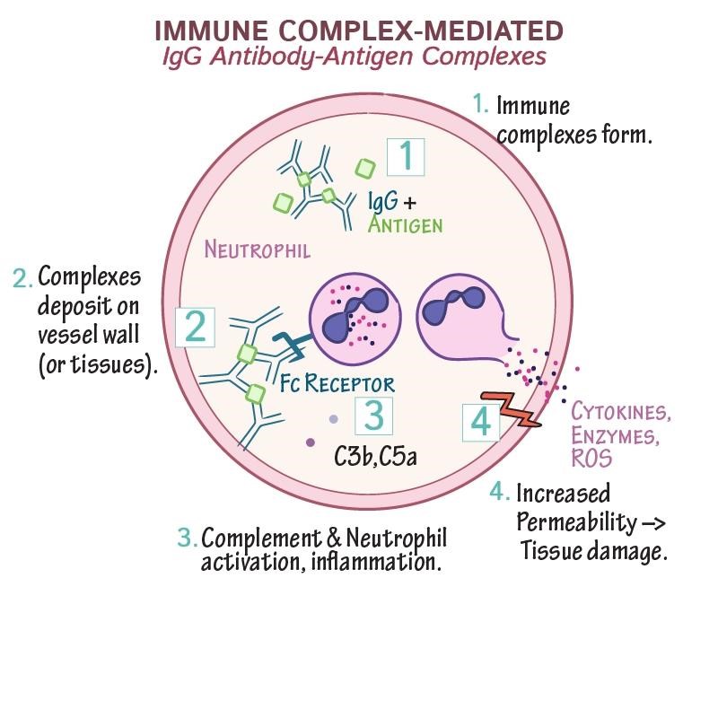
Like in type II hypersensitivity reactions, cell injury caused in the type III reaction is by complement system activation. But here, immune complexes are formed first when antibodies bind to antigens. The immune complexes can then precipitate in various tissues of joints, skin, blood vessel walls, the kidney’s glomerular basement membrane, and the brain’s choroid plexus. Such depositions trigger complement activation. It leads to the recruitment of inflammatory cells like monocytes and neutrophils that release lytic enzymes and free radicals at the site.
The whole process is described in three main steps: formation of immune complex, its deposition, and inflammation and tissue damage.
Immune complex formation
Exposure to exogenous as well as endogenous antigens triggers the formation of antibodies. Foreign proteins like microorganisms or pharmaceutical products are exogenous antigens, whereas self-antigens are endogenous antigens against which autoantibodies are generated (causing autoimmunity). The antigen can enter the body from ingestion, inhalation, or subcutaneous route. The antigens bind to antibodies and form circulating immune complexes. Such complexes migrate out of plasma later and deposit in host tissues.
Immune complex deposition
The harm an immune complex can do depends heavily on the antigen-antibody ratio, because the ratio decides the size of the complex.
When antibody is in excess, the complexes are large. Large complexes are efficiently caught and removed by macrophages in the spleen and lymph nodes, so they cause little damage.
When antigen is in slight excess, the complexes are small and soluble. These are the dangerous ones. They are too small to be cleared efficiently, so they keep circulating and eventually deposit in tissues where blood is filtered under pressure, such as the glomeruli of the kidney and the synovium of joints. This is why the kidney and joints are so often affected in Type III disease.
Complex size also decides where deposition happens. Larger deposited complexes tend to settle on the basement membrane of vessel walls and glomeruli, while smaller complexes can pass through and lodge beneath the epithelium.
| Site of deposition of immune complexes | Resulting disease |
|---|---|
| Blood vessels | Vasculitis |
| Kidney (glomerulus) | Glomerulonephritis |
| Joints | Arthritis |
 ### Inflammation and Tissue Damage
The final step of type III hypersensitivity reaction is activating the classical complement pathway. The complement split products, C3a and C5a, are released, which are anaphylactic, causing localized mast-cell degranulation and consequent increase in local vascular permeability.
The complexes and complement together draw a flood of neutrophils to the site. Here is the problem that makes Type III so destructive. The complex is deposited flat on a surface, such as a basement membrane, so the neutrophil cannot engulf it. It tries to phagocytose something it cannot surround. Frustrated in this way, the neutrophil instead spills its proteolytic enzymes, pro-inflammatory cytokines, and reactive oxygen species directly onto the tissue underneath. The C3b coating the complex acts as the opsonin that brings the neutrophil in. The result is that the tissue, not the complex, takes the damage. This is sometimes called frustrated phagocytosis.
Similarly, further activation of the membrane attack complex can also contribute to tissue destruction. In addition, it can induce aggregation of platelets, and the resulting release of clotting factors can lead to the formation of microthrombi.
These inflammatory reactions lead to tissue damage, producing the systemic and localized disease seen in Type III hypersensitivity. The damage is inflammatory, not infectious. No microbe is causing it. The harm comes from the deposited complexes and the complement and neutrophils they recruit.
Clinical Manifestation of Type III Hypersensitivity
The magnitude of the reaction depends on the quantity of immune complexes as well as their distribution within the body. A localized response occurs when immune complexes are deposited in tissue near the antigen entry. And when the complexes are formed in the blood, a reaction develops wherever the complexes are deposited. Several autoimmune diseases develop from circulating antigens or DNA-forming immune responses.
The reactions can be categorized as localized and systemic reactions.
Localized Reactions
- Local Arthus reaction: This is the localized form of Type III. It happens in a person who already has high levels of IgG against an antigen. When that antigen is injected into the skin, it meets the IgG right there in the tissue, forms immune complexes locally, activates complement, and recruits neutrophils. The result is local vasculitis with pain, swelling, redness, and in strong reactions, tissue necrosis. The reaction is not immediate. It develops over about 4 to 10 hours, which distinguishes it from the minutes-fast Type I reaction. An insect bite in a sensitized person can show both: a rapid Type I wheal within minutes, followed hours later by an Arthus reaction at the same site.
- Hypersensitivity pneumonitis (farmer’s lung): Pulmonary Arthus-type reaction can also be induced by bacterial spores, fungi (mold), hay dust, or dried fecal proteins. It causes pneumonitis or alveolitis.
- Others: A local Arthus-type reaction can occasionally follow a booster vaccine (such as tetanus toxoid) given to a person who already has very high antibody levels, producing swelling and pain at the injection site.
Systemic Reactions
- Serum sickness: It can be induced by older types of vaccines derived from antibodies from other species (foreign serum) or transfusions. For example, anti-venom reactions occur when anti-snake venom treatment is administered to a previously sensitized individual. Newer anti-venom treatments avoid this reaction. It can also happen by administrating therapeutic monoclonal antibodies.
It was described in soldiers given horse serum as protection against diphtheria and tetanus, when their own antibodies formed complexes with the still-circulating horse protein.
Antibodies specific to the foreign serum proteins form circulating immune complexes. Within days or weeks after exposure, fever, weakness, generalized vasculitis (rashes), lymphadenopathy, arthritis, and sometimes glomerulonephritis can be observed. - Systemic Lupus Erythematosus: SLE is an autoimmune disease with a genetic predisposition, not a simple single-gene genetic disorder. The body makes antibodies against its own DNA. These anti-DNA antibodies bind circulating DNA to form immune complexes, which deposit in the synovial membranes to cause arthritis and in the kidney's basement membrane to cause progressive kidney damage (lupus nephritis). It can also affect the digestive tract, heart, and skin. Sometimes, it shows Raynaud Phenomenon during cold, characterized by numbness and cyanosis (lacking oxygen), especially in fingers.
- Drug reactions: Allergies to penicillin and sulfonamides can also cause type III hypersensitivity reactions.
- Infectious diseases: Type III hypersensitivity reactions can cause several contagious diseases. Post-streptococcal glomerulonephritis (PSGN) occurs when circulating complexes of antibodies and streptococcal antigens are deposited in the kidney and damage glomeruli.
- Rheumatoid arthritis: Rheumatoid arthritis is another disease resulting from an autoimmune disorder due to the immune complex known as rheumatoid factor (rf). Immune complexes get deposited in joints and damage tissues in this disease.
Similarly, type III hypersensitivity reaction is also seen in other infectious diseases like meningitis, hepatitis, mononucleosis, malaria, and trypanosomiasis.
Diagnosis of Type III Hypersensitivity
Detection of a specific antigen causing type III hypersensitivity is difficult. The diagnosis is primarily based on the association of antigen exposure to clinical manifestations, such as fever, arthritis, and rash. The diagnosis includes clinical history or findings and laboratory tests, like:
Blood
A blood sample is tested for CBC (complete blood count), ESR, CRP, and complement levels. In active Type III disease the serum complement is often low, because complement is being consumed by the deposited immune complexes. This falling complement is a useful clue. In post-streptococcal glomerulonephritis, raised antibodies against streptococcal products (antistreptolysin O, anti-DNase B) point to a recent streptococcal infection as the antigen source.
Urine
Urinalysis with microscopy is carried out. Proteinuria and hematuria are observed in SLE, serum sickness, and PSGN.
Imaging
X-rays and CT scans are performed, especially for pneumonia and joint involvement.
Intradermal (Arthus-type) skin test, read at 4 to 10 hours, not the immediate wheal-and-flare read used for Type I
Biopsy
Renal biopsy, a skin biopsy, or a bronchoscopy
Culture
Blood, skin, throat culture.
Type I and II hypersensitivity reactions are antibody-mediated similar to type III hypersensitivity. Thus, their clinical features can overlap. In such cases, differential diagnosis is necessary.
Treatment and Management
- Avoidance of exposure
- Removal of the offending agent is essential.
- If an occupational hazard is a cause, appropriate precautions should be taken at the worksite, or one should change the nature of the work.
- Reviewing the drug allergy list and related side effects should be carried out for the sensitive patient.
- To avoid problems caused by older vaccines, antitoxins, or animal serum, current therapeutic antibodies are humanized or genetically engineered not to be recognized as foreign.
- Use of drugs and other treatments
- Disease treatment is based on the individual patient’s condition.
- Nonsteroidal anti-inflammatory drugs can relieve joint pain and fever. In more significant disease, corticosteroids and other immunosuppressive drugs are used to reduce the immune-complex-driven inflammation. Antihistamines have little role here, because the damage in Type III is driven by complement and neutrophils, not by histamine.
- The patient must be hospitalized in cases of hemodynamic instability, life-threatening symptoms, or unclear diagnosis.
- In some cases, dialysis or organ transplantation is to be carried out.
How to remember Type III
Floating complex, not fixed target. This is the one line that separates Type III from Type II. In Type II the antigen is stuck on a cell and the antibody comes to it. In Type III the antigen floats free, meets antibody in the blood, and they clump into a floating complex that deposits later. Fixed versus floating.
Small complexes at antigen excess are the killers. Not big complexes, not antibody excess. The dangerous complexes are small and soluble, formed when antigen is in slight excess, because those are the ones the body cannot clear. If you remember one fact for the exam, remember: small, soluble, antigen excess.
Three sites: vessels, kidney, joints. Immune complexes deposit where blood is filtered under pressure. Vasculitis (vessels), glomerulonephritis (kidney), arthritis (joints). If a disease hits those three, think immune complex.
Frustrated phagocytosis. The neutrophil cannot swallow a complex glued flat to a membrane, so it vomits its enzymes onto the tissue instead. The tissue takes the damage. The word "frustrated" is the memory hook: a frustrated neutrophil does the damage.
Serum sickness is the systemic prototype, Arthus is the local prototype. One antigen dose spread through the blood gives systemic serum sickness. One antigen injected into skin gives a local Arthus reaction. Same mechanism, different scale.
Key exam facts
| Point | Fact |
|---|---|
| Also called | Immune complex hypersensitivity |
| Antibodies | Mainly IgG (also IgM) |
| Antigen | Soluble and floating in the circulation (not fixed to a cell) |
| Pathogenic complex | Small, soluble complexes formed at slight antigen excess |
| Why small complexes deposit | Too small to be cleared efficiently by spleen and liver |
| Timing of serum sickness | Days to weeks after exposure (antibody must build up) |
| Timing of Arthus reaction | About 4 to 10 hours |
| Three deposition sites | Blood vessels, kidney glomeruli, joints |
| Complement products | C3a and C5a (anaphylatoxins, chemotactic) |
| Key effector cell | Neutrophil (via frustrated phagocytosis) |
| Local prototype | Arthus reaction |
| Systemic prototype | Serum sickness |
| Classic autoimmune example | Systemic lupus erythematosus (anti-DNA complexes) |
| Classic infectious example | Post-streptococcal glomerulonephritis |
| Occupational example | Hypersensitivity pneumonitis (farmer's lung) |
| Distinguishing test finding | Low serum complement (consumed), immune complexes on biopsy |
Where students get confused
"Bigger or more immune complexes cause more damage." No. Large complexes are cleared easily and cause little harm. The damaging ones are small and soluble, formed at slight antigen excess, because those escape clearance and deposit in tissue.
"Type III and Type II are the same because both use IgG and complement." They differ on where the antigen is. In Type II the antigen is fixed on a cell or tissue and the antibody binds it there. In Type III the antigen is soluble, the complex forms in the blood, and it deposits later. Fixed target versus floating complex.
"Type III is immediate like Type I." No. Type III is delayed. Serum sickness takes days to weeks because antibody must first build up. Even the local Arthus reaction takes several hours. Nothing about Type III is minutes-fast.
"The neutrophil eats the immune complex and clears it." It cannot. The complex is stuck flat on a membrane, so the neutrophil cannot surround it. It releases its destructive enzymes onto the tissue instead. This frustrated phagocytosis is what damages the tissue.
"Post-streptococcal glomerulonephritis is a direct streptococcal infection of the kidney." It is not. The kidney damage comes from immune complexes of streptococcal antigen and antibody depositing in the glomerulus, days to weeks after the throat or skin infection. The bacteria are not in the kidney.
"SLE is a single-gene genetic disease." SLE has a genetic predisposition, but it is a multifactorial autoimmune disease, not a simple inherited genetic disorder. The tissue damage comes from anti-DNA immune complexes.
References
- Punt J, Stranford SA, Jones PP, Owen JA. Kuby Immunology. 8th ed. New York: W. H. Freeman; 2019.
- Abbas AK, Lichtman AH, Pillai S. Cellular and Molecular Immunology. 10th ed. Philadelphia: Elsevier; 2022.
- Delves PJ, Martin SJ, Burton DR, Roitt IM. Roitt's Essential Immunology. 13th ed. Chichester: Wiley-Blackwell; 2017.
- Usman N, Annamaraju P. Type III Hypersensitivity Reaction. In: StatPearls. Treasure Island (FL): StatPearls Publishing; 2025.
Frequently Asked Questions
What is the main difference between Type II and Type III hypersensitivity?
What is the main difference between Type II and Type III hypersensitivity?
It comes down to where the antigen is. In Type II the antigen is fixed on a cell surface or in a tissue, and the antibody binds it there. In Type III the antigen is soluble and floating in the blood, the antibody meets it in the circulation, and they form immune complexes that deposit in tissues later. Fixed target means Type II; floating complex means Type III.
Why are small immune complexes more dangerous than large ones?
Why are small immune complexes more dangerous than large ones?
Large complexes are efficiently caught and removed by macrophages in the spleen and liver, so they rarely cause harm. Small soluble complexes, formed when antigen is in slight excess, slip past this clearance system. They keep circulating and eventually deposit in small vessels, the kidney, and joints, where they trigger inflammation.
Why does serum sickness take days to appear?
Why does serum sickness take days to appear?
Because the antibody has to build up first. When a foreign protein such as animal serum is given, it takes about a week to ten days for the body to produce enough antibody to form complexes with the still-circulating protein. The symptoms appear only once those complexes form and deposit, which is why the illness is delayed rather than immediate.
What is frustrated phagocytosis?
What is frustrated phagocytosis?
When an immune complex is deposited flat on a surface like a basement membrane, a neutrophil cannot surround and engulf it. Unable to complete phagocytosis, the neutrophil instead releases its digestive enzymes and reactive oxygen species directly onto the underlying tissue. This spilled content damages the tissue, and the process is called frustrated phagocytosis.
Why do immune complexes deposit in the kidney and joints so often?
Why do immune complexes deposit in the kidney and joints so often?
These are sites where blood is filtered under pressure. The glomerulus of the kidney and the synovium of joints both push fluid across membranes, which favors the trapping and deposition of small circulating complexes. This is why glomerulonephritis and arthritis are such common features of Type III disease.
How is post-streptococcal glomerulonephritis a Type III reaction?
How is post-streptococcal glomerulonephritis a Type III reaction?
After a streptococcal throat or skin infection, antibodies form against streptococcal antigens. These antigen-antibody complexes circulate and deposit in the glomeruli of the kidney, triggering inflammation and glomerulonephritis. The kidney injury is caused by deposited immune complexes, not by bacteria infecting the kidney directly.

Tankeshwar Acharya, MSc (Medical Microbiology)
Tankeshwar Acharya is an Assistant Professor in the Department of Microbiology at Patan Academy of Health Sciences (PAHS), Nepal, where he has been teaching and practicing clinical microbiology for over 14 years. He is the founder of Microbe Online, one of the leading free microbiology education resources on the web, covering bacteriology, mycology, parasitology, immunology, and clinical laboratory diagnostics written from direct experience in both the classroom and the diagnostic laboratory.
Comments
No comments yet. Be the first to share your thoughts.
Leave a comment
All comments are reviewed before they appear.