
C-reactive Protein (CRP) Test: Principle, Procedure, Interpretation, and Uses
CRP is a key hospital marker of inflammation. Learn how CRP is measured, how to read a result in mg/L, hs-CRP for heart risk, CRP versus ESR, and what a negative result does not rule out.

On this page
A patient is three days out from abdominal surgery. Her CRP was falling as expected, then this morning it climbed again. Nothing else has changed yet: her temperature is normal, her wound looks clean. The number moved before the patient did. A rising CRP after day three is one of the earliest signals that something is developing under the surface, often before the clinical picture catches up. Reading that number well is what this test is really about.
C-reactive protein (CRP) is an acute-phase protein made by the liver and released into the blood within hours of tissue injury, infection, or inflammation. It belongs to the pentraxin family of proteins. CRP binds to the surface of dead and dying cells and to some bacteria, activates the complement system, and helps phagocytes clear the target. It was named for its ability to bind the C-polysaccharide of Streptococcus pneumoniae.
 CRP is a nonspecific marker. A raised level tells you that inflammation is present somewhere in the body. It does not tell you where or why. That single fact governs almost everything about how the test is used and misused.
How CRP is measured
Modern hospital laboratories measure CRP as a number, in milligrams per liter (mg/L), using quantitative immunoassays. The two standard methods are immunoturbidimetry and nephelometry. In both, anti-CRP antibodies bind CRP in the serum to form antigen-antibody complexes, and the instrument measures how much these complexes scatter or block a beam of light. More CRP produces more complexes and a larger signal, which the analyzer converts to a concentration. These methods are fast, automated, and give the precise value that clinicians rely on.
A high-sensitivity version of the same assay, called hs-CRP, measures very low concentrations that the standard assay cannot resolve. hs-CRP is not a different protein. It is the same CRP measured with a more sensitive method, used for a different purpose (cardiovascular risk), explained below.
The older latex agglutination test is a qualitative or semi-quantitative slide method. It is cheap, needs no instrument, and is still valuable for teaching and for low-resource settings, so it is worth understanding in detail. It is described in full below as the worked example of how the antigen-antibody reaction is read by eye.
Principle of the CRP latex agglutination test
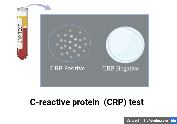
The latex agglutination test is based on the reaction between CRP in the patient's serum and anti-CRP antibodies. The antibodies are coated onto tiny latex particles. When serum containing CRP is mixed with the latex reagent, the CRP cross-links the antibody-coated particles into visible clumps. In a positive reaction, agglutination appears within two minutes. No clumping means no detectable CRP at the reagent's sensitivity level.
Materials required
- Serum sample
- Positive and negative controls
- Glass slide with reaction circles
- CRP latex reagent (suspension of latex particles coated with anti-CRP antibodies)
- Mixing sticks
- Micropipette or dropper
- Normal saline (for dilutions)
- Stopwatch
Procedure
Bring all reagents and serum to room temperature before starting, and mix the latex reagent gently before use. Do not dilute the controls.
Qualitative method
- Place one drop each of the serum, the positive control, and the negative control into separate reaction circles on the slide.
- Add one drop of CRP latex reagent to each.
- Mix each circle uniformly with a clean stick.
- Start the stopwatch and rock the slide gently back and forth for two minutes.
- Read agglutination by eye. Clumping is positive. A smooth, milky suspension is negative.
Semi-quantitative method
- Prepare serial dilutions of the serum in normal saline: 1:2, 1:4, 1:8, 1:16, 1:32, and so on.
- Place one drop of each dilution into a separate reaction circle, alongside the positive and negative controls.
- Add one drop of latex reagent to each circle and mix.
- Rock the slide gently for two minutes and read each circle for agglutination.
- The titer is the highest dilution that still shows visible agglutination.
Calculating the approximate concentration
Multiply the reagent sensitivity (printed on the latex reagent vial, for example 6 mg/L) by the titer to estimate the CRP concentration.
Approximate CRP (mg/L) = reagent sensitivity x titer
For example, with a reagent sensitivity of 6 mg/L, the dilutions give:
| Dilution | Approximate CRP (mg/L) |
|---|---|
| 1:1 | 6 |
| 1:2 | 12 |
| 1:4 | 24 |
| 1:8 | 48 |
| 1:16 | 96 |
| 1:32 | 192 |
How to interpret a CRP result
Read the number, not just positive or negative. The clinical meaning of CRP comes from how high it is and how it changes over time, not from a yes-or-no result. This is why quantitative methods have replaced the slide test in hospitals.
A rough guide to CRP levels in adults:
| CRP level | Common interpretation |
|---|---|
| Less than 10 mg/L | Normal or minor elevation |
| 10 to 50 mg/L | Mild inflammation, many viral infections, tissue injury, chronic low-grade inflammation |
| 50 to 100 mg/L | Moderate inflammation, active disease |
| Greater than 100 mg/L | Marked elevation, strongly suggests serious bacterial infection, but also seen in major trauma, surgery, and some autoimmune flares and cancers |
These bands are a guide, not a diagnosis. Reference ranges vary by laboratory, so always read the result against the range on the report.
What a negative or normal CRP does and does not mean. A CRP below 10 mg/L does not rule out infection or inflammation. Very early infection (before the liver has responded), localized infection, and some conditions can present with a normal CRP. In the first 6 to 8 hours after an insult, CRP may not have risen yet. A single normal CRP in a sick patient is reassuring only in context, never on its own.
What a raised CRP means. It confirms inflammation but not its cause. The magnitude narrows the possibilities: higher values shift the probability toward serious bacterial infection, but no single value is diagnostic. CRP is interpreted alongside the white cell count, the clinical picture, and often procalcitonin.
Trends matter more than single values. CRP has a short half-life of about 19 hours and falls by roughly half per day once the trigger is controlled. This makes it excellent for monitoring. After surgery, CRP normally peaks around day 2 to 3 and then declines. A CRP that keeps climbing after day 3 to 4, like the patient in the opening scenario, suggests a developing complication such as infection. During antibiotic treatment, a falling CRP suggests the treatment is working; a value that does not fall, or rises, suggests it is not.
Bacterial versus viral. Bacterial infections tend to drive CRP high, often above 100 mg/L in serious cases. Viral infections usually produce more modest rises, commonly in the 10 to 50 mg/L range, though severe viral illness can go higher. CRP alone cannot separate the two, but the degree of elevation contributes to the judgment.
Two important exceptions to remember. In systemic lupus erythematosus, CRP is often low or only mildly raised even during an active flare, so a markedly high CRP in a lupus patient should prompt a search for infection rather than being attributed to the flare. And IL-6 inhibitor drugs (for example, tocilizumab) directly suppress CRP production, so CRP can read low even with active infection in patients on these drugs.
hs-CRP and cardiovascular risk
High-sensitivity CRP measures the low-grade, chronic inflammation that contributes to atherosclerosis. It is used to help stratify cardiovascular risk in otherwise healthy people, not to diagnose acute illness. The American Heart Association and CDC framework uses three bands:
| hs-CRP level | Cardiovascular risk category |
|---|---|
| Less than 1 mg/L | Low risk |
| 1 to 3 mg/L | Average risk |
| Greater than 3 mg/L | High risk |
Important practical points. A value above 10 mg/L should not be used for cardiovascular risk, because it usually reflects an acute process; it should be repeated after the person has recovered, typically two to three weeks later. Because a single reading can be thrown off by a recent cold, hard exercise, or minor illness, the AHA and CDC recommend averaging two measurements taken at least two weeks apart to establish a baseline. hs-CRP is most useful in people at intermediate cardiovascular risk, where it can help decide whether to intensify prevention.
CRP versus ESR
CRP and the erythrocyte sedimentation rate (ESR) are both markers of inflammation and are often ordered together, but they behave differently. The difference is mostly about speed.
| Feature | CRP | ESR |
|---|---|---|
| What it measures | An acute-phase protein made by the liver | How fast red cells settle, driven mainly by fibrinogen |
| Speed of rise | Rises within hours | Rises over days |
| Speed of fall | Falls within days once inflammation resolves | Falls slowly, over days to weeks |
| Best for | Detecting and monitoring acute or rapidly changing inflammation | Tracking chronic inflammation; screening for giant cell arteritis and polymyalgia rheumatica |
| Affected by | Relatively few non-inflammatory factors | Age, sex, anemia, pregnancy, obesity |
In short, CRP is the faster, more responsive marker and is preferred when the question is about acute inflammation or monitoring a response to treatment. ESR is more useful for certain chronic conditions and remains part of the diagnostic criteria for giant cell arteritis and polymyalgia rheumatica.
Clinical uses of CRP
- Early detection and monitoring of bacterial infection across many settings.
- Helping distinguish bacterial from viral infection and, with other tests, conditions such as pyelonephritis versus cystitis.
- Monitoring disease activity and treatment response in chronic inflammatory diseases such as rheumatoid arthritis and inflammatory bowel disease.
- Monitoring infections that are hard to follow clinically, such as osteomyelitis and prosthetic joint infection.
- Detecting post-operative complications through the CRP trend.
- Cardiovascular risk stratification, using hs-CRP.
Limitations
- CRP is nonspecific. It confirms inflammation but does not identify the site or cause.
- A normal CRP does not rule out early, localized, or low-grade infection.
- In the latex agglutination method, reading later than two minutes can give a false positive as the drop dries.
- Only fresh serum should be used for the latex method. Plasma can cause nonspecific agglutination because of fibrinogen.
- False negatives can occur from the prozone effect, where a very high CRP concentration paradoxically gives a weak or negative reaction because antigen is in large excess and prevents lattice formation. If a strongly ill patient gives a negative or weak result, retest a diluted sample (for example, 1:10).
- Certain drugs, such as IL-6 inhibitors, suppress CRP and can mask real inflammation.
How to Remember
CRP is fast, ESR is slow. CRP rises in hours and falls in days. ESR rises in days and falls in weeks. If the clinical question is "what is happening right now," reach for CRP.
The hundred line. CRP above 100 mg/L points hard toward serious bacterial infection. Below that, the causes broaden.
Green light, red light for hs-CRP. Under 1 is low risk, over 3 is high risk, and 1 to 3 sits in between.
Lupus is the exception. In lupus, a high CRP means look for infection, not just a flare.
Key exam facts
| Item | Fact |
|---|---|
| Nature | Acute-phase protein, pentraxin family, made by the liver |
| Named for | Binding the C-polysaccharide of Streptococcus pneumoniae |
| Units | mg/L |
| Normal adult level | Less than 10 mg/L (lab dependent) |
| Half-life | About 19 hours |
| Rise after stimulus | Within 6 to 8 hours, peaks around 48 hours |
| Hospital method | Quantitative immunoturbidimetry or nephelometry |
| Teaching method | Latex agglutination (qualitative and semi-quantitative) |
| Titer calculation | Reagent sensitivity x titer = approximate mg/L |
| hs-CRP risk bands | Less than 1 low, 1 to 3 average, greater than 3 high |
| Prozone effect | Very high CRP gives false negative; retest diluted |
| Key pitfall | Normal CRP does not rule out early or localized infection |
Where Students Get Confused
"A negative CRP rules out infection." No. Early infection, localized infection, and the first several hours after an insult can all show a normal CRP. It is reassuring only alongside the clinical picture.
"hs-CRP is a different protein from CRP." No. It is the same protein measured with a more sensitive assay, used for cardiovascular risk rather than acute illness.
"Positive or negative is what matters." For the slide test, that is all you get. But clinically, the number and its trend carry the meaning. This is why hospitals report a quantitative value.
"A high CRP means bacterial infection." Not necessarily. High CRP means marked inflammation. Serious bacterial infection is one strong possibility, but major surgery, trauma, autoimmune flares, and some cancers also raise it.
"A very sick patient with a negative latex result must be CRP negative." Beware the prozone effect. A very high CRP can give a false negative on the slide test. Retest a diluted sample.
"The CRP is still high after two days of antibiotics, so treatment failed." Not on its own. CRP falls by about half per day, so a still-high but falling value early in treatment can be expected. A value that is not falling, or is rising, is the worrying pattern.
References and further reading
- Tille, P. M. (2022). Bailey and Scott's Diagnostic Microbiology (15th ed.). Elsevier.
- Mahmoud, F. A., and Rivera, N. V. (2002). The role of C-reactive protein as a prognostic indicator in advanced cancer. Current Oncology Reports.
- Pearson, T. A., et al. (2003). Markers of inflammation and cardiovascular disease: application to clinical and public health practice. A statement for healthcare professionals from the CDC and the AHA. Circulation, 107(3), 499–511.
- Vanderschueren, S., Deeren, D., Knockaert, D. C., Bobbaers, H., Bossuyt, X., and Peetermans, W. (2006). Extremely elevated C-reactive protein. European Journal of Internal Medicine, 17(6), 430–433.
Frequently Asked Questions
What does a positive CRP test mean?
What does a positive CRP test mean?
It means C-reactive protein is present at or above the test's detection level, which indicates inflammation somewhere in the body. It does not identify the cause or location. The result is interpreted with the clinical picture and other tests.
What does a negative or normal CRP mean?
What does a negative or normal CRP mean?
It means CRP is below the normal level, usually under 10 mg/L. This does not rule out infection or inflammation, because early, localized, or low-grade processes can occur with a normal CRP. In the first several hours after an insult, CRP may not have risen yet.
What is a normal CRP level?
What is a normal CRP level?
In most laboratories, less than 10 mg/L in adults. The exact cutoff varies by lab, so read the result against the range on the report.
What CRP level indicates a serious bacterial infection?
What CRP level indicates a serious bacterial infection?
There is no single cutoff, but values above 100 mg/L shift the probability strongly toward serious bacterial infection. Lower values do not exclude it, and high values also occur after surgery, trauma, and in some other conditions.
What is the difference between CRP and hs-CRP?
What is the difference between CRP and hs-CRP?
They measure the same protein. Standard CRP is used for acute illness. hs-CRP uses a more sensitive assay to detect very low levels and is used to estimate cardiovascular risk.
What is the difference between CRP and ESR?
What is the difference between CRP and ESR?
Both indicate inflammation. CRP rises and falls within hours to days, so it reflects current, changing inflammation. ESR changes over days to weeks, so it is better for chronic conditions. CRP is affected by fewer non-inflammatory factors.
What does CRP with dilution mean?
What does CRP with dilution mean?
In the semi-quantitative latex method, the serum is diluted in steps and the highest dilution that still shows agglutination is the titer. Multiplying the titer by the reagent sensitivity estimates the CRP concentration.
Why would a very sick patient have a negative CRP latex result?
Why would a very sick patient have a negative CRP latex result?
This can be the prozone effect. A very high CRP concentration can prevent the visible clumping reaction, giving a false negative. Retesting a diluted sample corrects it.

Tankeshwar Acharya, MSc (Medical Microbiology)
Tankeshwar Acharya is an Assistant Professor in the Department of Microbiology at Patan Academy of Health Sciences (PAHS), Nepal, where he has been teaching and practicing clinical microbiology for over 14 years. He is the founder of Microbe Online, one of the leading free microbiology education resources on the web, covering bacteriology, mycology, parasitology, immunology, and clinical laboratory diagnostics written from direct experience in both the classroom and the diagnostic laboratory.
Comments
No comments yet. Be the first to share your thoughts.
Leave a comment
All comments are reviewed before they appear.