Phagocytosis: Mechanism and Steps
Phagocytosis: Mechanism and Steps
Phagocytosis is the ingestion of extracellular particulate material such as invading pathogens or dead/dying cells by phagocytic cells and is one of the important innate defense mechanisms. It is primarily conducted by specialized cells, such as macrophages, neutrophils, and dendritic cells.
Phagocytosis is one type of endocytosis, others are, receptor-mediated endocytosis and pinocytosis.
Step 1: Activation of Phagocytic cells and Chemotaxis
In thefirst step of phagocytosis, phagocytes are attracted by and move toward a variety of substances generated in the immune response; this process is called chemotaxis.
Resting phagocytes are activated by inflammatory mediators (bacterial products, cytokines, prostaglandins, and complement proteins). Activation increases their metabolic and microbicidal activity. Activated cells also express more glycoprotein receptors which help them to reach the site of infections as well as to bind firmly with microorganisms. Neutrophils are the first to appear and are later replaced by macrophages.
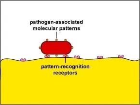 Figure: PRR binding with PAMPs(Image source:Gary E. Kaiser)
Step 2:Recognition of invading microbes
The next step in phagocytosis is the adherence of the antigen to the cell membrane of the phagocytic cells. Adherence induces membrane protrusions, called pseudopodia, to extend around the attached material and to ingest them.
Phagocytic cells contain various receptors which help them to attach with bacteria or viruses. Some of these receptors are:
- Pattern recognition receptors (PRR): Pattern recognition receptors recognize pathogen-associated molecular patterns (PAMPs). These include bacterial molecules such as peptidoglycan, teichoic acids, lipopolysaccharide, mannans, flagellin, pilin, and bacterial DNA. For example, scavenger receptors and toll-like receptors bind and internalize gram-positive and gram-negative bacteria after binding with PAMPs.
Opsonin is a molecule that binds to both antigen and macrophage and enhances phagocytosis.
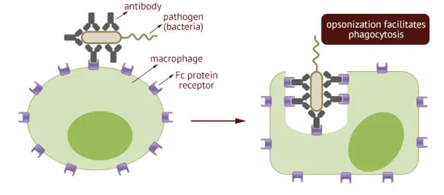 Figure: Fc receptor-mediated Opsonization(Image source: philpoteducation)
Fc receptors: Fc receptors (FcR) present on the surfaces of macrophages and neutrophils bind with the Fc portion of antibodies such as IgG and IgM complexed with antigens (bacterial cell or virus). This process, called opsonization, enhances phagocytosis.
Complement receptors (CR1): Complement receptors present on the phagocytic cells bind with complement proteins complexed with antigen-antibody complexes. For example, macrophages have receptors for C3b and so bind cells or complexes to which C3b has adhered, leading to phagocytosis. Mannose-binding lectins (MBL) also help to enhance phagocytosis.
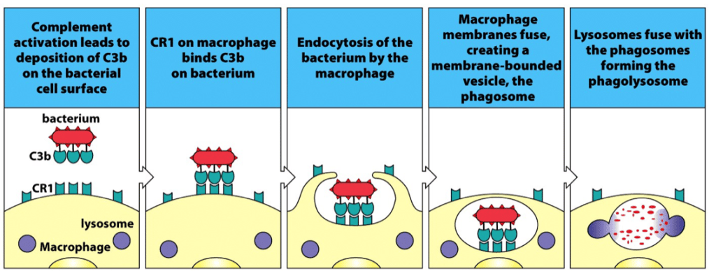 Figure: Complement mediated phagocytosis
Step 3:Ingestion and formation of phagosomes
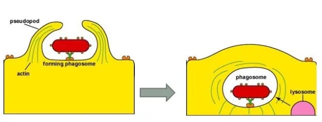 Figure: Formation of phagosome(Image source: Gary E. Kaiser)
Following attachment, polymerization and then depolymerization of actin filaments send pseudopods out to engulf the microbe. Fusion of the pseudopodia encloses the material within an endocytic vesicle called a phagosome, which then enters the endocytic processing pathway.
Step 4:Formation of phagolysosome
In this pathway, a phagosome moves toward the cell interior, where it fuses with a lysosome to form a phagolysosome.
Step 5:Microbial killing and formation of residual bodies
Lysosomes contain lysozyme and a variety of antimicrobial and cytotoxic substances that can destroy phagocytosed microorganisms and cells. Microorganisms are killed either by oxygen-dependent or by oxygen-independent mechanisms.
- Oxygen-Dependent Killing: Activated phagocytes produce a number of reactive oxygen intermediates (ROIs) and reactive nitrogen intermediates that have potent antimicrobial activity. A metabolic process known as respiratory burst occurs in phagocytic cells that activate membrane-bound oxidase forming superoxide anion, hydroxyl radicals, and hydrogen peroxide. Other potent antimicrobial substances such as hypochlorite, nitric oxide, etc are also formed inside the phagolysosome. All of these substances showed marked antimicrobial activity against bacteria, fungi, parasitic worms, and protozoa.
- Oxygen Independent Killing: Activated phagocytic cells also synthesize lysozyme and various hydrolytic enzymes (for example, cathepsin G, elastase, collagenase, cathelicidins and bactericidal permeability inducing protein) whose degradative activities do not require oxygen. In addition, activated macrophages produce a group of antimicrobial and cytotoxic peptides, commonly known as defensins. Defensins can kill a variety of bacteria, including Staphylococcus aureus, Streptococcus pneumoniae, Escherichia coli, Pseudomonas aeruginosa, and Haemophilus influenzae. Activated macrophages also secrete tumor necrosis factor α (TNF-α), a cytokine that has a variety of effects and is cytotoxic for some tumor cells.
Step 6:Elimination or exocytosis
The digested contents of the phagolysosome are then eliminated in a process called exocytosis.
References and further readings
- Punt, Jenni, Stranford, Sharon, Jones, Patricia, & Owen, Judy. (2018). Kuby Immunology (Eighth edition). W. H. Freeman.
- The Innate Immune System: Phagocytosis—The Process of Phagocytosis. Retrieved April 17, 2020.

Tankeshwar Acharya, MSc (Medical Microbiology)
Tankeshwar Acharya is an Assistant Professor in the Department of Microbiology at Patan Academy of Health Sciences (PAHS), Nepal, where he has been teaching and practicing clinical microbiology for over 14 years. He is the founder of Microbe Online, one of the leading free microbiology education resources on the web, covering bacteriology, mycology, parasitology, immunology, and clinical laboratory diagnostics written from direct experience in both the classroom and the diagnostic laboratory.