
Structure and Components of DNA
The structure of DNA has three levels: the nucleotide sequence, the double helix, and its 3D forms. Learn the components, base pairing, and the A, B, and Z forms, with clear diagrams.

On this page
DNA is often called the instruction manual of life, but a manual is only useful because of how it is built. The way DNA is structured, a long chain of just four building blocks, twisted into a double helix, is what lets it store an enormous amount of information, copy itself accurately, and pass traits from parent to offspring. Understanding the structure is the key to understanding everything DNA does.
This article builds that structure up in clear levels. First the components, the small parts DNA is made from. Then the primary structure, how those parts link into a strand. Then the secondary structure, how two strands pair into the famous double helix. And finally the tertiary structure, the different 3D forms the helix can take. Each level sits on the one before it, so by the end the whole molecule makes sense as one connected picture.
DNA (deoxyribonucleic acid) is a hereditary material commonly found in the nucleus of a cell. It is also present in mitochondria, which is called mitochondrial DNA (mtDNA). DNA is a polymer made of many deoxyribonucleotides, covalently linked by phosphodiester bonds. It is a double-stranded molecule in which two strands wind around each other and form a double helix. Whereas some viruses like Parvovirus have a single-stranded DNA molecule.
DNA consists of deoxyribose sugar, nitrogenous bases (purine and pyrimidine), and phosphoric acid. Information is encoded in the DNA as the sequences of ATGC. Adenine (A) pairs with thymine (T) with the double hydrogen bonds, whereas guanine (G) pairs with cytosine (C) with the triple hydrogen bonds. This is the base-pairing rule, and it never changes: A always pairs with T, and G always pairs with C. A purine always pairs with a pyrimidine, which is what keeps the two strands an even distance apart along the whole helix. The arrangement of the nucleotides (ATGC) in two strands gives rise to the spiral structure of DNA, commonly known as the double helix.
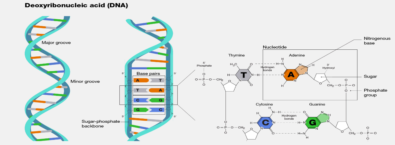 Figure: Structure of DNA ,Courtesy: National Human Genome Research Institute
Components of DNA
The essential components of DNA are acid (phosphoric acid), pentose sugar (deoxyribose sugar), and nitrogen bases; purine (adenine and guanine) and pyrimidine (cytosine and thymine).
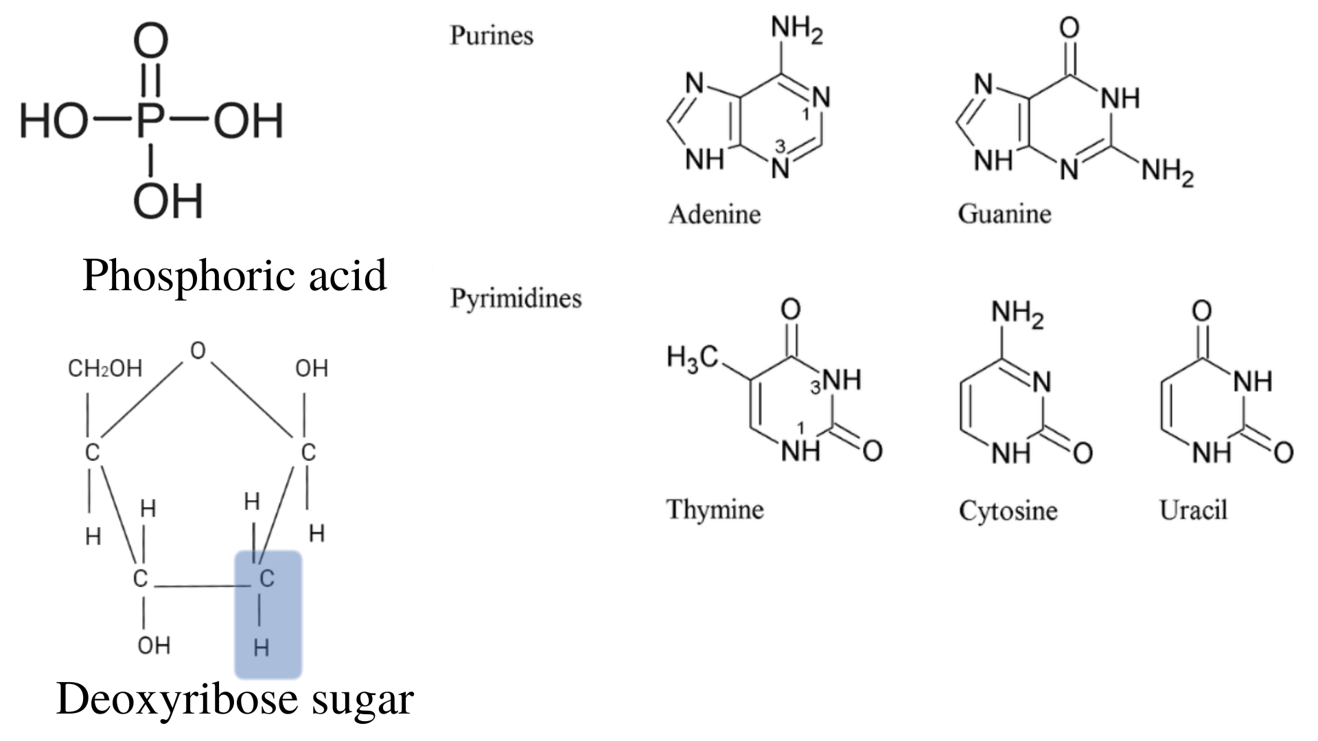 Figure: Phosphoric acid, deoxyribose sugar, and nitrogenous bases
Phosphoric acid
It contains the three monovalent hydroxyl groups and one divalent oxygen atom linked to the pentavalent phosphorus atom. The molecular formula of phosphoric acid is H3PO4.
Pentose sugar
Deoxyribose sugar is the pentose sugar present in the DNA. Since it lacks one oxygen atom in carbon-2, it is known as deoxyribose. Pentose sugar forms the ester with phosphoric acid and forms the 3’-5′ phosphodiester bond.
3′-5′ phosphodiester bonds
A phosphodiester bond links the 3′ carbon of one sugar to the 5′ carbon of the next, through a phosphate group. The phosphate group joins these hydroxyl groups in the adjacent nucleotides. In the DNA, the sequences are encoded in the 5′ end to the 3′ end of the chain. E.g., 5’-ATGC-3′
Nitrogenous bases
There are two types of nitrogenous bases: purine and pyrimidines. The purine derivatives present in the DNA are adenine (A) and guanine (G). The pyrimidine derivatives present in the DNA are thymine (T) and cytosine ( C). In the case of the RNA, thymine (T) is absent, which is replaced by the uracil (U).
Base-compositions of DNA (Chargaff rule)
The Chargaff rule was given by Erwin Chargaff and his colleagues in the 1940s and has given the following conclusions:
- The base composition of DNA generally varies from one species to another.
- DNA specimens isolated from the different tissues of the same species have the same base composition.
- The base composition of DNA in a given species does not change with the organism’s age, nutritional state, or changing environment.
- The number of adenine is equal to thymine (A=T), and guanine is equal to cytosine (G=C), i.e., A+G=T+C.
Proportion of the nitrogenous bases in human and E.coli are:
- Human: A-30.9 T-29.4 G-19.9 C-19.8
- E.coli: A 24.7, T 23.6, G 26.0, C 25.7 (percentages)
The three levels of DNA structure
DNA structure is described at three levels, each built on the one before it. The primary structure is the order of nucleotides in a single strand. The secondary structure is how two strands pair and twist into the double helix. The tertiary structure is the 3D form the helix takes, such as the A, B, and Z forms. The sections below take them in order.
Primary structure of DNA
The primary structure of DNA consists of the linear sequence of nucleotides linked together by phosphodiester bonds. Nucleotides is composed of three components; nitrogenous bases, 5-carbon (pentose) sugar, and phosphate groups.
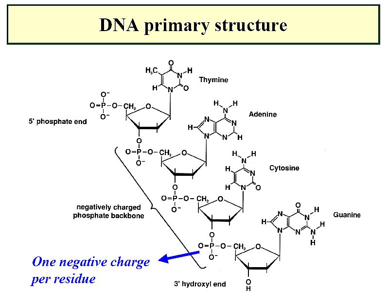 Figure: Primary structure of DNA
Secondary structure of DNA
The secondary structure of DNA is the double helix: two strands held together and wound around each other. The two strands are joined by hydrogen bonds between the paired bases (A with T, G with C). Note the difference from the primary structure: within a single strand, nucleotides are linked by phosphodiester bonds, while between the two strands, the bases are held by hydrogen bonds. This pairing is what gives DNA its double-helical shape.
Tertiary structure of DNA
The tertiary structure of DNA is the most common double helix structure which includes the 3-forms of the DNA; A, B, and Z form. The handedness (left or right) of the DNA, length of helix turn, and the number of bases per turn are the basis for the different forms. It is the location of the atom in the 3-dimensional space.
Double helix: Watson and Crick’s model
Watson and Crick’s model of double helical DNA was postulated by James D. Watson and Francis H.C. Crick in 1953. According to this model, the two helical DNA chains are wound around each other in the same axis of symmetry.
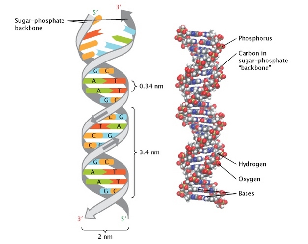 Figure: Watson and Crick’s model of double-helical DNA, source: Nature
The purine and the pyrimidine bases in both the strands are hydrophobic and stacked inside the double helix. Deoxyribose and phosphate groups are the backbone of the DNA outside the double helix, which are hydrophilic.
When the two strands wind around each other, the helix surface has two kinds of grooves: a wider major groove and a narrower minor groove. These grooves matter because proteins that read or bind DNA often do so by fitting into them, especially the major groove, where the base edges are more exposed.
For the common B-form of DNA: the helix is about 20 Å (2 nm) wide, neighboring base pairs are 3.4 Å apart along the axis, and each complete turn of the helix is 34 Å long and contains 10 base pairs. The 34 Å turn and the 10 base pairs per turn are worth remembering, as they come up often.
The hydrogen bonds between paired bases give the double helix its stability. Adenine and thymine are joined by two hydrogen bonds, while guanine and cytosine are joined by three. Because G-C pairs have an extra hydrogen bond, DNA with more G-C pairs is more stable and needs more heat to separate the strands. The B-form of DNA (Watson-Crick structure of DNA) is the most stable form of DNA.
- A-DNA: right-handed helix, about 23 Å in diameter, with 11 base pairs per turn. It appears in dehydrated conditions.
- B-DNA: right-handed helix, about 20 Å in diameter, with 10 base pairs per turn. This is the common form found in cells and the one Watson and Crick described.
- Z-DNA: left-handed helix, about 18 Å in diameter, with 12 base pairs per turn. It has a zig-zag backbone, which is where the "Z" comes from.
The key contrast to remember: B is the normal right-handed form, and Z is the unusual left-handed one.
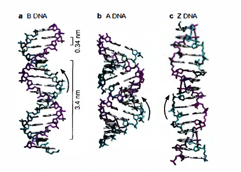 Figure: Different forms of DNA
Some viruses like Parvoviruses contain single-stranded DNA.
Single-stranded versus double-stranded DNA
Most DNA is double-stranded: two strands paired into the double helix. This double-stranded form is stable and behaves like a rigid rod. Some viruses, such as the parvoviruses, instead carry their genome as single-stranded DNA. A single strand has no complementary partner, so it is more flexible and tends to fold back on itself into loops and coils. The chemical building blocks are the same; the difference is the number of strands and the shape that results.
Functions of DNA
The DNA carries and transfers the genetic material from the parent cell to its offspring during replication. DNA also carries all the genetic codes for synthesizing RNA. The RNA then codes proteins essential for the growth, development, and proper functioning of any cell. So, DNA can be termed an instruction manual for cells.
How to Remember
The three levels, built up. Primary is the sequence (one strand's order of bases). Secondary is the shape (two strands as a double helix). Tertiary is the form (A, B, Z in 3D). "Sequence, shape, form" climbs the three levels in order.
Base pairing, locked. A-T and G-C, always. Two ways to hold it: the letters that look "straight" go together in the alphabet grouping students use, but the reliable version is the bond count. A-T = 2 bonds, G-C = 3 bonds. A useful phrase: "GC is the stronger glue" (three bonds), which is why GC-rich DNA is harder to melt apart.
Purine with pyrimidine, every time. A big base always pairs with a small one (a two-ring purine with a one-ring pyrimidine). This keeps the helix the same width all the way down. If someone offers you A-G (two purines) or T-C (two pyrimidines), it is wrong.
The B-form numbers. For normal B-DNA: 10 base pairs per turn, 34 Å per turn, 3.4 Å between bases, 20 Å wide. Notice 34 = 10 × 3.4, which ties three of the numbers together.
A, B, Z handedness. A and B are Right-handed. Z is the odd one, left-handed, with a Zig-zag backbone. "Z is left and zig-zag" separates it from the two right-handed forms.
Key exam facts in one table
| Fact | Detail |
|---|---|
| Building block | Nucleotide (phosphate + deoxyribose sugar + nitrogenous base) |
| Purines | Adenine (A), Guanine (G) — two rings |
| Pyrimidines | Thymine (T), Cytosine (C) — one ring |
| Base pairing | A-T (2 hydrogen bonds), G-C (3 hydrogen bonds) |
| Within a strand | Nucleotides linked by phosphodiester bonds (primary structure) |
| Between strands | Bases held by hydrogen bonds (secondary structure) |
| Chargaff's rule | A = T and G = C |
| Common form | B-DNA: right-handed, 10 bp per turn, 34 Å per turn, ~20 Å wide |
| Other forms | A-DNA (right-handed), Z-DNA (left-handed, zig-zag) |
| Backbone | Sugar-phosphate, on the outside, hydrophilic |
| Bases | Stacked inside the helix, hydrophobic |
| Model | Watson and Crick, 1953 |
Where Students Get Confused
Phosphodiester bonds versus hydrogen bonds. These hold DNA together in two different ways. Phosphodiester bonds link nucleotides along a single strand (they build the backbone). Hydrogen bonds hold the two strands together by joining paired bases. Mixing these up is the most common structural error. Backbone = phosphodiester; base pairs = hydrogen.
A-T has two bonds, G-C has three (not the other way round). A common slip is to reverse them. The pair with three bonds is G-C, which is why GC-rich DNA is more stable. If you remember "G-C is the stronger pair," you will not reverse it.
A purine never pairs with a purine. A pairs with T and G pairs with C, always a big base with a small base. A-G or T-C pairings are wrong because two large purines would not fit and two small pyrimidines would leave a gap.
Primary structure is not the same as the double helix. The primary structure is just the order of bases in one strand. The double helix is the secondary structure. A single strand has a primary structure even before it pairs with another.
A, B, and Z are all still DNA. They are different 3D shapes of the same molecule, not different molecules. B-DNA is the usual form in cells; A and Z appear under particular conditions.
References
- Watson JD, Baker TA, Bell SP, Gann A, Levine M, Losick R. Molecular Biology of the Gene. 7th ed. Pearson; 2013.
- Alberts B, Heald R, Johnson A, et al. Molecular Biology of the Cell. 7th ed. W.W. Norton; 2022.
- Nelson DL, Cox MM. Lehninger Principles of Biochemistry. 8th ed. W.H. Freeman; 2021.
- National Human Genome Research Institute. Deoxyribonucleic Acid (DNA). genome.gov.
Frequently Asked Questions
What are the three components of DNA?
What are the three components of DNA?
Each DNA building block (a nucleotide) has three parts: a phosphate group, a five-carbon sugar called deoxyribose, and one of four nitrogenous bases (adenine, thymine, guanine, or cytosine).
What is the primary structure of DNA?
What is the primary structure of DNA?
The primary structure is the linear sequence of nucleotides in a single strand, joined together by phosphodiester bonds between their sugars and phosphates. In other words, it is the order of bases (such as 5′-ATGC-3′) along one strand.
What is the difference between the primary, secondary, and tertiary structure of DNA?
What is the difference between the primary, secondary, and tertiary structure of DNA?
The primary structure is the sequence of bases in one strand. The secondary structure is the double helix formed when two strands pair through hydrogen bonds. The tertiary structure is the 3D form the helix takes, such as the A, B, or Z form.
How do the DNA bases pair, and how many hydrogen bonds hold them?
How do the DNA bases pair, and how many hydrogen bonds hold them?
Adenine pairs with thymine using two hydrogen bonds, and guanine pairs with cytosine using three. A purine always pairs with a pyrimidine, which keeps the helix an even width.
Why is G-C base pairing stronger than A-T?
Why is G-C base pairing stronger than A-T?
Because G-C pairs are held by three hydrogen bonds, while A-T pairs have only two. DNA with a high proportion of G-C pairs is more stable and needs more heat to separate the two strands.
What are the A, B, and Z forms of DNA?
What are the A, B, and Z forms of DNA?
They are three 3D forms of the double helix. B-DNA is the common right-handed form found in cells and the one Watson and Crick described. A-DNA is also right-handed and appears in dehydrated conditions. Z-DNA is a left-handed form with a zig-zag backbone.
What holds the two strands of DNA together?
What holds the two strands of DNA together?
Hydrogen bonds between the paired bases hold the two strands together. This is different from the phosphodiester bonds, which link the nucleotides within a single strand to form the backbone.
What is Chargaff's rule?
What is Chargaff's rule?
Chargaff's rule states that in DNA the amount of adenine equals the amount of thymine, and the amount of guanine equals the amount of cytosine (A = T and G = C). This reflects the fact that A always pairs with T and G always pairs with C.

Tankeshwar Acharya, MSc (Medical Microbiology)
Tankeshwar Acharya is an Assistant Professor in the Department of Microbiology at Patan Academy of Health Sciences (PAHS), Nepal, where he has been teaching and practicing clinical microbiology for over 14 years. He is the founder of Microbe Online, one of the leading free microbiology education resources on the web, covering bacteriology, mycology, parasitology, immunology, and clinical laboratory diagnostics written from direct experience in both the classroom and the diagnostic laboratory.
Comments
No comments yet. Be the first to share your thoughts.
Leave a comment
All comments are reviewed before they appear.