
MUG (Beta-Glucuronidase) Test for E. coli
The MUG test detects beta-glucuronidase to rapidly identify E. coli by blue fluorescence under UV light. Learn how to read the result, why E. coli O157:H7 is MUG-negative, and which organisms give false positives.

On this page
A stool culture from a child with bloody diarrhea grows an organism confirmed as Escherichia coli. Under UV light, the MUG test shows no fluorescence. For almost any other E. coli that would be a surprise, because nearly all of them light up. Here the absence of light is the clue. A MUG-negative E. coli raises the possibility of the dangerous O157:H7 strain, and that single dark tube is what tells the technologist to look harder.
MUG stands for 4-methylumbelliferyl-beta-D-glucuronide. Because roughly 94 to 97% of Escherichia coli strains produce the enzyme beta-D-glucuronidase, the MUG test is a fast way to help identify E. coli. The enzyme hydrolyzes the substrate and releases a product that fluoresces under ultraviolet light.
The most useful exception is E. coli O157:H7, the main Shiga toxin-producing (verotoxin-producing) strain, which is characteristically beta-glucuronidase-negative and therefore MUG-negative. This makes a MUG-negative E. coli a signal to consider O157:H7, explained in its own section below. A few strains of Salmonella, Shigella, and Yersinia can also produce beta-glucuronidase and give a positive result, so the test is interpreted with the organism's other features in mind.
Beta-D-glucuronidase hydrolyzes beta-D-glucopyranosiduronic derivatives into an aglycon and D-glucuronic acid.
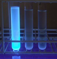 Figure: E. coli use MUG as a substrate, releasing a fluorogenic compound
The substrate, 4-methylumbelliferyl-beta-D-glucuronide, is colorless and non-fluorescent as supplied. When beta-D-glucuronidase is present, it cleaves the substrate and releases the 4-methylumbelliferyl group, which fluoresces bright blue under long-wavelength ultraviolet light. The fluorescence is the positive signal, so the test is read in the dark under a UV lamp, not in ordinary light.
Significance of MUG Test
The U.S. Environmental Protection Agency has approved 4-methylumbelliferyl-beta-D-glucuronide Escherichia coli broth medium (EC-MUG) as an effective, rapid method for detecting and verifying E. coli in food, water, and environmental samples.
Standard analysis of water includes the most probable number (MPN) for the presumptive and quantitative detection of coliform and fecal coliform bacteria in water samples. According to ASM, EC broth and agar media with MUG are best suited for confirmatory testing of the presence of E. coli after a presumptive positive result for fecal coliform bacteria.
MUG test can be used to separate potential verotoxin-producing E. coli from other E. coli strains (usually MUG positive) in gastrointestinal specimens, once the isolate has been identified as E. coli.
E. coli O157:H7 and how to interpret a MUG-negative result
The MUG test is most valuable not when it is positive, but when it is unexpectedly negative in a confirmed E. coli.
Why O157:H7 is MUG-negative. Nearly all ordinary E. coli produce beta-glucuronidase and fluoresce. E. coli O157:H7, the main cause of Shiga toxin-producing (enterohemorrhagic) infection, characteristically does not produce this enzyme, so it is MUG-negative. It also characteristically fails to ferment sorbitol, or does so only slowly. These two unusual features, no beta-glucuronidase and no sorbitol fermentation, are what set O157:H7 apart from the roughly 96% of other E. coli that are positive for both.
How this is used at the bench. Suspected O157:H7 is first screened on sorbitol-MacConkey agar, where it forms colorless (non-sorbitol-fermenting) colonies while most other E. coli are pink. A MUG-negative result on an isolate already confirmed as E. coli adds weight to the suspicion. The combination of a sorbitol-negative, MUG-negative E. coli from a patient with bloody diarrhea is a classic trigger to pursue O157.
Why MUG-negative does not confirm O157. MUG-negativity is a clue, not a diagnosis. Rare non-O157 E. coli can be beta-glucuronidase-negative, and rare O157 strains can behave atypically. A MUG-negative E. coli must be confirmed as O157:H7 by detecting the O157 and H7 antigens, and Shiga toxin or its genes should be sought directly. Do not report O157 on the basis of a dark tube alone.
False positives from other enterics. A positive MUG result is not unique to E. coli. Some strains of Salmonella, Shigella, and Yersinia also produce beta-glucuronidase. For this reason, the test should not be used to identify E. coli from abdominal or blood sources without other confirmatory features, because a MUG-positive Salmonella or Shigella could be misread as E. coli.
The two-way caution. Put simply: a MUG-negative E. coli may be O157:H7, and a MUG-positive enteric organism may not be E. coli. Both errors are avoided by interpreting MUG alongside the organism's other identification results, never in isolation.
Microorganisms Tested
Fresh colonies on blood agar plate of possible E. coli that are indole-positive, oxidase-negative, Gram-negative rods, whether they are lactose positive or negative.
Procedure for MUG test
MUG tube method
- Prepare a dense milky suspension of the organism in a small tube containing 0.25 ml of saline. Colonies from MacConkey agar can be used for the tube method, because the dye does not interfere with the tube test (unlike the disk test).
- Add a MUG disk for detection of beta-glucuronidase activity.
- Place a stopper in the tube and agitate vigorously for a few seconds.
- Incubate the tube at 35-37 °C for 2 hours.
- Observe fluorescence using long-wave UV light in a dark room.
MUG disk method
- Place MUG disk in a sterile empty petri dish and wet with 1 drop of water. If excess water is used, test may be falsely negative.
- Using a wooden stick or bacteriological loop, roll a colony of the suspected E. coli from a blood agar plate onto the disk.
- Incubate at 35°C for a minimum of 2 hours.
- Observe the disk using long-wave UV light in a darkened room.
How to read the result
Read the tube or disk in a dark room under a long-wave UV lamp. Bright blue (electric blue) fluorescence is positive. No fluorescence is negative.
Positive means beta-glucuronidase is present, which supports E. coli, provided the organism's other features fit.
Negative does not exclude E. coli. About 3 to 6% of E. coli are naturally MUG-negative, and O157:H7 is characteristically negative. A negative result on a confirmed E. coli should prompt thought about O157, not a conclusion that the organism is not E. coli.
Watch for false fluorescence. Some organisms, such as Pseudomonas aeruginosa, can fluoresce under UV for reasons unrelated to beta-glucuronidase and can mimic a positive. Do not run MUG on oxidase-positive organisms for this reason.
Avoid dye-containing media for the disk method. Dyes in media such as MacConkey or EMB can interfere with the disk test and cause a false negative. The dyes do not interfere with the tube method.
Give it the full time. Incubate at least 2 hours at 35 to 37°C before reading. A dark tube read too early may simply not have developed.
Quality control of MUG Test
- Perform quality control on each new lot or shipment of disks or liquid reagent prior to use.
- Organisms
- E. coli ATCC 25922: blue fluorescence (positive)
- Klebsiella pneumoniae ATCC 13883 or ATCC 27736: no fluorescence (negative)
Limitations
- Not all E. coli are MUG positive. A negative MUG test does not mean the organism is not E. coli.
- Do not use media that contain dyes (for example, EMB agar or MacConkey agar) for the disk test. Dyes do not interfere with the tube test.
- Do not perform the MUG test on oxidase-positive organisms. Some organisms, such as Pseudomonas aeruginosa, fluoresce under UV for other reasons and can resemble a positive result.
How to Remember
Light means E. coli, dark means look harder. Most E. coli glow blue under UV. When a confirmed E. coli stays dark, think O157:H7.
O157 breaks two rules. Ordinary E. coli ferment sorbitol and make beta-glucuronidase. O157:H7 does neither, which is exactly why it stands out.
Dark is a clue, not a verdict. MUG-negative suggests O157 but never confirms it. Antigen and toxin testing confirm it.
Positive is not proof of E. coli. Salmonella, Shigella, and Yersinia can also glow, so read MUG with the other results.
Key exam facts
| Item | Fact |
|---|---|
| Full form | 4-methylumbelliferyl-beta-D-glucuronide |
| Detects | Beta-D-glucuronidase |
| Positive | Bright blue fluorescence under long-wave UV |
| Negative | No fluorescence |
| Positive supports | Escherichia coli (about 94 to 97% are positive) |
| Key negative exception | E. coli O157:H7 (beta-glucuronidase-negative and sorbitol-negative) |
| False positives | Some Salmonella, Shigella, Yersinia strains |
| False fluorescence | Pseudomonas aeruginosa and other UV-fluorescing organisms |
| Read under | Long-wave UV in a dark room |
| Incubation | 35 to 37°C for at least 2 hours |
| Do not use disk test with | Dye-containing media (MacConkey, EMB) |
| Main uses | Rapid E. coli ID; water and food testing (EC-MUG); flagging possible O157 |
Where Students Get Confused
"A negative MUG test means the organism is not E. coli." No. About 3 to 6% of E. coli are MUG-negative, and O157:H7 is characteristically negative. A negative result on a confirmed E. coli is a reason to consider O157, not to rule out E. coli.
"A MUG-negative E. coli confirms O157:H7." No. It is a strong clue, especially with sorbitol-negativity and bloody diarrhea, but confirmation requires O157 and H7 antigen testing and detection of Shiga toxin or its genes.
"A positive MUG test proves the organism is E. coli." No. Some Salmonella, Shigella, and Yersinia strains also produce beta-glucuronidase. MUG is interpreted with the organism's other features, especially for abdominal or blood isolates.
"Any blue glow under UV is a positive." No. Some organisms, such as Pseudomonas aeruginosa, fluoresce for other reasons. This is why MUG is not run on oxidase-positive organisms.
"MacConkey agar is fine for the disk test." No. Dyes in MacConkey and EMB interfere with the disk method and can cause a false negative. Use a non-dye medium for the disk test, or use the tube method.
References and further readings
- Leber, A. L. (Ed.). (2016). Clinical Microbiology Procedures Handbook (4th ed.). ASM Press. https://doi.org/10.1128/9781555818814
- Tille, P. M. (2022). Bailey and Scott's Diagnostic Microbiology (15th ed.). Elsevier.
Frequently Asked Questions
What does the MUG test detect?
What does the MUG test detect?
It detects the enzyme beta-D-glucuronidase. The substrate, 4-methylumbelliferyl-beta-D-glucuronide, releases a blue-fluorescing product when the enzyme is present.
What is a positive MUG test?
What is a positive MUG test?
Bright blue fluorescence under long-wave UV light in a dark room. It indicates beta-glucuronidase activity and supports identification of E. coli.
What does a negative MUG test mean?
What does a negative MUG test mean?
No fluorescence. It does not rule out E. coli, because a small percentage of E. coli are MUG-negative, and E. coli O157:H7 is characteristically negative.
Why is E. coli O157:H7 MUG-negative?
Why is E. coli O157:H7 MUG-negative?
O157:H7 characteristically does not produce beta-glucuronidase, and it also does not ferment sorbitol. These two features distinguish it from the roughly 96% of other E. coli that are positive for both.
Does a MUG-negative E. coli confirm O157:H7?
Does a MUG-negative E. coli confirm O157:H7?
No. It is a useful clue, but confirmation requires detecting the O157 and H7 antigens and testing for Shiga toxin or its genes.
Can organisms other than E. coli give a positive MUG test?
Can organisms other than E. coli give a positive MUG test?
Yes. Some strains of Salmonella, Shigella, and Yersinia produce beta-glucuronidase, so a positive result is not unique to E. coli, especially for abdominal or blood isolates.
Why is the test read under UV light?
Why is the test read under UV light?
The product released by the enzyme fluoresces blue only under long-wave ultraviolet light, so the test must be read in a dark room with a UV lamp.
Why should dye-containing media be avoided for the disk test?
Why should dye-containing media be avoided for the disk test?
Dyes in media such as MacConkey and EMB interfere with the disk method and can cause a false negative. The tube method is not affected.

Tankeshwar Acharya, MSc (Medical Microbiology)
Tankeshwar Acharya is an Assistant Professor in the Department of Microbiology at Patan Academy of Health Sciences (PAHS), Nepal, where he has been teaching and practicing clinical microbiology for over 14 years. He is the founder of Microbe Online, one of the leading free microbiology education resources on the web, covering bacteriology, mycology, parasitology, immunology, and clinical laboratory diagnostics written from direct experience in both the classroom and the diagnostic laboratory.
Comments
No comments yet. Be the first to share your thoughts.
Leave a comment
All comments are reviewed before they appear.