
Lysine Iron Agar (LIA): Principle, Composition, Results, and Interpretation
LIA differentiates enteric bacteria by lysine decarboxylation, lysine deamination, and H₂S production. Learn how to read LIA results alongside TSI and KIA to identify Salmonella, Shigella, Proteus, and Providencia.

On this page
Two enteric isolates both show K/A (alkaline slant, acid butt) on KIA, both grow on XLD agar as pink colonies without black centers. Both could be Shigella. LIA differentiates them: the first shows K/K with purple butt (lysine decarboxylation positive), i.e., Salmonella (which decarboxylates lysine). The second shows R/A (red-purple slant, acid butt, lysine deamination), i.e., Proteus or Providencia (which deaminates lysine, producing a different alkaline reaction on the slant with a distinctly different color).
LIA is the medium that resolves the KIA ambiguity. Used in combination with KIA or TSI, it dramatically narrows the identification before any formal panel is run.
Lysine Iron Agar (LIA) is a combination medium used for the differentiation of gram-negative bacilli (enterics) based on decarboxylation or deamination of lysine and the formation of hydrogen sulfide (H₂S). In combination with Triple Sugar Iron (TSI) agar, LIA is used to identify members of Salmonella and Shigella.
Composition of Lysine Iron Agar
Lysine iron agar (LIA) contains peptone and yeast extract to support bacterial growth. The amino acid lysine is used for detecting deamination and decarboxylation reactions. A small amount of glucose (0.1%) is a fermentable carbohydrate.
| Ingredients | Gram/Liter |
|---|---|
| Enzymatic digest of gelatin | 5 g |
| yeast extract | 3 g |
| Dextrose | 1 g |
| L-Lysine | 10 g |
| Ferric ammonium citrate | 0.5 g |
| Sodium thiosulfate | 0.04 g |
| Bromocresol purple | 0.02 g |
| Agar | 13.5 g |
| pH: 6.5 |
Sodium thiosulfate is a source of reducible sulfur. Ferric ammonium citrate is included as a sulfur reduction indicator. The H₂S producing organism produces black color in the medium due to black precipitation of ferrous sulfide (FeS). Bromocresol purple, the pH indicator, is yellow at or below pH 5.2 and purple at or above pH 6.8.
Principle
Lysine iron agar contains an aerobic slant and an anaerobic butt. The test organism is stabbed in the butt and streaked on the slant in a fishtail streak. The tube is then tightly capped and incubated for 18 to 24 hours before reading the results.
 Figure: Breakdown of Lysine
In the anaerobic butt, organisms capable of glucose fermentation produce acid, resulting in yellow color. If the organism produces lysine decarboxylase, it removes the carboxyl group from L-lysine, releasing carbon dioxide and forming cadaverine, an alkaline product. Cadaverine neutralizes the organic acids formed by glucose fermentation, and the butt of the medium reverts to the original alkaline state (purple). The purple color throughout indicates lysine decarboxylation. The purple color in the slant with a yellow (acidic) butt indicates no lysine decarboxylation.
Deamination reactions require the presence of oxygen. Therefore, any evidence of deamination will be seen only in the slant. If the organism produces lysine deaminase, the resulting deamination reaction will produce compounds that react with the ferric ammonium citrate and a coenzyme, flavin mononucleotide (FMN), forming a burgundy (dark red color) on the slant. A red slant with a yellow (acidic) butt indicates lysine deamination.
Hydrogen sulfide (H₂S) is produced in LIA by the anaerobic reduction of thiosulfate. Ferric ions in the medium react with the H₂S to form a black precipitate in the butt.
The difference between decarboxylation and deamination, why it matters:
- Lysine decarboxylation (K/K result, purple butt): The organism removes the carboxyl group from lysine, producing cadaverine which is a strongly alkaline amine. The entire butt turns purple. This is the Salmonella pattern.
- Lysine deamination (R/A result, red/burgundy slant): The organism removes the amino group from lysine under aerobic conditions on the slant, producing alpha-keto acid. The slant turns a distinctive red or burgundy color, different from the standard alkaline pink or red. This is the Proteus, Providencia, and Morganella pattern. These two reactions produce different colors on the slant and help separate organisms that look similar on KIA.
Procedure
Preparation of the medium
- Suspend 33 grams of the medium in 1000 mL of demineralized water.
- Heat to boiling with agitation to completely dissolve
- Dispense into tubes and sterilize by autoclaving at 121°C for 15 minutes.
- Cool in a slanted position so that deep butts are formed.
Inoculation of the medium
- With a straight inoculating needle, inoculate LIA by stabbing through the center of the medium to approximately within 3 mm of the bottom of the tube.
- Streak the slant of the medium while removing the inoculating loop from the stab.
- Cap the tube tightly and incubate at 35°C to 37°C in ambient air for 18 to 24 hours.
Results
Proteus spp. are capable of deaminating lysine in the presence of oxygen, resulting in a red color change on the slant of the medium.
| Color | Result | Interpretation |
|---|---|---|
| Purple slant/ purple butt | Alkaline slant/alkaline butt (K/K) | Lysine deaminase negative; Lysine decarboxylase positive |
| Purple slant/ yellow butt | Alkaline slant/acid butt (K/A) | Lysine deaminase negative; Lysine decarboxylase negative; Glucose fermentation |
| Red slant / yellow butt | Red slant/acid butt (R/A) | Lysine deaminase positive; Lysine decarboxylase negative; Glucose fermentation |
| Black precipitate | H₂S production | Sulfur reduction |
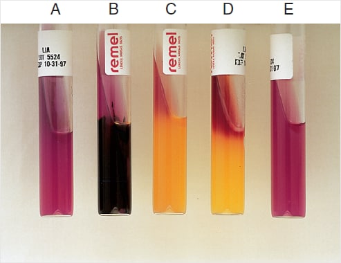 Figure: A: Alkaline slant and alkaline butt (K/K) B: Alkaline slant/alkaline butt, H₂S positive (K/K, H₂S) C: Alkaline slant/acid butt (K/A) D: Red slant/acid butt (R/A) E: Uninoculated tube
LIA + KIA Combined Interpretation
Used together, LIA and KIA (or TSI) provide complementary information for enteric identification:
| KIA Result | LIA Result | Most Likely Organism |
|---|---|---|
| K/A, H₂S−, Gas− | K/K (purple butt, decarboxylation+) | Salmonella Typhi (weak H₂S on KIA but K/K on LIA) |
| K/A, H₂S+, Gas+ | K/K (purple butt) | Salmonella (non-Typhi) |
| K/A, H₂S−, Gas− | R/A (red slant, deamination+) | Proteus mirabilis, Providencia, Morganella |
| K/A, H₂S variable | K/A (no decarboxylation) | Salmonella Paratyphi A (exception: lysine decarboxylase negative, so it does NOT give the usual Salmonella K/K) |
| A/A, H₂S−, Gas+ | K/K (purple butt) | E. coli (lysine decarboxylation positive) |
| A/A, H₂S−, Gas− | K/A (no decarboxylation, no deamination) | Shigella (K/A on LIA, no lysine reaction) |
| K/A, H₂S−, Gas− | K/A | Shigella, confirmed by K/A on both |
| K/K | K/K or variable | Non-fermenter. Investigate separately |
Key distinguishing use: LIA differentiates Salmonella (K/K, decarboxylation positive) from Citrobacter (K/A, decarboxylation negative, may show H₂S on KIA), a common confusion in enteric workups.
Limitations of Lysine Iron Agar
- Proteus spp. that produces hydrogen sulfide but does not produce lysine decarboxylase will not blacken the LIA medium, since the acid in the butt suppresses H₂S formation. Additional testing, such as triple sugar iron (TSI) agar, should be used as a follow-up identification method.
- LIA is not a substitute for TSI.
Quality Control
- Salmonella Typhimurium (ATCC 14028): purple slant, purple butt, H₂S positive (K/K, decarboxylase positive)
- Escherichia coli (ATCC 25922): purple slant, purple butt, H₂S negative (K/K, decarboxylase positive)
- Citrobacter freundii (ATCC 8090): purple slant, acid (yellow) butt, H₂S positive (K/A, decarboxylase negative)
- Proteus mirabilis (ATCC 12453): red slant, acid (yellow) butt (R/A, deaminase positive)
How to Remember: LIA
"LIA completes what KIA starts": KIA tells you carbohydrate fermentation and H₂S. LIA tells you what the organism does with the amino acid lysine. Together they answer four questions: Does it ferment glucose? Lactose? Produce H₂S? Decarboxylate or deaminate lysine?
The color memory, "Purple = decarboxylation, Red = deamination":
- Purple butt = cadaverine produced = lysine DEcarboxylation (Salmonella, E. coli)
- Red/burgundy slant = alpha-keto acid = lysine DEamination (Proteus, Providencia)
The Salmonella pattern: K/A on KIA + K/K (purple) on LIA = Salmonella until proven otherwise.
Key Exam Facts in One Table
| Feature | Detail |
|---|---|
| Reactions tested | Lysine decarboxylation, lysine deamination, H₂S production |
| pH indicator | Bromocresol purple (yellow = acid, purple = alkaline) |
| H₂S indicator | Sodium thiosulphate + ferric ammonium citrate → black FeS |
| K/K result (purple butt) | Lysine decarboxylation positive: Salmonella, E. coli |
| R/A result (red/burgundy slant) | Lysine deamination: Proteus, Providencia, Morganella |
| K/A result | No lysine reaction, Shigella |
| Salmonella Typhi pattern | K/K on LIA + K/A with weak H₂S on KIA |
| Shigella pattern | K/A on LIA + K/A no H₂S on KIA |
| Citrobacter vs Salmonella | Citrobacter: K/A on LIA (no decarboxylation); Salmonella: K/K (decarboxylation positive) |
| Read time | 18–24h |
| Used with | KIA or TSI for complete enteric workup |
Where Students Get Confused
1. LIA is capped tightly; TSI is capped loosely. This is the most common mix-up between the two tubes. LIA needs an anaerobic butt for lysine decarboxylation to show, so the cap goes on tight. TSI needs an aerobic slant for the alkaline reversion, so its cap is left loose. Using the wrong cap position breaks the reaction the medium depends on.
2. Not every Salmonella gives K/K on LIA. The rule "K/A on KIA plus K/K on LIA equals Salmonella" has an important exception: Salmonella Paratyphi A does not produce lysine decarboxylase, so it gives K/A on LIA, the same as Shigella. A K/A result does not rule out Salmonella. Confirm with serology when the clinical picture fits.
3. A red slant is deamination, not just "alkaline." The burgundy or red slant of Proteus, Providencia, and Morganella is a specific deamination color, not the ordinary alkaline pink of a non-reacting slant. Read it as a positive deaminase result, which points away from Salmonella and toward the Proteus group.
4. Citrobacter can mimic Salmonella on KIA but separates on LIA. Citrobacter freundii is H₂S positive and can look like Salmonella on KIA. On LIA it is decarboxylase negative, giving K/A (purple slant, yellow butt), whereas Salmonella gives K/K (purple butt). This is one of LIA's most useful discriminations, and it is why LIA is run alongside KIA.
5. A K/K alkaline butt can hide H₂S. H₂S only precipitates as black FeS in an acidic environment. If decarboxylation makes the butt strongly alkaline, weak H₂S production can be suppressed and read falsely negative. LIA is less sensitive for H₂S than KIA or SIM, so confirm weak H₂S on a more sensitive medium rather than reporting it negative.
References
- Tille PM. Bailey and Scott's Diagnostic Microbiology. 15th ed. St. Louis: Elsevier; 2022.
- Procop GW, Church DL, Hall GS, Janda WM, Koneman EW, Schreckenberger PC, Woods GL. Koneman's Color Atlas and Textbook of Diagnostic Microbiology. 7th ed. Philadelphia: Wolters Kluwer; 2017.
- Murray PR, Rosenthal KS, Pfaller MA. Medical Microbiology. 9th ed. Elsevier; 2020.
- Leber AL, editor. Clinical Microbiology Procedures Handbook. 4th ed. Washington, DC: ASM Press; 2016. doi:10.1128/9781683670438.CMPH
- Edwards PR, Fife MA. Lysine-iron agar in the detection of Arizona cultures. Applied Microbiology. 1961;9(5):478-480.

Tankeshwar Acharya, MSc (Medical Microbiology)
Tankeshwar Acharya is an Assistant Professor in the Department of Microbiology at Patan Academy of Health Sciences (PAHS), Nepal, where he has been teaching and practicing clinical microbiology for over 14 years. He is the founder of Microbe Online, one of the leading free microbiology education resources on the web, covering bacteriology, mycology, parasitology, immunology, and clinical laboratory diagnostics written from direct experience in both the classroom and the diagnostic laboratory.
Comments
No comments yet. Be the first to share your thoughts.
Leave a comment
All comments are reviewed before they appear.