
Common Fungal Culture Media and Their Uses in Clinical Mycology

On this page
One additive, two opposite jobs
Fungal culture has a built-in conflict that bacterial culture mostly avoids. Fungi grow slowly, over weeks, which gives faster saprophytic molds and bacteria plenty of time to overrun a plate before the pathogen you want ever appears. The standard solution is cycloheximide, which suppresses those fast-growing contaminants and lets a slow pathogen come up cleanly.
The catch is that cycloheximide does not distinguish between a contaminant and a pathogen that happens to be susceptible to it. Cryptococcus neoformans, Aspergillus, Fusarium, and several Candida species are inhibited by it just as effectively as the molds it is meant to hold back. The single additive that makes fungal culture manageable is also capable of suppressing some of the most important organisms it is used to find.
This is why fungal media are chosen as a battery rather than a single plate, and why the rules that follow, selective paired with non-selective, cycloheximide-containing paired with cycloheximide-free, the right temperature for the phase you expect, are not bureaucratic caution but the direct consequence of that one tradeoff. Get the combination right and slow pathogens grow while contaminants are held back. Get it wrong and the plate stays clean while the pathogen never had a place to grow.
Fungal infections range from superficial skin conditions caused by dermatophytes to life-threatening invasive mycoses caused by dimorphic fungi and opportunistic pathogens in immunocompromised patients. Recovering the causative organism in culture is essential for definitive diagnosis, species identification, and antifungal susceptibility testing and each category of fungal pathogen requires different culture media and incubation conditions to grow reliably.
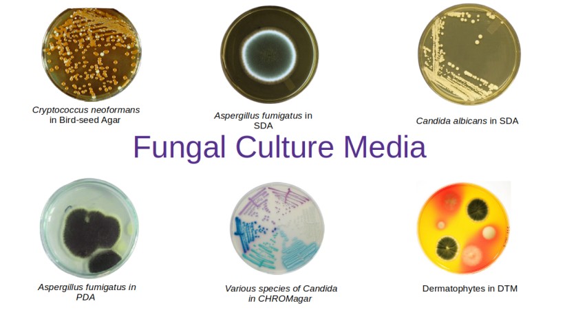 Figure: Common Fungal Culture Media
Unlike bacterial culture, where a blood agar plate recovers most clinically significant organisms within 24–48 hours, fungal culture is slower (most fungi require 1–4 weeks), more fastidious, and more susceptible to bacterial overgrowth from mixed specimens. This makes media selection critical.
Core principles of fungal culture media selection
1. Always use a battery of media
No single medium recovers all clinically significant fungi. The minimum recommended battery for most clinical specimens includes:
- One non-selective enriched medium: for recovery of all fungi including fastidious dimorphic pathogens
- One selective medium with cycloheximide: for dermatophytes and suppression of saprophytic contaminants
- One selective medium without cycloheximide: for cycloheximide-sensitive pathogens
2. Cycloheximide; critical clinical consideration
Cycloheximide (actidione) is added to selective fungal media to inhibit rapidly growing saprophytic contaminating molds. However, several clinically important pathogenic fungi are also sensitive to cycloheximide and will be completely inhibited on media containing it:
| Cycloheximide-sensitive pathogens | Clinical significance |
|---|---|
| Cryptococcus neoformans | Meningitis, especially in HIV patients |
| Aspergillus spp. | Invasive aspergillosis in immunocompromised |
| Fusarium spp. | Invasive fusariosis; keratitis |
| Pseudallescheria boydii | Mycetoma; invasive disease |
| Trichosporon spp. | Disseminated infection in immunocompromised |
| Some Candida spp. (C. krusei, C. tropicalis) | Candidemia; mucosal infections |
Bottom line: Media containing cycloheximide (Mycosel, Mycobiotic agar, DTM) must never be used as the only medium when these organisms are clinically suspected. Always pair with a cycloheximide-free medium.
3. Incubation temperatures
Fungal media require careful temperature selection:
| Temperature | Purpose | Examples |
|---|---|---|
| 25–30°C | Mold (saprophytic) phase; dermatophytes; identification of colony morphology and sporulation | SDA, DTM, Czapek-Dox, PDA |
| 35–37°C | Yeast phase of dimorphic fungi; Candida spp.; rapid recovery | BHI agar, SABHI, CHROMagar Candida |
| Both simultaneously | Dimorphic fungi — 25°C for mold phase, 37°C for yeast phase conversion | BHI agar, IMA |
Incubate all fungal cultures for a minimum of 4 weeks before reporting as negative, as some dimorphic fungi (Histoplasma capsulatum, Blastomyces dermatitidis) may take 2–4 weeks to appear.
4. Antibacterial agents
Bacterial overgrowth rapidly kills slow-growing fungi in mixed specimens. Antibacterial agents commonly added to fungal media include:
- Chloramphenicol: broad-spectrum; most common
- Gentamicin: gram-negative coverage
- Ciprofloxacin: broad-spectrum alternative
For specimens from sterile sites (CSF, blood, bone marrow), antibacterial agents are unnecessary and non-selective enriched media are preferred.
Fungal Culture Media: A Complete Reference Table
| Medium | Type | Cycloheximide | Antibacterial | Incubation temp | Primary use |
|---|---|---|---|---|---|
| Sabouraud dextrose agar (SDA) | Non-selective | No | Optional | 25–30°C | General recovery; maintenance; subculture |
| Brain-heart infusion (BHI) agar | Non-selective enriched | No | No (sterile sites) | 25–30°C and 35–37°C | Dimorphic fungi; fastidious pathogens |
| SABHI agar | Non-selective enriched | No | Optional | 25–30°C and 35–37°C | Fastidious and slow-growing dimorphic fungi |
| Inhibitory mold agar (IMA) | Non-selective enriched | No | Yes (chloramphenicol ± gentamicin) | 25–30°C | Dimorphic pathogens; non-dermatophyte molds |
| Potato flake agar | Non-selective | No | Optional | 25–30°C | Saprophytic and dimorphic fungi; sporulation |
| Mycosel / Mycobiotic agar | Selective | Yes | Yes | 25–30°C | Dermatophytes from mixed specimens |
| Dermatophyte test medium (DTM) | Selective/differential | Yes | Yes (multiple) | 25–30°C | Presumptive dermatophyte identification |
| CHROMagar Candida | Selective/differential | No | Yes | 35–37°C | Species-level presumptive ID of Candida |
| Niger seed agar (Birdseed agar) | Selective/differential | No | Yes | 35–37°C | Cryptococcus neoformans identification |
| Cornmeal agar + Tween 80 | Differential | No | No | 25–30°C | C. albicans chlamydospore; yeast morphology |
| Potato dextrose agar (PDA) | Non-selective | No | Optional | 25–30°C | Slide culture; dermatophyte sporulation; pigment |
| Czapek-Dox agar | Differential | No | No | 25–30°C | Aspergillus and Penicillium identification |
| Rice starch agar | Differential | No | No | 25–30°C | C. albicans chlamydospore production |
Individual Media
1. Sabouraud Dextrose Agar (SDA)
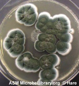
Sabouraud dextrose agar is the most widely used and universally recognised fungal culture medium. It contains peptone and a high concentration of glucose (2–4%), with a low pH (5.6) that inhibits most bacteria and favors fungal growth.
 Key features:
- Low pH (5.6) provides a degree of natural bacterial suppression without added antibiotics
- High glucose concentration supports growth of a broad range of fungi
- Available with or without chloramphenicol and/or cycloheximide
Limitations:
- Not suitable as the primary isolation medium when fastidious dimorphic fungi (Histoplasma, Blastomyces, Coccidioides) are suspected; the medium is insufficiently rich for their primary recovery
- Most useful as a subculture medium to enhance sporulation and provide characteristic colony morphology after initial recovery on enriched media
Primary use: General maintenance and subculture of fungi; recovery of dermatophytes from skin, hair, and nail specimens; recovery of yeasts from genital cultures.
2. Brain-Heart Infusion (BHI) Agar
BHI agar is a rich, non-selective medium originally developed for the culture of fastidious bacteria, but it is also an excellent primary fungal isolation medium — particularly for dimorphic fungi that require extra nutrients for primary recovery.
Key features:
- Supplemented with hemin, L-cysteine, and sheep blood in some formulations for enhanced recovery
- Supports both mold phase (25–30°C) and yeast phase (35–37°C) of dimorphic fungi
- No cycloheximide — safe to use when Cryptococcus, Aspergillus, or cycloheximide-sensitive pathogens are suspected
- Incubating duplicate plates at both 25°C and 35–37°C simultaneously maximizes recovery of all fungal types
Primary use: Primary isolation of dimorphic fungi (Histoplasma capsulatum, Blastomyces dermatitidis, Coccidioides immitis, Sporothrix schenckii) from tissue, blood, and bone marrow specimens
Biosafety caution: Coccidioides immitis and C. posadasii are among the most hazardous organisms in the clinical mycology laboratory. The mold-phase culture produces arthroconidia that are highly infectious by inhalation and have caused numerous laboratory-acquired infections. Whenever coccidioidomycosis is suspected, cultures must be handled inside a biosafety cabinet under BSL-3 practices, and any mold suspected of being Coccidioides should be sealed and referred to a reference laboratory rather than examined openly on the bench.
3. Sabouraud Heart Infusion Agar (SABHI)
SABHI combines the selective low pH of Sabouraud dextrose agar with the nutritional richness of brain-heart infusion. This hybrid formulation provides the best of both media — broad-spectrum fungal recovery with enough nutritional content for fastidious dimorphic pathogens.
Key features:
- Richer than standard SDA: better supports slow-growing and fastidious fungi
- Can be supplemented with chloramphenicol for bacterial suppression
- Used at both 25–30°C and 35–37°C for maximum recovery
Primary use: Primary recovery of saprophytic and dimorphic fungi, particularly fastidious strains that fail to grow on standard SDA; an excellent all-purpose primary plating medium for most clinical specimens
4. Inhibitory Mold Agar (IMA)
IMA is an enriched medium containing tryptone, beef extract, yeast extract, starch, dextrin, and a saline buffer, with chloramphenicol and gentamicin for antibacterial suppression. Despite the name, IMA does not inhibit molds — it supports a wide range of fungi while inhibiting bacterial overgrowth.
Key features:
- No cycloheximide: supports cycloheximide-sensitive pathogens
- Enriched formulation supports dimorphic fungi and other fastidious organisms
- Does not support dermatophytes well: not a replacement for Mycosel for skin specimens
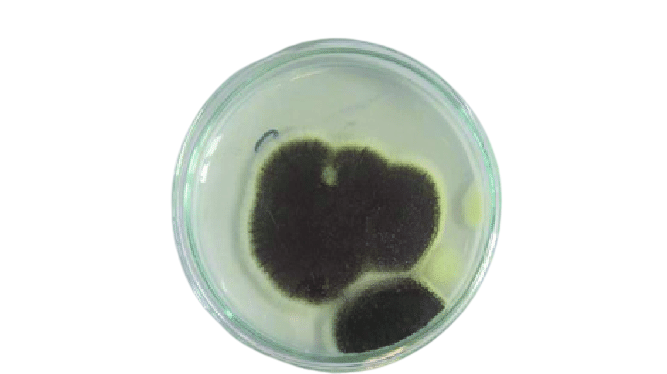 Primary use: Primary recovery of dimorphic pathogenic fungi (Histoplasma, Blastomyces, Coccidioides) and other pathogenic molds from respiratory and tissue specimens; used alongside a cycloheximide-containing medium in the primary battery
5. Mycosel / Mycobiotic Agar
Mycosel (BD) and Mycobiotic agar (Remel) are equivalent formulations — both are Sabouraud dextrose agar with cycloheximide (0.4 mg/mL) and chloramphenicol added.
Key features:
- Cycloheximide suppresses rapidly growing saprophytic contaminating molds
- Chloramphenicol suppresses bacteria
- Must never be used alone — cycloheximide inhibits Cryptococcus, Aspergillus, Fusarium, and some Candida spp.
- Highly selective — only dermatophytes and some other slow-growing pathogenic fungi survive
Primary use: Selective isolation of dermatophytes from skin, hair, and nail specimens containing heavy bacterial and saprophytic mold contamination; always used in combination with a cycloheximide-free medium
6. Dermatophyte Test Medium (DTM)
DTM is a selective and differential medium containing cycloheximide, chloramphenicol, and gentamicin for suppression of non-dermatophyte organisms, plus phenol red as a pH indicator.
Differential mechanism: Dermatophytes metabolize protein preferentially, producing alkaline metabolites that raise the pH and turn the phenol red indicator from yellow to red. Saprophytic fungi that do grow on DTM ferment carbohydrates first, producing acid and turning the medium yellow — the opposite color change.
Reading DTM:
- Red color change within 10–14 days = presumptive dermatophyte (positive)
- Yellow color change = saprophytic contaminant (negative)
- No color change = no growth or very slow growth
Limitations: False positives can occur if the plate is held beyond 14 days, as even saprophytic colonies will eventually exhaust carbohydrates and begin metabolizing protein, producing a late red color change. DTM is presumptive only — species identification requires subculture and microscopy.
Primary use: Office-based or point-of-care presumptive identification of dermatophytes from skin, hair, and nail specimens; useful in dermatology practices without full mycology laboratory support → Full article: Dermatophyte Test Medium
7. CHROMagar Candida
CHROMagar Candida is a selective and differential chromogenic medium that allows presumptive species-level identification of Candida directly from primary culture based on colony color — without waiting for additional biochemical testing.
Colony colors on CHROMagar Candida:
| Organism | Colony color |
|---|---|
| Candida albicans | Green / teal |
| Candida tropicalis | Blue-grey to metallic blue |
| Candida krusei | Pink, flat, dry, spreading |
| Candida glabrata | variable, often white to mauve; not reliably distinguished on the classic medium |
| Candida parapsilosis | Pink-white, cream |
| Other Candida spp. | White/cream (non-chromogenic) |
Key features:
- Chloramphenicol suppresses bacteria
- No cycloheximide: Candida krusei and other cycloheximide-sensitive species are recovered
- Mixed infections (two or more Candida spp.) are immediately detected by different colony colors
- Results in 48 hours vs 5–7 days for conventional biochemical identification
Primary use: Primary isolation and presumptive identification of Candida spp. from urine, genital swabs, and other specimens where Candida is expected; particularly valuable in high-volume urine culture settings and for detecting mixed Candida infections → Full article: Candida albicans
8. Niger Seed Agar (Birdseed Agar)
Niger seed agar (also called Staib agar or Birdseed agar) is a selective and differential medium specifically used for the identification of Cryptococcus neoformans and Cryptococcus gattii.
Mechanism: Cryptococcus neoformans produces the enzyme laccase (phenoloxidase), which oxidises caffeic acid compounds in Guizotia abyssinica (niger/birdseed) seeds to produce melanin. Melanin deposits in the cell wall, producing characteristic brown to dark brown colonies. Other yeasts and fungi do not produce laccase and therefore remain white or cream-colored.
Key features:
- Highly specific for Cryptococcus spp. — no other common clinical yeast produces brown colonies
- Cryptococcus neoformans and C. gattii both produce brown colonies but can be differentiated by canavanine-glycine-bromothymol blue (CGB) agar
- Chloramphenicol suppresses bacteria
- No cycloheximide: Cryptococcus is cycloheximide-sensitive
Primary use: Definitive identification of Cryptococcus neoformans from CSF, blood, and respiratory specimens; confirms urease-positive, encapsulated yeast isolates → Full article: Cryptococcus neoformans
9. Cornmeal Agar with Tween 80
Cornmeal agar is a low-nutrient medium made from corn meal infusion and agar. The addition of Tween 80 (polysorbate 80) reduces surface tension and enhances the formation of microscopic morphological structures in yeasts and some molds.
Uses in yeast identification:
- Chlamydospore production: Candida albicans and C. dubliniensis produce large, thick-walled terminal chlamydospores (blastoconidia) on cornmeal-Tween 80 agar that are not produced by other Candida species. Chlamydospore production is a presumptive identification of C. albicans
- Pseudohyphae and true hyphae: the pattern of pseudohyphae, blastoconidia, and hyphae production differs between Candida species and aids identification
Primary use: Identification of Candida albicans by chlamydospore production; microscopic morphology of yeasts for species-level identification
10. Potato Dextrose Agar (PDA)
PDA is made from potato extract and dextrose. The nutrients from potato infusion promote the development of aerial mycelia and conidia, making it particularly useful for inducing sporulation in fungi that fail to sporulate on richer media.
Key features:
- Promotes excellent sporulation of dermatophytes — Trichophyton rubrum produces its characteristic red/wine-red pigment on the reverse of PDA colonies
- Ideal base medium for slide culture preparation — its low nutrient content encourages sporulation rather than hyphal overgrowth
- Can be acidified (pH 3.5 with lactic acid or tartaric acid) or supplemented with antibiotics to reduce bacterial contamination
Primary use: Slide culture preparation for microscopic identification of mold sporulation patterns; inducing sporulation in non-sporulating isolates; demonstrating T. rubrum red pigment → Full article: Slide Culture for Fungi
11. Czapek-Dox Agar
Czapek-Dox agar is a chemically defined (synthetic) medium containing inorganic salts and sodium nitrate as the sole nitrogen source, with glucose as the carbon source.
Key features:
- Completely defined composition: allows observation of colony characteristics under controlled, reproducible conditions
- Supports characteristic pigment production and colonial morphology for Aspergillus and Penicillium species
- Sodium nitrate as sole nitrogen source creates nutritional conditions that accentuate species-specific pigmentation
Colony characteristics on Czapek-Dox:
- Aspergillus fumigatus: blue-green colonies with a white border
- Aspergillus niger: black/brown-black conidial heads
- Aspergillus flavus: yellow-green to yellow colonies
- Penicillium spp.: blue-green colonies with brush-like conidiophore arrangement
Primary use: Species-level identification of Aspergillus spp. and Penicillium spp. by colonial morphology and pigment production; part of the standard mold identification battery in reference mycology laboratories → Full article: Czapek-Dox Agar
12. Potato Flake Agar
Similar in principle to PDA but made from commercial potato flakes rather than potato infusion. It is richer than standard PDA while still promoting sporulation.
Primary use: Primary recovery of saprophytic and dimorphic fungi, particularly fastidious slow-growing strains; promotes sporulation for microscopic identification.
13. Rice Starch Agar (Cream of Rice Agar)
A very low-nutrient medium made from commercial rice cereal (cream of rice) and Tween 80. The near-absent nutrient conditions strongly stimulate chlamydospore production in Candida albicans.
Primary use: Alternative to cornmeal-Tween 80 agar for chlamydospore production in Candida albicans; particularly reliable for strains that fail to produce chlamydospores on cornmeal agar.
Specimen-Based Media Selection Guide
| Specimen type | Recommended media | Incubation |
|---|---|---|
| Skin scrapings, hair, nails | Mycosel or Mycobiotic agar + SDA or DTM | 25–30°C, 4 weeks |
| Vaginal/genital swabs | SDA + CHROMagar Candida | 35–37°C, 48–72 hours |
| Urine | CHROMagar Candida + SDA | 35–37°C, 48–72 hours |
| Sputum / respiratory | BHI or SABHI + IMA + Mycosel | 25–30°C and 35–37°C, 4 weeks |
| CSF (suspected Cryptococcus) | Niger seed agar + BHI (no cycloheximide) | 35–37°C, 2 weeks |
| Blood / bone marrow (dimorphic fungi) | BHI blood agar + SABHI | 25–30°C and 35–37°C, 4 weeks |
| Tissue biopsy | BHI + SABHI + Mycosel + SDA | Both temperatures, 4 weeks |
| Wound / abscess (mycetoma) | BHI + SDA | 25–30°C, 4 weeks |
References and Further Reading
- Tille, P. M. (2022). Bailey and Scott's Diagnostic Microbiology (15th ed.). St. Louis: Elsevier.
- Larone, D. H., Walsh, T. J., Hayden, R. T., & Larone, D. H. (2018). Larone's Medically Important Fungi: A Guide to Identification (6th ed.). Washington, DC: ASM Press.
- Procop, G. W., Church, D. L., Hall, G. S., Janda, W. M., Koneman, E. W., Schreckenberger, P. C., & Woods, G. L. (2017). Koneman's Color Atlas and Textbook of Diagnostic Microbiology (7th ed.). Philadelphia: Wolters Kluwer.
- Leber, A. L. (Ed.). (2016). Clinical Microbiology Procedures Handbook (4th ed.). Washington, DC: ASM Press.
Frequently Asked Questions
Why must cycloheximide-containing media never be used alone?
What is the difference between SDA and BHI for fungal isolation?
How do you identify Candida species on CHROMagar?
Why is Niger seed agar specific for Cryptococcus?
Why do fungi require longer incubation?
What are the limitations of DTM?
What is the purpose of Tween 80 in cornmeal agar?
Which media for CSF with suspected Cryptococcus meningitis?

Tankeshwar Acharya, MSc (Medical Microbiology)
Tankeshwar Acharya is an Assistant Professor in the Department of Microbiology at Patan Academy of Health Sciences (PAHS), Nepal, where he has been teaching and practicing clinical microbiology for over 14 years. He is the founder of Microbe Online, one of the leading free microbiology education resources on the web, covering bacteriology, mycology, parasitology, immunology, and clinical laboratory diagnostics written from direct experience in both the classroom and the diagnostic laboratory.
Comments
No comments yet. Be the first to share your thoughts.
Leave a comment
All comments are reviewed before they appear.