
Serratia marcescens: The Red-Pigment Hospital Pathogen and How to Identify It
Why Serratia marcescens produces a red pigment, how it causes hospital-acquired infections through biofilm, and how the laboratory identifies it (late lactose fermenter, DNase positive, VP positive).

On this page
For centuries, people reported bread and communion wafers that appeared to be spotted with fresh blood, and treated it as a miracle or an omen. The cause turned out to be a bacterium that grows a bright red pigment: Serratia marcescens. For a long time it was thought harmless, and scientists even sprayed it over cities to track how germs spread, assuming it could not cause disease.
That assumption was wrong. Serratia marcescens is now a well-known cause of hospital-acquired infection, especially in intensive care units and in patients with catheters and breathing tubes. This page is about the organism behind the red color: how it causes infection, why it is so hard to treat, and how the laboratory identifies it.
Overview
Serratia is a genus of Gram-negative rods in the family Enterobacteriaceae. The genus has more than 15 species, but one matters most in medicine: Serratia marcescens, the species found most often in clinical specimens and the main cause of Serratia infections. A few other species (S. liquefaciens, S. odorifera) turn up in clinical material occasionally.
S. marcescens is best known for two things: some strains produce a striking red pigment, and it is an important cause of hospital-acquired (nosocomial) infection. It is an opportunistic pathogen, meaning it rarely troubles healthy people but readily infects patients who are already vulnerable, especially those with medical devices in place.
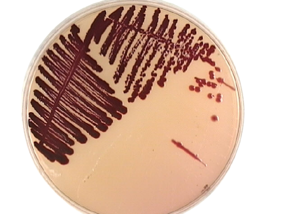 Figure: Dark red colonies of Serratia marcescens in MacConkey Agar
The red pigment: prodigiosin
Some strains of S. marcescens produce prodigiosin, a red pigment. This is the organism's signature and the reason for its long, strange history.
Two practical points about the pigment matter in the laboratory:
- It is temperature-dependent. Prodigiosin is produced best at around room temperature (25 to 28°C) and is often not produced at 37°C. So a Serratia growing from a patient at body temperature may look ordinary and pale, while the same strain left at room temperature turns red. A colorless clinical isolate does not rule out Serratia.
- Not all strains make it. Many clinical S. marcescens strains are non-pigmented. The red color, when present, is a strong clue, but its absence means nothing.
The pigment is not thought to be a major cause of disease. Its importance is as an identification clue and as the reason the organism was noticed for centuries.
Virulence factors and how Serratia causes disease
S. marcescens is not aggressive in the way Salmonella or Shigella are. It succeeds by living on surfaces, spreading in hospitals, and resisting antibiotics. Each factor below explains part of that.
Biofilm formation (the key factor). This is the heart of Serratia pathogenesis. The organism attaches to plastic and metal surfaces, catheters, breathing tubes, contact lenses, and the inside of medical equipment, and builds a biofilm, a community of bacteria wrapped in a protective slime layer. Inside a biofilm, the organism is shielded from both the immune system and antibiotics. This is why Serratia infections cluster around devices, and why they are hard to clear: the biofilm protects the bacteria and is also a reservoir that keeps re-seeding the infection.
Attachment (pili/fimbriae). Serratia uses surface pili to stick to host cells and to surfaces, the necessary first step before biofilm can form.
Endotoxin (LPS). As a Gram-negative organism, its lipopolysaccharide can trigger the inflammatory response of sepsis and endotoxic shock when the organism reaches the blood.
Secreted enzymes. Serratia releases several enzymes (including a protease, lipase, and chitinase) that break down host tissue and help it obtain nutrients. These contribute to tissue damage in wound and eye infections.
How it spreads and causes infection
S. marcescens lives in wet places: tap water, sinks, soap solutions, and moist medical equipment such as nebulizers, ventilator tubing, and bronchoscopes. It spreads from these reservoirs, and from patient to patient, mainly on the hands of hospital staff. Once it reaches a vulnerable site, usually via a catheter, a breathing tube, or a surgical wound, it attaches, forms biofilm, and establishes infection. This is why hand hygiene and equipment decontamination are central to preventing Serratia outbreaks.
Diseases it causes
S. marcescens causes urinary tract infection (often catheter-related), wound and surgical-site infection, pneumonia (often ventilator-associated), bloodstream infection and endotoxic shock, and endocarditis, which is classically seen in people who inject drugs. Eye infections linked to contact lenses also occur. Most of these appear in hospitalized or otherwise vulnerable patients.
Identifying Serratia in the laboratory
Gram stain. Serratia is a Gram-negative rod. It is motile.
On the plate.
- Blood agar and MacConkey: grows well on both. On MacConkey it is a late (slow) lactose fermenter, so it usually looks pale at 24 hours and may turn slightly pink on longer incubation. Do not expect the bright pink of E. coli.
- Nutrient agar or TSA at room temperature: pigmented strains produce the red prodigiosin color, best seen at 25 to 28°C, not at 37°C.
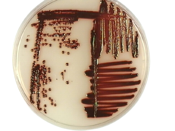 Figure: Serratia marcescens grew as bright, glossy red colonies on the TSA plate
What distinguishes Serratia from other coliforms. Three enzyme tests are the classic Serratia markers and separate it from most other Enterobacteriaceae: it is DNase-positive, lipase-positive, and gelatinase-positive. Among the Enterobacteriaceae, this trio is unusual and is the practical way to confirm Serratia when the pigment is absent.
The identification panel.
| Test | Serratia marcescens result | What it tells you |
|---|---|---|
| Oxidase | Negative | Places it in the Enterobacteriaceae. |
| Catalase | Positive | Consistent with the family. |
| Lactose | Late (slow) fermenter | Often pale early; not a strong fermenter like E. coli. |
| DNase | Positive | Key Serratia marker; unusual in the family. |
| Gelatinase / lipase | Positive | Together with DNase, distinguishes Serratia. |
| Voges-Proskauer (VP) | Positive | Like Klebsiella/Enterobacter, not E. coli. |
| Citrate | Positive | Can use citrate as sole carbon source. |
| Indole | Negative | Helps separate from indole-positive coliforms. |
| Urease | Variable/weak | Not a strong urease producer like Proteus. |
| ONPG | Positive | Has beta-galactosidase (fits the slow-lactose pattern). |
| Lysine decarboxylase | Positive | Part of the coliform panel. |
| Motility | Motile | Separates from non-motile Klebsiella. |
| TSI | A/A (or K/A), no gas or little, no H₂S | Ferments glucose; sugar pattern varies by strain. |
Antibiotic resistance
Serratia is often resistant to multiple antibiotics, which is a large part of why it is a problem in hospitals. Two things drive this. First, it carries an inducible chromosomal AmpC beta-lactamase, so it can appear susceptible to some cephalosporins at first and then become resistant during treatment. For this reason Serratia is one of the organisms for which many clinicians avoid relying on third-generation cephalosporins even when the isolate tests susceptible. It is also intrinsically resistant to polymyxins (colistin). Second, its biofilm shields it from antibiotics that would otherwise work.
The practical result: Serratia infections often need carbapenems or other agents guided by susceptibility testing, and device removal is frequently part of clearing a biofilm-associated infection.
How to remember
Red pigment, room temperature. Serratia makes red prodigiosin, but only in the cold (around 25°C), not at body temperature. Picture the "miracle" bleeding bread sitting out at room temperature turning red.
The device organism. Serratia lives on wet hospital surfaces and builds biofilm on catheters and tubes. If you picture Serratia, picture it as a slime layer on a plastic tube, not a free-swimming invader. That one image explains its infections (device-related), its spread (staff hands, wet equipment), and its resistance (biofilm shields it).
DNase-positive coliform. The bench trick: a slow-lactose Gram-negative rod that is DNase, gelatinase, and lipase positive is Serratia. Most Enterobacteriaceae are DNase-negative, so a DNase-positive coliform is a strong pointer.
AmpC, like the rest of the SPACE group. Serratia is one of the organisms that can turn on AmpC and defeat cephalosporins mid-treatment. It sits with Enterobacter, Citrobacter, Providencia, and Morganella in that group.
Key exam facts in one table
| Fact | Detail |
|---|---|
| Organism / family | Serratia marcescens, Gram-negative rod, family Enterobacteriaceae |
| Signature feature | Red pigment (prodigiosin), produced best at ~25°C, not at 37°C; not all strains |
| Lactose | Late (slow) fermenter |
| Distinguishing tests | DNase, gelatinase, and lipase positive (unusual among Enterobacteriaceae) |
| Oxidase / VP / citrate | Oxidase −, VP +, citrate + |
| Motility | Motile (separates from Klebsiella) |
| Main clinical setting | Hospital-acquired infection (ICU, devices) |
| Key virulence factor | Biofilm formation on devices |
| Diseases | Catheter UTI, wound/surgical infection, ventilator pneumonia, bloodstream infection, endocarditis (injection drug use) |
| Reservoir / spread | Wet environments (water, equipment); staff hands |
| Resistance | Inducible AmpC (SPACE group); intrinsic polymyxin resistance; biofilm-protected |
Where students get confused
The pigment is not reliable. Two traps: many clinical strains make no pigment at all, and even pigmented strains often do not show red at 37°C. So a plain-looking colony can still be Serratia. Use the biochemical tests, especially DNase, not the color, to identify it.
Lactose fermentation. Serratia is a late lactose fermenter, not a brisk one and not a true non-fermenter. It often reads pale at 24 hours.
It is an opportunist, not an aggressive pathogen. Serratia rarely infects healthy people. Its danger is in hospitals, in vulnerable patients with devices. Understanding it as a biofilm-and-device organism explains its whole clinical behavior.
AmpC and cephalosporins. Serratia can test susceptible to a cephalosporin and then become resistant during treatment, because it can switch on its AmpC enzyme. This is a classic trap and the reason cephalosporins are often avoided for serious Serratia infection.
Was it ever really harmless? Serratia was historically treated as non-pathogenic and used as a tracer organism sprayed into the environment. That was a mistake; it is a genuine opportunistic pathogen.
References
- Tille, P. M. (2022). Bailey & Scott's Diagnostic Microbiology (15th ed.). Elsevier.
- Procop, G. W., et al. (2017). Koneman's Color Atlas and Textbook of Diagnostic Microbiology (7th ed.). Wolters Kluwer.
- Mahlen, S. D. (2011). Serratia infections: from military experiments to current practice. Clinical Microbiology Reviews, 24(4), 755–791. https://doi.org/10.1128/CMR.00017-11
- Gupta, V., Sharma, S., Pal, K., Goyal, P., Agarwal, D., & Chander, J. (2021). Serratia, no longer an uncommon opportunistic pathogen. Infectious Disorders Drug Targets, 21(7), e300821191666. https://doi.org/10.2174/1871526521666210222125215
Frequently Asked Questions
Why does Serratia marcescens produce a red color?
Why does Serratia marcescens produce a red color?
Some strains make a red pigment called prodigiosin. It is produced best at room temperature (around 25°C) and often not at body temperature (37°C). Not all strains make it, so a colorless isolate can still be Serratia.
Is Serratia marcescens a lactose fermenter?
Is Serratia marcescens a lactose fermenter?
It is a late (slow) lactose fermenter. On MacConkey agar it usually looks pale at 24 hours and may turn slightly pink with longer incubation, so it does not show the strong early pink of E. coli.
How is Serratia distinguished from other coliforms?
How is Serratia distinguished from other coliforms?
Its most useful markers are that it is DNase-positive, gelatinase-positive, and lipase-positive, which is unusual among the Enterobacteriaceae. It is also Voges-Proskauer positive and citrate positive. These tests identify it even when the red pigment is absent.
What infections does Serratia marcescens cause?
What infections does Serratia marcescens cause?
Mostly hospital-acquired infections: catheter-related urinary tract infection, wound and surgical-site infection, ventilator-associated pneumonia, bloodstream infection, and endocarditis (classically in people who inject drugs). It rarely infects healthy people.
Why is Serratia so hard to treat?
Why is Serratia so hard to treat?
Two reasons. It forms biofilms on devices that shield it from antibiotics and the immune system, and it carries an inducible AmpC enzyme that can make it resistant to cephalosporins during treatment. It is also naturally resistant to polymyxins. Treatment is guided by susceptibility testing, and infected devices often have to be removed.
Where does Serratia marcescens live in the hospital?
Where does Serratia marcescens live in the hospital?
In wet places: tap water, sinks, soap and antiseptic solutions, and moist equipment such as nebulizers, ventilator tubing, and bronchoscopes. It spreads mainly on the hands of staff, which is why hand hygiene is central to preventing outbreaks.
Was Serratia marcescens once thought to be harmless?
Was Serratia marcescens once thought to be harmless?
Yes. It was considered non-pathogenic and was even sprayed into the environment as a tracer to study how germs spread, because its red color made it easy to follow. It is now recognized as a genuine opportunistic pathogen.

Tankeshwar Acharya, MSc (Medical Microbiology)
Tankeshwar Acharya is an Assistant Professor in the Department of Microbiology at Patan Academy of Health Sciences (PAHS), Nepal, where he has been teaching and practicing clinical microbiology for over 14 years. He is the founder of Microbe Online, one of the leading free microbiology education resources on the web, covering bacteriology, mycology, parasitology, immunology, and clinical laboratory diagnostics written from direct experience in both the classroom and the diagnostic laboratory.
Comments
No comments yet. Be the first to share your thoughts.
Leave a comment
All comments are reviewed before they appear.