

A district hospital laboratory in the Terai receives 40 stool samples in a single morning. Watery stool, no blood, no fever. The clinicians are asking one question: is this cholera?
The technician plates on TCBS. Overnight, yellow colonies appear, exactly as expected for a sucrose fermenter. She picks a colony straight from the TCBS plate, rubs it onto oxidase reagent paper, and waits.
Nothing. No purple. Not at 10 seconds, not at two minutes.
Vibrio cholerae is oxidase positive. Every textbook says so. The colony in front of her is oxidase negative. She writes "not Vibrio" and moves on.
She is wrong, and the reason is not the organism. It is the plate. V. cholerae ferments the sucrose in TCBS and floods its immediate surroundings with acid. Below roughly pH 5.1, the cytochrome c oxidase reaction simply will not run, no matter how much enzyme the organism has. The colony was positive. The test could not say so.
The oxidase test is the first branch point in every Gram-negative identification you will ever do. It is also one of the easiest tests in microbiology to break. This article is about the second half of that sentence.
The oxidase test asks one question of a Gram-negative organism: does your respiratory chain end in a cytochrome c oxidase? A purple color within 10 seconds says yes, and in that moment the organism has left the Enterobacteriaceae. A colorless paper says no, or it says the test was set up in a way that made a positive result impossible. Learning to tell those two situations apart is most of what this test actually requires.
The enzyme detected is cytochrome c oxidase, also called cytochrome aa3 or complex IV of the electron transport chain. Pseudomonas, Vibrio, Neisseria, Campylobacter, and Helicobacter are positive. Escherichia, Klebsiella, Salmonella, Shigella, Proteus, and every other member of the Enterobacteriaceae are negative.
Oxidase Test Principle
The oxidase test is used to identify bacteria that produce cytochrome c oxidase, an enzyme of the bacterial electron transport chain. When present, the cytochrome c oxidase oxidizes the reagent (tetramethyl-p-phenylenediamine dihydrochloride) to indophenols, a purple or dark blue color end product. When the enzyme is not present, the reagent remains reduced and is colorless.
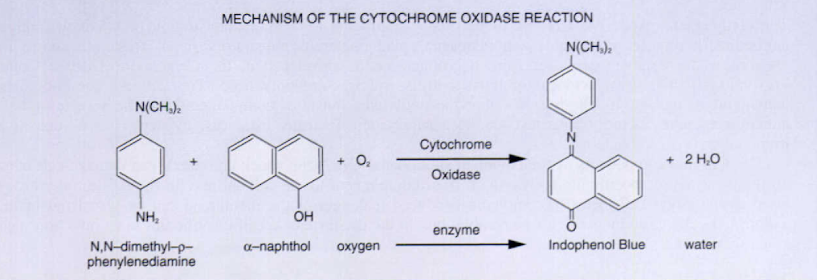 Figure: Mechanism of the Cytochrome Oxidase Reaction
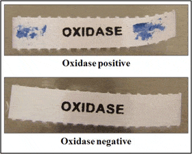 Figure: Oxidase test result
Why Enterobacteriaceae are negative: the branched respiratory chain
A common misreading is that oxidase-negative organisms cannot respire aerobically. E. coli respires aerobically perfectly well. It grows on nutrient agar in air. So why is it oxidase negative?
Because bacterial respiratory chains are branched, and they do not all terminate the same way.
The test reagent, TMPD, is an artificial electron donor. It does not enter the chain at the beginning. It donates electrons at the level of cytochrome c. For the reagent to be oxidized to the purple indophenol, the organism must possess a terminal oxidase that accepts electrons from cytochrome c. That is cytochrome c oxidase.
Enterobacteriaceae terminate their chain in quinol oxidases instead: cytochrome bo3 under high oxygen, cytochrome bd-I under low oxygen. These accept electrons from the quinone pool, upstream of where TMPD sits. The reagent has nothing to hand its electrons to. It stays reduced. It stays colorless.
So the oxidase test does not measure "can you use oxygen." It measures "does your chain pass through cytochrome c on the way to oxygen." Every confusing result in this article follows from that one sentence.
All bacteria that are oxidase-positive are aerobic and can use oxygen as a terminal electron acceptor in respiration. This does NOT mean that they are strict aerobes. Bacteria that are oxidase-negative may be anaerobic, aerobic, or facultative; the oxidase negative result just means that these organisms do not have the cytochrome c oxidase that oxidizes the test reagent. They may respire using other oxidases in electron transport.
Oxidase test vs catalase test
Students frequently confuse these two tests. Both are rapid enzyme detection tests used early in bacterial identification, but they detect completely different enzymes and serve different diagnostic purposes:
| Feature | Oxidase test | Catalase test |
|---|---|---|
| Enzyme detected | Cytochrome c oxidase | Catalase |
| Reaction | Oxidation of test reagent by cytochrome c oxidase | Decomposition of H₂O₂ → H₂O + O₂ |
| Positive result | Purple/dark blue color within 10 seconds | Vigorous bubbling within 5–10 seconds |
| Reagent | TMPD (tetramethyl-p-phenylenediamine) | 3% hydrogen peroxide (H₂O₂) |
| Primary use | Differentiates oxidase-positive gram-negative rods (Pseudomonas, Neisseria, Vibrio, Campylobacter) from Enterobacteriaceae (oxidase-negative) | Differentiates Staphylococcus (positive) from Streptococcus and Enterococcus (negative) |
| Applied primarily to | Gram-negative organisms | Gram-positive cocci |
| Can use nichrome loop? | No (false positive) | Yes, acceptable |
| Can test from MacConkey? | No (false result) | Yes, acceptable |
In a typical gram-negative identification workflow, the oxidase test is performed first, before TSI, SIM, urease, and other biochemical tests because a positive oxidase result immediately removes the organism from the Enterobacteriaceae and directs testing toward non-fermenters. See more: Catalase Test: Principle, Procedure, Results
Test requirements for Oxidase test
- Moist filter paper with the substrate (1% tetramethyl-p-phenylenediamine dihydrochloride), or commercially prepared paper disk, wooden applicator stick, or platinum wire.
- Kovács oxidase reagent (1% tetramethyl-p-phenylenediamine dihydrochloride in water). Store refrigerated in a dark bottle for no longer than 1 week.
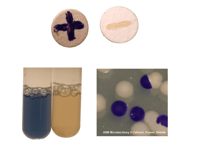 Figure: Various types of oxidase test procedure
Procedure of Oxidase test
Oxidase test can be performed in various ways. These include, but are not limited to, the filter paper test, filter paper spot test, direct plate method, and test tube method.
Filter Paper Test Method
- Soak a small piece of filter paper in 1% Kovács oxidase reagent and let it dry.
- Use a sterile loop to pick a well-isolated colony from a fresh (18- to 24- hour culture) bacterial plate and rub it onto treated filter paper.
- Observe for color changes.
Note: Nickel, steel and other wire loop give false-positive results, so one should use platinum or inert transfer loop. The alternative options are glass rods, wooden sticks, sterile plastic loops, sterile toothpicks and sterile swabs.
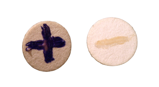 Figure: Oxidase-positive Pseudomonas aeruginosa (left) and oxidase-negative Escherichia coli (right).
Filter Paper Spot Method
- Use a loop to pick a well-isolated colony from a fresh bacterial plate and rub it onto a small piece of filter paper.
- Place 1 or 2 drops of 1% Kovács oxidase reagent on the organism smear.
- Observe for color changes.
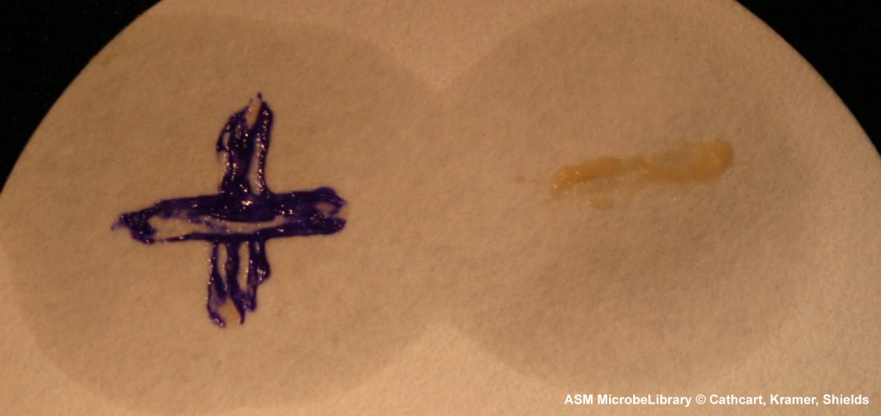 Figure: Oxidase-positive Pseudomonas aeruginosa (left) and oxidase-negative Escherichia coli (right).
Direct Plate Method
- Grow a fresh culture (18 to 24 hours) of bacteria on nutrient agar or trypticase soy agar using the streak plate method so that well-isolated colonies are present.
- Place 1 or 2 drops of 1% Kovács oxidase reagent on the organisms.
- Do not invert or flood plate.
- Observe for color changes.
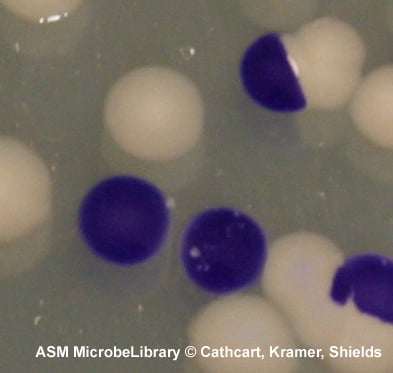 Figure: Oxidase-positive Vibrio cholerae showing purple colonies, and oxidase-negative Escherichia coli with lack of color change
Test Tube Method
- Grow a fresh culture (18 to 24 hours) of bacteria in 4.5 ml of nutrient broth (or standard media that does not contain a high concentration of sugar).
- Add 0.2 ml of 1% α-naphthol, then add 0.3 ml of 1% p-aminodimethylaniline oxalate (Gaby and Hadley reagents).
- Observe for color changes.
 Figure: Oxidase positive Neisseria sicca (left) and oxidase negative Staphylococcus aureus (right)
Reading the result
Kovács reagent (filter paper, spot, and direct plate methods)
- Purple in 10 seconds: oxidase positive.
- Purple at 60 to 90 seconds: delayed positive. See below.
- No color, or color only after 2 minutes: oxidase negative.
Gaby and Hadley reagent (test tube method)
- Blue in 15 to 30 seconds: oxidase positive.
- Purple at 2 to 3 minutes: delayed positive.
- No color: oxidase negative.
Purple in 10 seconds
This is the only unambiguous positive. The reaction is fast because cytochrome c oxidase is abundant and TMPD is a small molecule that reaches it immediately. Speed is diagnostic information, not just a stopwatch detail. A Pseudomonas aeruginosa colony goes purple almost as the loop lifts.
Delayed oxidase reaction
Purple appearing at 60 to 90 seconds is reported as a delayed positive, but it should also make you suspicious of your own technique. Genuine delayed positives are uncommon and are seen with some Pasteurella and Aeromonas isolates. Far more often, a delay means the culture is older than 24 hours and metabolically slow, or the inoculum was too light.
Color developing after 2 minutes is not a delayed positive. It is autooxidation of the reagent by air, and it happens on a blank paper with no organism on it at all. Run a reagent-only paper alongside your test and you will see this for yourself.
False negative oxidase test
The three causes, in order of how often they occur:
- Acidic growth medium. Sugar fermentation drives local pH below 5.1 and the reaction stops. Lactose on MacConkey, sucrose on TCBS. This is why the V. cholerae colony in the opening reads negative. Subculture to nutrient agar or 5% sheep blood agar and repeat.
- Old culture. Beyond 24 hours, enzyme activity falls. Use an 18 to 24 hour culture.
- Reagent age or exposure. Kovács reagent oxidizes in light and air. If the bottle or the paper is already purple, throw it out.
A false negative is far more dangerous clinically than a false positive, because it silently reclassifies a non-fermenter as a member of the Enterobacteriaceae and sends the entire downstream workup in the wrong direction.
Uses of oxidase test
- Oxidase test is most helpful in screening colonies suspected of being a member of the Enterobacteriaceae family; all the members of the Enterobacteriaceae family including E. coli are oxidase negative.
- To avoid misidentification, perform an oxidase test on all Gram-negative rods. Oxidase test is especially important in separating Aeromonas from Enterobacteriaceae.
- Note: Swarming growth on blood agar is a strong presumptive clue for Proteus species, which are oxidase negative like all Enterobacteriaceae. The oxidase test is still worth performing, because it costs seconds and it protects you against the one organism that swarming does not rule out. Pick from the edge of the swarm, not the center, where the film of growth is too thin to give a readable smear.
- Oxidase test is used as a major characteristic for the identification of Gram-negative rods that are not in the Enterobacteriaceae family. Colonies suspected of belonging to other genera Aeromonas, Pseudomonas, Neisseria, Campylobacter, and Pasteurella are oxidase positive.
- Gram-negative diplococci give a positive reaction. All members of the genus Neisseria are oxidase positive. Moraxella spp. that are either Gram-negative diplococci or coccobacilli are also oxidase-positive.
Oxidase-Positive and Oxidase-Negative Organisms
Oxidase-positive organisms
| Organism | Gram stain | Clinical significance |
|---|---|---|
| Pseudomonas aeruginosa | Gram-negative rod | Nosocomial pneumonia, UTI, wound infections, cystic fibrosis; key distinction from Enterobacteriaceae |
| Pseudomonas fluorescens | Gram-negative rod | Rare clinical significance; environmental contaminant; causes transfusion-associated septicemia |
| Burkholderia cepacia complex | Gram-negative rod | Important pathogen in cystic fibrosis; intrinsically resistant to aminoglycosides and polymyxins, usually susceptible to meropenem |
| Neisseria gonorrhoeae | Gram-negative diplococci | Gonorrhea, PID, disseminated gonococcal infection |
| Neisseria meningitidis | Gram-negative diplococci | Bacterial meningitis, meningococcemia |
| Moraxella catarrhalis | Gram-negative diplococci | COPD exacerbations, otitis media, sinusitis |
| Haemophilus influenzae | Gram-negative coccobacilli | Meningitis (type b), pneumonia, epiglottitis, otitis media |
| Campylobacter jejuni | Gram-negative curved rod | Most common bacterial cause of gastroenteritis worldwide |
| Campylobacter coli | Gram-negative curved rod | Gastroenteritis; similar presentation to C. jejuni |
| Helicobacter pylori | Gram-negative curved rod | Peptic ulcer disease, gastric carcinoma; part of triple test (oxidase + urease + catalase all positive) |
| Vibrio cholerae | Gram-negative curved rod | Cholera (rice-water diarrhea); important in outbreak investigation |
| Vibrio parahaemolyticus | Gram-negative rod | Gastroenteritis associated with raw seafood |
| Vibrio vulnificus | Gram-negative rod | Severe wound infection and septicemia from seawater exposure |
| Aeromonas hydrophila | Gram-negative rod | Wound infections, diarrhea, septicemia in immunocompromised |
| Aeromonas caviae | Gram-negative rod | Gastroenteritis, wound infections |
| Plesiomonas shigelloides | Gram-negative rod | Diarrheal illness; associated with seafood and travel |
| Brucella spp. | Gram-negative coccobacilli | Brucellosis (undulant fever); zoonotic infection |
| Pasteurella multocida | Gram-negative coccobacilli | Wound infections from animal bites (dog, cat); bacteremia |
| Legionella pneumophila | Gram-negative rod | Legionnaires' disease; weakly and variably oxidase positive, so the test has no diagnostic role here; does not grow on routine media and requires BCYE agar |
| Alcaligenes faecalis | Gram-negative rod | Rare opportunistic infections; environmental organism |
| Bordetella pertussis | Gram-negative coccobacilli | Whooping cough; laboratory diagnosis requires specific media and DFA |
Oxidase-negative organisms
| Organism | Gram stain | Notes |
|---|---|---|
| Escherichia coli | Gram-negative rod | Prototype oxidase-negative; UTI, diarrhea, septicemia |
| Klebsiella pneumoniae | Gram-negative rod | Nosocomial pneumonia, UTI, bacteremia |
| Salmonella spp. | Gram-negative rod | Typhoid fever, gastroenteritis, bacteremia |
| Shigella spp. | Gram-negative rod | Bacillary dysentery |
| Proteus mirabilis | Gram-negative rod | UTI, wound infections; swarming on blood agar |
| Enterobacter spp. | Gram-negative rod | Nosocomial infections; AmpC beta-lactamase |
| Serratia marcescens | Gram-negative rod | Nosocomial infections; red pigment at room temperature |
| Morganella morganii | Gram-negative rod | Nosocomial UTI and wound infections |
| Yersinia spp. | Gram-negative rod | Y. enterocolitica causes gastroenteritis; Y. pestis causes plague |
| Acinetobacter baumannii | Gram-negative coccobacilli | The trap. A non-fermenting Gram-negative coccobacillus, so it looks like it should be oxidase positive. It is not. Oxidase-negative plus non-fermenting is the pairing that identifies it. |
| Stenotrophomonas maltophilia | Gram-negative rod | The other trap. Non-fermenter, oxidase negative. Distinguished from Acinetobacter by DNase production and maltose oxidation. |
| Staphylococcus spp. | Gram-positive cocci | Oxidase-negative (use modified oxidase/microdase test to differentiate from Micrococcus) |
| Streptococcus spp. | Gram-positive cocci | Oxidase-negative |
| Enterococcus spp. | Gram-positive cocci | Oxidase-negative |
The exceptions that matter for exams
Three organisms break the pattern students are taught, and examiners know it.
- Acinetobacter baumannii: non-fermenter, but oxidase negative.
- Stenotrophomonas maltophilia: non-fermenter, but oxidase negative.
- Vibrio metschnikovii: a Vibrio, but oxidase negative. The genus rule has an exception, and this is it.
Note what the first two have in common. If you have been taught "oxidase positive means non-fermenter," you have been taught the converse of a true statement. The true statement is: oxidase positive rules out Enterobacteriaceae. It does not rule in non-fermenters, and oxidase negative does not rule them out.
Clinical Identification Workflow: How the Oxidase Test Is Used
The oxidase test is one of the first tests performed on gram-negative organisms and immediately divides them into two major groups:
Gram-negative organism identification- decision pathway
GRAM-NEGATIVE ORGANISM
│
├── OXIDASE POSITIVE → Not Enterobacteriaceae
│   │
│   ├── Cocci or diplococci → Neisseria spp., Moraxella catarrhalis
│   │   └── Further: sugar fermentation, superoxol test
│   │
│   ├── Coccobacilli → Haemophilus, Brucella, Bordetella, Pasteurella, Francisella
│   │   └── Further: X and V factor requirements, growth characteristics
│   │
│   ├── Curved rods → Campylobacter, Helicobacter, Vibrio
│   │   └── Further: oxidative-fermentative test, growth temperature, microaerophily
│   │
│   └── Straight rods (non-fermenter) → Pseudomonas, Burkholderia, Alcaligenes
│       └── Further: O-F test, pigment, cetrimide agar, motility
│
└── OXIDASE NEGATIVE → Likely Enterobacteriaceae (or Acinetobacter / Stenotrophomonas)
    │
    ├── Fermenter (O-F positive) → Enterobacteriaceae
    │   └── Further: TSI, SIM, urease, citrate, IMViC battery
    │
    └── Non-fermenter (O-F negative/alkaline) → Acinetobacter, Stenotrophomonas
        └── Further: growth at 44°C (A. baumannii), DNase and maltose oxidation (S. maltophilia)
Three clinically important oxidase-positive identifications
1. Pseudomonas aeruginosa: Oxidase positive + non-fermenter on O-F test + blue-green pyocyanin pigment + fruity grape-like odor + growth on cetrimide agar = presumptive P. aeruginosa. Confirmed by MALDI-TOF or biochemical panel.
2. Neisseria gonorrhoeae: Gram-negative intracellular diplococci from urethral/cervical discharge + oxidase positive + glucose fermentation only (not maltose) = presumptive N. gonorrhoeae. N. meningitidis ferments both glucose and maltose.
3. Campylobacter jejuni: Oxidase positive + gram-negative curved rod + growth at 42°C in microaerophilic conditions + hippurate positive = C. jejuni. Hippurate-negative oxidase-positive curved rods suggest C. coli.
Quality Control
Bacterial species showing positive and negative reactions should be run as controls at frequent intervals.
The following are suggested:
- Oxidase positive: Pseudomonas aeruginosa ATCC 27853
- Oxidase negative: Escherichia coli ATCC 25922
Precautions and Limitations
- Timing is critical to accurate testing.
- Use fresh reagents, no older than 1 week, older reagents can autooxidize thus giving erroneous results. Do not use if the reagent or filter paper is purple.
- Do not test organisms growing on media that contain glucose or dyes (e.g., MacConkey agar or EMB agar).
- Do not use nickel-base alloy wires containing chromium and iron (nichrome) to pick the colony and make a smear as this may give false-positive results.
- Bacteria grown on media-containing dyes may give aberrant results.
- Older cultures are less metabolically active so may give false-negative results within the mentioned observation time.
The oxidase test must be performed from 5% sheep blood agar or another medium without fermentable sugar. Fermentation of carbohydrates results in acidification of the medium (e.g., lactose in MacConkey Agar or Sucrose in TCBS), and a false negative oxidase test may result if the surrounding pH is below 5.1. Subinoculation on nutrient agar is required before the oxidase test can be performed. During the identification of suspected Vibrio cholerae isolate, it is not possible to perform an oxidase test directly from a TCBS culture because the acid produced by the sucrose fermenting colonies will inhibit the oxidase reaction.
How to remember
Mnemonics for oxidase positive organisms-PVNCH (It’s just an acronym inspired by the famous mnemonic for urease positive organisms-PUNCH)
PVNCH, and why it is not enough
The five you will be asked for first:
- P: Pseudomonas
- V: Vibrio
- N: Neisseria
- C: Campylobacter
- H: Helicobacter and Haemophilus
This mnemonic is deliberately incomplete. It covers the Gram-negative rods and cocci you will meet on a ward. It leaves out Aeromonas, Plesiomonas, Moraxella, Pasteurella, Brucella, Bordetella, and Alcaligenes. If you find yourself reaching for PVNCH and the organism is not in it, that is the mnemonic working correctly. It is telling you the answer is one of the environmental or zoonotic organisms, and those are worth learning by name.
The gate, not the fork
Picture the oxidase reagent as a gate with a guard who only accepts a ticket handed over by cytochrome c. Pseudomonas walks up holding that ticket. E. coli arrives holding a different ticket, one issued upstream at the quinone pool, and the guard cannot read it. E. coli is not turned away from respiring. It is turned away from this particular door.
Ask yourself, before you look anything up: if E. coli respires aerobically, why is it oxidase negative? If you can answer that in one sentence, you understand this test.
Key exam facts
| Question | Answer | The reason behind it |
|---|---|---|
| What enzyme is detected? | Cytochrome c oxidase, also called cytochrome aa3 or complex IV | It is the terminal oxidase that accepts electrons from cytochrome c |
| What is the reagent? | 1% TMPD (tetramethyl-p-phenylenediamine dihydrochloride), Kovács reagent | TMPD is an artificial electron donor that enters the chain at cytochrome c |
| What is the positive result? | Purple within 10 seconds | TMPD is oxidized to indophenol blue |
| What is a delayed positive? | Purple at 60 to 90 seconds | Slow or aged culture, or genuinely slow organisms such as some Pasteurella |
| Color after 2 minutes? | Negative. This is reagent autooxidation, not the organism | Air oxidizes TMPD on its own; a blank paper will do the same |
| Which family is uniformly negative? | Enterobacteriaceae, all members | They terminate in quinol oxidases (cytochrome bo3, bd-I), upstream of where TMPD donates |
| Does negative mean anaerobe? | No | E. coli is a facultative anaerobe that respires aerobically and is oxidase negative |
| Five to name fast | Pseudomonas, Vibrio, Neisseria, Campylobacter, Helicobacter (PVNCH) | The clinically commonest positives |
| The two non-fermenter traps | Acinetobacter baumannii, Stenotrophomonas maltophilia: both oxidase negative | Oxidase positive rules out Enterobacteriaceae. It does not rule in non-fermenters |
| The genus exception | Vibrio metschnikovii is oxidase negative | Genus-level rules in microbiology almost always have one |
| Why never test from MacConkey or TCBS? | Sugar fermentation drops local pH below 5.1 and the reaction cannot proceed | Gives a false negative on a truly positive organism |
| Correct medium to test from | Nutrient agar, trypticase soy agar, or 5% sheep blood agar | No fermentable sugar, no dyes |
| Why not a nichrome loop? | Nickel and iron oxidize TMPD directly | Gives a false positive with no organism involved |
| Acceptable transfer tools | Platinum loop, wooden applicator stick, sterile plastic loop, glass rod, sterile toothpick, sterile swab | All are inert toward TMPD |
| Culture age? | 18 to 24 hours | Older colonies are metabolically slow and read false negative |
| Reagent shelf life | 1 week, refrigerated, in a dark bottle | Autooxidation. Discard if the reagent or paper has turned purple |
| Positive QC organism | Pseudomonas aeruginosa | Rapid, unambiguous purple |
| Negative QC organism | Escherichia coli | Prototype Enterobacteriaceae |
| Where does oxidase sit in the workflow? | First, before TSI, SIM, urease, citrate | A positive result removes the organism from the Enterobacteriaceae and redirects the entire panel |
Where students get confused
"Oxidase negative means the organism is anaerobic." It does not. E. coli respires aerobically and is oxidase negative. The test asks whether the chain terminates in cytochrome c oxidase, not whether the organism uses oxygen. This is common misconception about this test.
"Oxidase positive means non-fermenter." The converse of a true statement is not a true statement. Oxidase positive rules out Enterobacteriaceae. Vibrio and Aeromonas are oxidase positive and are fermenters. Acinetobacter and Stenotrophomonas are non-fermenters and are oxidase negative. Use the O-F test to establish fermentation, not the oxidase test.
Testing straight from MacConkey, EMB, or TCBS. The most consequential error on this list. A lactose fermenter on MacConkey or a sucrose fermenter on TCBS acidifies its own surroundings. Below pH 5.1 the reaction is chemically blocked. You will call a Vibrio negative. Always subculture to nutrient agar or blood agar first.
Using a nichrome loop. The nickel and iron in the alloy oxidize TMPD without any enzyme present. You will get a purple that means nothing. Platinum, wood, or plastic only. Notice that this error and the previous one push in opposite directions: the wrong medium gives you a false negative, the wrong loop gives you a false positive. Students often remember that both are forbidden and forget which does what.
Waiting too long. Read at 10 seconds. Read again at 90. Stop. Anything that appears after 2 minutes is the reagent reacting with air, and it will appear on a blank paper too. If you are unsure, run a reagent-only control alongside. This takes 5 seconds and settles the question permanently.
Reading a purple reagent bottle as a good sign. If the Kovács reagent or the impregnated paper is already purple before the organism touches it, the reagent has autooxidized. It is dead. Discard it. Reagent that has been left on the bench in daylight will do this within a day.
Confusing the catalase and oxidase tests. They are both rapid, both enzyme tests, both done early. Beyond that they share nothing. Catalase separates Staphylococcus from Streptococcus using hydrogen peroxide, and a nichrome loop is fine. Oxidase separates non-Enterobacteriaceae from Enterobacteriaceae using TMPD, and a nichrome loop ruins it. If you remember one distinction, remember the loop.
Testing a swarming Proteus and skipping the confirmation. Swarming is a strong clue, not a result. Pick from the edge of the swarm where the growth is thick enough to smear, and run the test. It takes seconds and it protects you.
References and further readings
- Kovács N. Identification of Pseudomonas pyocyanea by the oxidase reaction. Nature. 1956;178(4535):703. doi:10.1038/178703a0
- Shields P, Cathcart L. Oxidase test protocol. American Society for Microbiology, 2010 (updated 2017).
- Leber AL, editor. Clinical Microbiology Procedures Handbook. 4th ed. Washington, DC: ASM Press; 2016. doi:10.1128/9781555818814
- Procop GW, Church DL, Hall GS, Janda WM, Koneman EW, Schreckenberger PC, Woods GL. Koneman's Color Atlas and Textbook of Diagnostic Microbiology. 7th ed. Philadelphia: Wolters Kluwer; 2017.
- Tille PM. Bailey and Scott's Diagnostic Microbiology. 15th ed. St. Louis: Elsevier; 2022.
- World Health Organization. Laboratory Methods for the Diagnosis of Epidemic Dysentery and Cholera. Geneva: WHO; 1999.
- Borisov VB, Gennis RB, Hemp J, Verkhovsky MI. The cytochrome bd respiratory oxygen reductases. Biochim Biophys Acta. 2011;1807(11):1398-1413. doi:10.1016/j.bbabio.2011.06.016

Comments
No comments yet. Be the first to share your thoughts.
Leave a comment
All comments are reviewed before they appear.