
Shigella: Identification, Pathogenesis, and Bacillary Dysentery

On this page
A child is brought in a Hospital with the complaints of frequent, painful stools that contain blood and mucus, and a constant urge to pass more even when little comes out. She has a fever and stomach cramps. The stool is scanty, not the large watery kind. This picture, small bloody stools with straining, points to dysentery, and one of its two main causes is the bacterium Shigella.
Shigella is remarkable for how little it takes to make someone ill. As few as a hundred organisms can cause disease, which is why it spreads so easily from person to person. This page is about how Shigella invades the gut lining and spreads inside it, and how the laboratory tells it apart from the other pale colony it most resembles, Salmonella.
Shigella is a Gram-negative, non-motile, non-spore-forming rod of the family Enterobacteriaceae. It is one of the two main causes of dysentery, an infection of the large intestine that produces small, bloody, mucoid stools. Shigella is closely related to E. coli; in fact they are genetically almost the same organism, which is worth knowing because Shigella behaves like an invasive E. coli in the gut.
There are four species, and each forms one serogroup:
| Serogroup | Species | Note |
|---|---|---|
| Group A | Shigella dysenteriae | Most severe disease; produces Shiga toxin |
| Group B | Shigella flexneri | Common in developing countries |
| Group C | Shigella boydii | Less common |
| Group D | Shigella sonnei | Mildest illness; most common worldwide, especially in industrialized countries |
Two facts to hold together: S. dysenteriae type 1 causes the most severe disease (it makes Shiga toxin and can cause hemolytic uremic syndrome), while S. sonnei is the most commonly isolated species overall but causes the mildest illness, often just watery diarrhea. Severity and frequency run in opposite directions here.
Disease: bacillary dysentery
Shigella causes shigellosis, also called bacillary dysentery. The illness ranges from mild watery diarrhea (typical of S. sonnei) to severe dysentery with small, frequent, bloody, mucoid stools and painful straining (tenesmus), typical of S. dysenteriae. The severe form reflects what the organism does to the gut: it invades and destroys the lining of the colon.
The most serious complication is caused by S. dysenteriae type 1, which produces Shiga toxin and can lead to hemolytic uremic syndrome (HUS), a combination of kidney failure, low platelets, and destruction of red blood cells.
Dysentery has two main causes, bacterial (mainly Shigella) and amoebic (Entamoeba histolytica), and telling them apart matters for treatment. That comparison is covered in a separate article: amoebic vs bacillary dysentery.
How Shigella spreads between people
Humans are the only host of Shigella; there is no animal reservoir. It spreads by the fecal-oral route, remembered as the five Fs: Feces, Fingers, Flies, Food, and Fomites (contaminated objects such as taps and toilet seats).
The reason it spreads so easily is its very low infectious dose. As few as 10 to 200 organisms can cause disease, far fewer than most gut pathogens need. This is why Shigella passes readily from person to person, spreads in households and daycare centers, and causes outbreaks where hygiene is difficult. Most gut pathogens need thousands or millions of organisms; Shigella needs a handful.
Virulence factors and how Shigella causes disease
Shigella does something unusual: it invades the cells of the colon and then spreads from one cell to the next without ever going back outside. Each step below explains part of the illness.
It survives the stomach. Shigella tolerates acid, so a small number of organisms can pass through the stomach alive. This is part of why the infectious dose is so low.
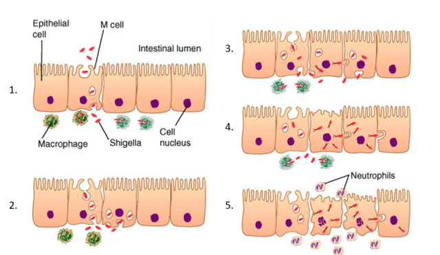 It invades through M cells. In the colon, Shigella is taken up by specialized M cells in the gut lining and delivered to the immune cells beneath. A macrophage engulfs it, but instead of being killed, Shigella kills the macrophage and escapes.
It forces its way into epithelial cells. Shigella then enters the colon lining cells from underneath, tricking them into taking it in. Once inside, it breaks out of the vacuole and sits free in the cell's cytoplasm.
It moves by building an actin tail. This is the signature. Shigella hijacks the cell's own actin to build a tail that pushes it sideways directly into the next cell. Because it spreads cell to cell from the inside, it largely avoids the antibodies and immune defenses waiting in the tissue outside.
It destroys the colon lining. Infected cells die and slough off, leaving ulcers. This triggers intense inflammation with neutrophils, bleeding, and mucus. That tissue destruction is exactly what you see in the stool: blood, mucus, and pus cells.
Shiga toxin (S. dysenteriae type 1). The most virulent species also makes Shiga toxin, which stops protein synthesis in host cells and damages the lining of small blood vessels. When it damages vessels in the kidney, the result can be hemolytic uremic syndrome.
Putting it together
The illness follows the mechanism. Shigella survives the stomach, invades the colon lining through M cells, escapes being killed by macrophages, then spreads cell to cell using actin tails while destroying the epithelium. The dying cells and intense inflammation produce the small, bloody, mucoid stools and the painful straining of dysentery. Because the organism stays in the colon lining and rarely enters the blood, dysentery is a local, destructive gut infection, not a systemic fever like typhoid. That contrast, Shigella stays local and destructive while Salmonella Typhi goes systemic, explains why one is diagnosed from stool and the other from blood.
Identifying Shigella in the laboratory
Specimen. Freshly passed stool is preferred. It should reach the laboratory within about 2 hours, or go into a transport medium (Cary-Blair, buffered glycerol saline) if any delay is likely, kept cool. Speed matters because Shigella is fragile and dies off quickly in stool.
Looking at the stool.
- Macroscopic: scanty stool, bright red with blood, and mucus, rather than a large watery volume.
- Microscopic: many pus cells (neutrophils) and red cells, against a background with no motile bacteria. That absence of motile organisms is itself a clue, because Shigella is non-motile.
On the plate.
- MacConkey and DCA: pale, non-lactose-fermenting colonies. Note S. sonnei is a late lactose fermenter and may look pale at first, then turn slightly pink on longer incubation.
- XLD agar: red-pink colonies without black centers. The absence of black centers is important: Salmonella makes black centers from H₂S, Shigella does not.
- SS agar: despite its name, SS agar is inhibitory to many Shigella strains and is not a good choice for isolating it. See the SS agar article.
The identification panel. Each result states what it means; follow the link for the method.
| Test | Shigella result | What it tells you |
|---|---|---|
| Oxidase | Negative | Places it in the Enterobacteriaceae. |
| Catalase | Positive (except S. dysenteriae type 1) | Consistent with the family; the exception is worth noting. |
| Lactose (MacConkey) | Non-fermenter (pale); S. sonnei late fermenter | Groups it with Salmonella; separates from E. coli. |
| Motility | Non-motile | The key split from Salmonella, which is motile. Shigella has no flagella and no H antigen. |
| H₂S | Negative | The other key split from Salmonella. No black centers on selective media. |
| TSI | K/A, no gas, no H₂S | Alkaline slant, acid butt (glucose only), and crucially no gas and no black color. |
| Urease | Negative | Separates from Proteus. |
| Citrate | Negative | Cannot use citrate as sole carbon source. |
| ONPG | Negative (except S. sonnei, positive) | Reflects the S. sonnei late-lactose exception. |
How Shigella and Salmonella differ on TSI. Both give an alkaline slant over an acid butt. The separators are gas and H₂S: Salmonella often makes H₂S (black) and gas; Shigella makes neither. A K/A tube with no gas and no black is the Shigella pattern.
Confirmation. A suspected isolate is confirmed and grouped by slide agglutination using antisera against its O antigen. The four polyvalent antisera (A, B, C, D) identify the group, then monovalent antisera pin down the serotype.
Serotyping
Shigella is serotyped using its O antigen only. Because it is non-motile, it has no H antigen, which simplifies typing compared with Salmonella. A K antigen may be present on the surface and can interfere with O-antigen typing, so it is sometimes removed by heating before the test. After isolation, the four polyvalent antisera (groups A to D) are used to find the group, then monovalent antisera identify the exact serotype. Which species predominates depends on geography; groups A and B are more common in developing countries.
The S. sonnei exceptions
S. sonnei breaks several of the rules above, and it is the most common species, so its exceptions are worth collecting in one place. Insert after the panel:
Why S. sonnei is the exception
Shigella sonnei is the species you are most likely to isolate, and it disagrees with the usual Shigella pattern on three tests:
- It is a late lactose fermenter, so it may look pale early and slightly pink later, unlike the other species which stay pale.
- It is ONPG-positive, because it has the enzyme for lactose even though it ferments slowly.
- It is ornithine decarboxylase (ODC)-positive, unlike the other Shigella species.
If an isolate looks like Shigella but is ONPG-positive and ODC-positive, think S. sonnei rather than assuming the tests are wrong.
Treatment
Most mild shigellosis is self-limited and needs mainly fluid and electrolyte replacement. Antibiotics are used for severe disease, for S. dysenteriae type 1, and in vulnerable patients, and they shorten the illness and reduce spread. Because resistance to older agents (ampicillin, co-trimoxazole) is now widespread, the choice depends on local susceptibility, with fluoroquinolones, third-generation cephalosporins, or azithromycin used depending on the setting. Susceptibility testing matters.
One safety point worth remembering: drugs that stop gut movement (anti-motility agents) should be avoided in dysentery, because slowing the gut can prolong the infection and worsen the illness.
How to remember
No black, no gas, no movement: that's Shigella. On a stool workup, Shigella is the pale colony that does nothing showy: no H₂S (no black on XLD or SS), no gas on TSI, and no motility. Compare it to Salmonella, which makes black centers, often gas, and swims. Ask yourself: two pale non-lactose colonies, which is Shigella? The quiet one that makes no black and does not move.
The actin tail is the whole story. Picture Shigella inside a colon cell, building a tail out of the cell's own actin to shoot itself sideways into the next cell. It never steps outside, so antibodies cannot reach it. That one image explains why it spreads so fast and why the disease destroys the colon lining.
A handful is enough. As few as 10 to 200 organisms cause disease. Most gut pathogens need thousands. That is why Shigella spreads person to person so easily and why handwashing matters so much.
Sonnei is the rule-breaker. The most common species is the one that breaks the pattern: late lactose fermenter, ONPG-positive, ODC-positive. If a Shigella seems to fail the usual tests, suspect S. sonnei.
Key exam facts in one table
| Fact | Detail |
|---|---|
| Gram reaction / shape | Gram-negative rod, non-motile, non-spore-forming, Enterobacteriaceae |
| Relationship to E. coli | Genetically almost identical; behaves like invasive E. coli |
| Species / serogroups | S. dysenteriae (A), S. flexneri (B), S. boydii (C), S. sonnei (D) |
| Most severe | S. dysenteriae type 1 (Shiga toxin, HUS) |
| Most common | S. sonnei (mildest illness) |
| MacConkey | Pale (non-lactose); S. sonnei late fermenter |
| Motility | Non-motile (key split from Salmonella); no H antigen |
| H₂S | Negative (no black centers; key split from Salmonella) |
| TSI | K/A, no gas, no H₂S |
| Infectious dose | Very low, 10 to 200 organisms |
| Transmission | Fecal-oral, five Fs; humans only, no animal reservoir |
| Signature mechanism | Invades colon cells, spreads cell to cell via actin tails |
| Disease | Bacillary dysentery (bloody mucoid stools, tenesmus) |
| S. sonnei exceptions | Late lactose fermenter, ONPG-positive, ODC-positive |
| Serotyping | By O antigen only (no H antigen) |
| Anti-motility drugs | Avoid in dysentery |
Where students get confused
Shigella vs Salmonella on a pale plate. Both are pale non-lactose fermenters. The separators are motility (Salmonella motile, Shigella non-motile) and H₂S (Salmonella often positive with black centers, Shigella negative). On TSI, Shigella makes no gas and no black; Salmonella often makes both.
S. sonnei looks like it fails the tests. The most common species is a late lactose fermenter and is ONPG- and ODC-positive, against the usual Shigella pattern. This is not a lab error; it is S. sonnei being itself.
SS agar for Shigella. The name suggests it is ideal, but SS agar inhibits many Shigella strains and is a poor choice for isolating it. This trips students because of the name.
Severity vs frequency. S. dysenteriae causes the worst disease but is not the most common; S. sonnei is the most common but causes the mildest illness. They run in opposite directions.
Dysentery is not always Shigella. Dysentery has a bacterial cause (Shigella) and an amoebic cause (Entamoeba histolytica). Bloody stool alone does not confirm Shigella; the two are distinguished by stool findings and are covered separately.
Non-motile means no H antigen. Because Shigella has no flagella, it has no H antigen, so serotyping uses the O antigen only. Students sometimes look for an H type that does not exist.
References
- Tille, P. M. (2022). Bailey & Scott's Diagnostic Microbiology (15th ed.). Elsevier.
- Procop, G. W., et al. (2017). Koneman's Color Atlas and Textbook of Diagnostic Microbiology (7th ed.). Wolters Kluwer.
- Niyogi, S. K. (2005). Shigellosis. Journal of Microbiology, 43(2), 133–143.
- The, H. C., Thanh, D. P., Holt, K. E., Thomson, N. R., & Baker, S. (2016). The genomic signatures of Shigella evolution, adaptation and geographical spread. Nature Reviews Microbiology, 14(4), 235–250. https://doi.org/10.1038/nrmicro.2016.10
Frequently Asked Questions
Is Shigella motile or non-motile?
Is Shigella motile or non-motile?
Non-motile. Shigella has no flagella, so it does not move and has no H antigen. This is one of the main features that separates it from Salmonella, which is motile.
Does Shigella produce H₂S?
Does Shigella produce H₂S?
No. Shigella does not produce hydrogen sulfide, so it forms no black centers on selective media such as XLD agar. This is a key difference from Salmonella, which usually does produce H₂S.
What are the TSI results for Shigella?
What are the TSI results for Shigella?
An alkaline slant over an acid butt (K/A), with no gas and no H₂S. It ferments glucose but not lactose or sucrose. The absence of both gas and black color helps separate it from Salmonella.
How do you tell Shigella from Salmonella?
How do you tell Shigella from Salmonella?
Both are pale, non-lactose-fermenting colonies on MacConkey agar. Shigella is non-motile and H₂S-negative; Salmonella is motile and usually H₂S-positive with black centers on selective media.
Which Shigella species causes the most severe disease?
Which Shigella species causes the most severe disease?
Shigella dysenteriae type 1. It produces Shiga toxin and can cause hemolytic uremic syndrome. However, it is not the most common species; S. sonnei is the most commonly isolated but causes the mildest illness.
Why does Shigella spread so easily?
Why does Shigella spread so easily?
Because its infectious dose is very low. As few as 10 to 200 organisms can cause disease, so it passes readily from person to person, especially where handwashing and sanitation are difficult.
Why is SS agar not ideal for Shigella?
Why is SS agar not ideal for Shigella?
Despite its name (Salmonella-Shigella agar), SS agar inhibits many Shigella strains, so it can fail to grow them. Other media such as XLD or MacConkey are more reliable for isolating Shigella.
What is the difference between bacillary and amoebic dysentery?
What is the difference between bacillary and amoebic dysentery?
Bacillary dysentery is caused by bacteria, mainly Shigella; amoebic dysentery is caused by the parasite Entamoeba histolytica. They differ in stool findings and treatment, and are compared in a separate article.

Tankeshwar Acharya, MSc (Medical Microbiology)
Tankeshwar Acharya is an Assistant Professor in the Department of Microbiology at Patan Academy of Health Sciences (PAHS), Nepal, where he has been teaching and practicing clinical microbiology for over 14 years. He is the founder of Microbe Online, one of the leading free microbiology education resources on the web, covering bacteriology, mycology, parasitology, immunology, and clinical laboratory diagnostics written from direct experience in both the classroom and the diagnostic laboratory.
Comments
No comments yet. Be the first to share your thoughts.
Leave a comment
All comments are reviewed before they appear.