
Pus Sample: Collection, Processing, Staining and Culture
Why an abscess aspirate beats a swab, the correct fallback when you cannot aspirate, how much pus to collect, how to protect anaerobes in transport, and how the lab stains and cultures a pus sample.

On this page
The most important decision in a pus sample happens before the lab ever sees it: aspirate or swab. An aspirate from an undrained abscess gives volume, keeps anaerobes alive, and skips the skin surface. A swab gives a small sample, dries out, and drags in skin flora that muddy the result. Almost everything the laboratory can tell you is decided at that moment of collection. The rest of this page is how to collect well, and what to do when the ideal is not possible.
Skin is the body’s largest and thinnest organ which serves as an anatomical barrier between the sterile internal organs and the external environment, which is teemed with microorganisms. Break in the skin surface may result in skin and soft tissue infections. Wound infection can also occur as a complication of surgery, trauma, and bites or diseases that interrupt mucosal or skin surfaces.
Wound infections may be caused by one or many organisms depending on the site of the infection. For example, dermatophytes are responsible for infections in the keratinized layer; superficial skin wounds are often caused by aerobes only, while anaerobes are commonly isolated from abscesses of the perineal, inguinal, and buttock area, whereas mixed facultative aerobic organisms cause non-perineal infections.
Similarly, postoperative wounds are often infected with a mixture of aerobes and anaerobes. In contrast, deep wound infections, such as internal body or organ infections, can be caused by one or several aerobes and/or anaerobes.
Abscesses are accumulations of pus in tissue and any organism isolated from them may be of significance.
Wounds, especially postoperative wounds, may become colonized with potential pathogens. A gram stain is a useful diagnostic tool in determining colonization versus infection. A gram stain showing few or no polymorphonuclear cells with relatively large amounts of normal skin flora are consistent with colonization. However, wound gram stains showing moderate to many polymorphonuclear cells usually indicate infection.
Possible pathogens in Pus
This article focuses on the isolation and identification of common bacterial isolates (aerobes and facultative anaerobes) from pus aspirate/swab.
| Gram-positive | Gram-negative |
|---|---|
| Staphylococcus aureus | Pseudomonas aeruginosa |
| Streptococcus pyogenes | Escherichia coli |
| Enterococcus species | Proteus species |
| Anaerobic streptococci | Klebsiella species |
| Other streptococci | Bacteroides species |
| Clostridium perfringens and other clostridia | Acinetobacter species |
| Actinomycetes | Other enteric bacilli |
| Mycobacterium tuberculosis | |
| Others | |
| Fungi: Histoplasma, Candida, and fungi that cause mycetoma. | |
| Parasites: Entamoeba histolytica (in pus aspirated from an amoebic liver abscess) | |
| Viruses: Pox viruses and herpes viruses |
Specimen Collection
As far as possible, collect specimens before antimicrobial therapy and/or before applying the antiseptic dressing. The ideal specimen is an aspirate from a previously undrained abscess or a tissue biopsy. Ideally, a minimum volume of 1mL (up to 5 mL) of pus should be collected. Large volumes of purulent material maintain the viability of anaerobes for longer.
The aspirate should be collected in a sterile syringe; any air bubbles should be expelled. Needle safely and tightly capped (needles should NOT be sent).
A tissue specimen should be placed in a sterile universal bottle (or any sterile leakproof container) and sent to the lab for immediate processing if anaerobes are suspected. If there is a delay in transporting, the tissue should be placed in an anaerobic transport system.
Aspirate or swab, and what to do when you cannot aspirate
The ideal pus specimen is an aspirate from an undrained abscess or a piece of tissue. Swabs sit at the bottom of the ranking, but they are what most wards send, so knowing how to make a swab as good as it can be matters as much as knowing why it is second best.
| Rank | Sample | When to use it | How to collect it | What you trade off |
|---|---|---|---|---|
| 1 (ideal) | Pus aspirate from an undrained abscess | Any closed collection you can reach with a needle | Aspirate 1 to 5 mL into a sterile syringe, expel air, cap tightly, remove and discard the needle safely | Needs an intact abscess; not possible once drained or for a superficial spreading wound |
| 1 (ideal, equal) | Tissue biopsy | Deep or chronic wounds, suspected mycetoma or mycobacterial or fungal infection | Sterile leakproof container; anaerobic transport system if anaerobes are suspected | Invasive; needs a clinician |
| 2 (compromise) | Two swabs from the wound depth | Aspirate not obtainable, for example an open or already-drained wound | Clean off superficial slough first; sample the deepest part; one swab for culture, one for the Gram smear; soak both well in pus | Small sample, dries fast, catches more surface flora; interpret with the Gram film |
| 3 (last resort) | A single swab | Only one swab available | Same as above; use the swab to inoculate culture media first, then make the smear | You must choose between culture and smear; culture is prioritized |
Two swabs, not one, is the practical rule when you cannot aspirate. One swab is used for culture and the second to make the Gram film. With a single swab you have to inoculate the plates first (culture is the priority) and only then roll out a smear from what remains, which gives a poorer film. If anaerobes matter, a swab is a weak choice at any count, so push for an aspirate.
Superficial versus deep sampling changes the answer. A swab dragged across the surface reports skin colonizers, not the pathogen. Whatever the sample, clear the superficial debris and sample the advancing edge or the depth of the wound. This single habit separates a useful swab from a misleading one.
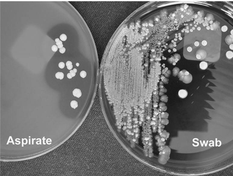 Figure: Comparison of aspirated pus and swab culture from an infected joint site. Image source: Koneman’s Color Atlas and Textbook of Diagnostic Microbiology
Swabs are less desirable because of the smaller amount of sample specimen and the fact that they are often contaminated with normal skin flora, making interpretation of results difficult. When using swabs, the deepest part of the wound should be sampled, avoiding the superficial microflora. Swabs should be well soaked in pus.
Collecting pus correctly sits within the wider rules for specimen collection and transport. For those general principles, see Specimen Collection and Transport in Microbiology.
Specimen Transport
Label the specimen and deliver it to the laboratory as soon as possible with a completed request form. The specimen volume and the suspected organism decide the acceptable transport time. Anaerobe recovery falls off once transport exceeds about 3 hours.
The best protection for anaerobes is the collection method itself: pus aspirated into a sterile syringe with the air expelled and the syringe capped, or tissue in a dedicated anaerobic transport system. These keep oxygen away far better than any swab medium.
If only a swab could be taken and transport will be delayed, place it in Amies transport medium to stop it drying out. This limits drying and slows the loss of organisms, but it is a compromise, not a true anaerobic transport method. A swab in Amies does not preserve strict anaerobes the way an aspirate or an anaerobic transport system does. (Cary-Blair is for enteric specimens and is not the medium for pus; use Amies if a pus swab must be held.)
If processing is delayed, refrigeration is preferable to leaving the sample at room temperature for routine aerobic culture. One exception: do not refrigerate when anaerobes or fastidious organisms are suspected, since cold reduces their recovery. For those, transport fast at room temperature instead.
Laboratory examination of Pus sample
Handling clinical specimens, especially pus samples that may contain unknown pathogens, invokes all the foundational microbiology laboratory safety practices. Before collecting, transporting, or processing any specimen, review microbiology laboratory safety rules for specimen safety, universal precautions, and exposure protocols
- Describe the appearance of the specimen: Describe the presence or absence of sulfur granules (needed only for the suspected cases of mycetoma or actinomycosis, when requested).
Preparation of the Smear
If a pus swab is sent:
- If only one swab was submitted: inoculate the culture media first, then use the same swab to make the Gram smear. Culture takes priority over the smear.
- If two or more swabs were submitted: use one for culture and a second to make the Gram smear.
If tissue sample is submitted: make a Gram stain from ground tissue.
If pus aspirate is sent, place one drop of pus onto a clean microscope slide using a sterile pipette. Spread this using a sterile loop to make a thin smear for Gram staining.
Gram Staining: Make an evenly spread smear of the specimen on a clean, grease-free slide. Allow the smear to air-dry in a safe place. Heat fix the specimen and stain by Gram staining technique. Examine the smear for the presence of bacteria and pus cells (PMNs) using 100x objective lens and look especially for:
Gram-negative rods (possible pathogens are E.coli, Klebsiella pneumoniae, Pseudomonas aeruginosa, Proteus or Bacteroides species) Gram-positive cocci in pairs, chains or clusters (possible pathogens are Staphylococcus aureus, Streptococcus pyogenes, anaerobic streptococci or enterococci).
Gram positive large rods with square ends (possible pathogens are Clostridium perfringens or Bacillus anthracis). If Bacillus anthracis is suspected on clinical grounds, notify the laboratory and handle the specimen under appropriate biosafety precautions, as this changes processing and reporting.
In the case of anaerobic infections large number of pleomorphic bacteria (streptococci, Gram positive and Gram negative rods of various sizes and fusiform bacteria) may be seen. Sometimes, Gram-positive yeast cells with pseudohyphae may be seen, which can be Candida albicans.
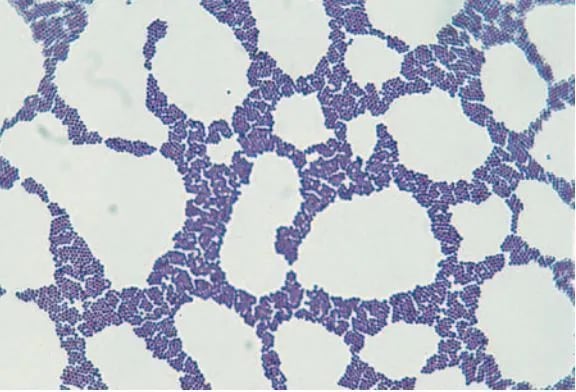 Figure: Staphylococcus in Gram Stain
Pus Culture
Culture Media: Wound specimens collected on aerobic swabs or pus aspirate should be plated on to the following media:
- Sheep blood agar (to isolate S. aureus and Streptococcus pyogenes or other streptococci)
- MacConkey agar (to isolate Gram-negative rods)
Incubation Condition:
- Temperature: 35ºC -37ºC
- Atmosphere: Blood Agar plate in carbon dioxide enriched atmosphere (e.g. 5% CO₂ incubator or in a candle jar) and MacConkey agar plate in ambient air (normal incubator)
- Time: Up to 48 hours (observe the plate after 24 hours of incubation, if growth is seen, do further processing, if not, reincubate for additional 24 hours.)
Examination and Reporting the Culture results
If the growth is seen after 24/48 hours of culture, the colony morphology and identification of the isolates should be examined.
In the Blood Agar plate, look for hemolysis. Staphylococcus aureus and Streptococcus pyogenes give beta-hemolysis in Blood Agar (Some S. aureus isolates may not show hemolysis).
- S. aureus gives yellow to cream or white colonies. Colonies are slightly raised and easily emulsified.
- S. pyogenes produces beta-hemolytic colonies. Colonies are usually small, colorless, dry, shiny, or mucoid.
- Enterococci give non-hemolytic colonies in blood agar.
We can differentiate between streptococci and staphylococci by a very simple and rapid test-Catalase test (Staphylococcus-positive, Streptococcus-negative). For identification of suspected S. aureus colonies, perform coagulase test (to differentiate coagulase-negative Staphylococci from S. aureus) and for suspected Group A Streptococci (S. pyogenes) perform bacitracin sensitivity test (can be added in the blood agar plate with other antibiotics). If enterococci is suspected perform bile esculin test.
Look for the growth of lactose fermenter colonies (pink) or non-lactose fermenter colonies (pale) in the MacConkey Agar plate. Lactose fermenter colonies can be of Escherichia coli, Klebsiella spp, or Enterobacter spp and non-lactose fermenter colonies can be of Pseudomonas aeruginosa, Acinetobacter spp, Proteus spp etc.
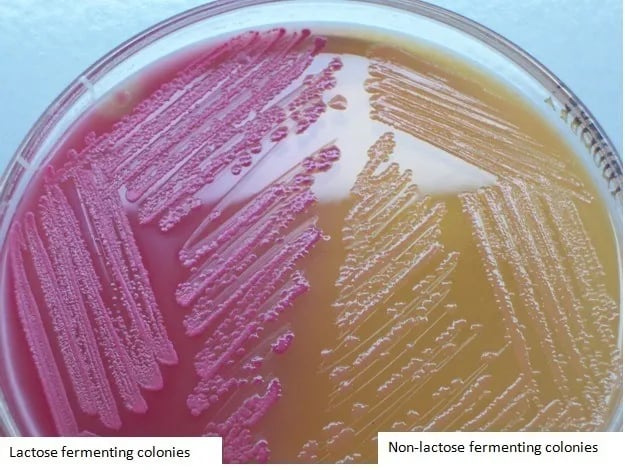 Figure: LF and NLF colonies in MacConkey Agar
Member of the family Enterobacteriaceae can be differentiated from other Gram-negative bacilli by performing two rapid tests (catalase test +ve, and oxidase test –ve). Identifications of the enteric bacteria can be done by using biochemical tests such as citrate utilization test, Triple Sugar Iron (TSI) Agar test, Sulphite-Indole-Motility (SIM) test, and urease test.
Pseudomonas aeruginosa gives large, flat, spreading pale-colored colonies in MacConkey Agar. It is oxidase positive and can be identified by its pigments and/or distinctive smell (characteristics of fruity smell).
Depending on the facilities available in the diagnostic laboratories, organisms can be identified using enterotube test or API-20E test or other newer diagnostics tests available for identifying isolates.
Antimicrobial Sensitivity Testing
S. aureus and the gram-negative bacilli are tested on plain Mueller-Hinton Agar (MHA). Streptococcus pyogenes and enterococci are tested on Mueller-Hinton Agar supplemented with sheep blood.
The selection of the antibiotics panel depends on the isolated organism. Unless indicated routinely used (or first line), antibiotics should be used. If the patient is in an intensive care unit (SICU, PICU, NICU) or is receiving particular antibiotic, or the isolate is resistant to first-line antibiotics, sensitivity testing should include requested antibiotics and/or second-line antibiotics.
How to Remember
Aspirate beats swab for three reasons: volume, oxygen, and skin. An aspirate gives more sample (volume), keeps air out so anaerobes survive (oxygen), and skips the surface colonizers (skin). A swab loses on all three.
Two swabs when you cannot aspirate. One for culture, one for the Gram stain. If you have only one, plate first, smear second.
Sample the depth, not the surface. The surface is where skin flora live. The pathogen is at the advancing edge. A swab rolled across the top reports the wrong organisms.
Anaerobes hate three things: air, drying, and time. Capped syringe (no air), transport medium if you are stuck with a swab (no drying), under 3 hours (no time). Refrigeration is the fourth enemy for anaerobes, so keep those warm.
Key exam facts in one table
| Point | Fact |
|---|---|
| Ideal specimen | Aspirate from an undrained abscess, or tissue biopsy |
| Volume of pus | 1 to 5 mL; larger volumes keep anaerobes viable longer |
| Aspirate handling | Sterile syringe, expel air, cap; do not send the needle |
| Swab (compromise) | Two swabs from the wound depth, well soaked; avoid the surface |
| Single swab rule | Inoculate culture first, then make the smear |
| Best anaerobe protection | Capped aspirate or anaerobic transport system, not a swab |
| Swab-in-Amies | Prevents drying only; not true anaerobic transport |
| Cary-Blair | Enteric specimens, not pus |
| Transport time | As soon as possible; anaerobe recovery falls after about 3 h |
| Refrigeration | Fine for routine aerobes; NOT for anaerobes or fastidious organisms |
| Colonization vs infection | Few PMNs plus skin flora suggests colonization; many PMNs suggests infection |
| Routine culture media | Sheep blood agar and MacConkey agar |
| Incubation | 35 to 37 C, blood agar in CO₂, up to 48 h |
| Quick GPC split | Catalase: Staphylococcus positive, Streptococcus negative |
| Sulfur granules | Look for them when mycetoma or actinomycosis is suspected |
Where Students Get Confused
"Why is a swab so much worse than an aspirate?" Three reasons at once: a swab holds far less sample, it dries and lets air reach anaerobes, and rolled over a wound it collects skin colonizers. An aspirate avoids all three. The Koneman figure on this page shows the difference in what actually grows.
"How does a Gram stain separate colonization from infection?" By counting pus cells. Few or no polymorphonuclear cells with plenty of normal skin flora points to colonization. Moderate to many pus cells points to infection. The bugs alone do not decide it; the host response does.
"If anaerobes are suspected, is a swab in Amies good enough?" No. Amies stops the swab drying, but it is not a true anaerobic transport method. Strict anaerobes still die. The real answer is an aspirate in a capped syringe with the air expelled, or tissue in an anaerobic transport system. Swab in Amies is a last resort.
"Why keep anaerobe samples warm when refrigeration protects most specimens?" Cold protects routine aerobic cultures by slowing overgrowth, but it lowers recovery of anaerobes and fastidious organisms. When those are in the differential, speed and room temperature beat the fridge.
"Only one swab came, do I stain or culture?" Culture. Inoculate the plates first because the culture is the result that guides treatment, then make the smear from what is left on the swab.
References and further reading
- Procop GW, Church DL, Hall GS, et al. Koneman's Color Atlas and Textbook of Diagnostic Microbiology. 7th ed. Philadelphia: Wolters Kluwer; 2017.
- Tille PM. Bailey & Scott's Diagnostic Microbiology. 15th ed. St. Louis: Elsevier; 2022.
- Leber AL, editor. Clinical Microbiology Procedures Handbook. 4th ed. Washington, DC: ASM Press; 2016. DOI: 10.1128/9781683670438.CMPH
- Cheesbrough M. District Laboratory Practice in Tropical Countries, Part 2. 2nd ed. Cambridge: Cambridge University Press; 2006.
Frequently Asked Questions
Why is an aspirate preferred over a swab for pus?
Why is an aspirate preferred over a swab for pus?
An aspirate provides more sample, keeps air out so anaerobes survive, and avoids the skin surface where colonizing flora live. A swab loses on all three counts, which is why an aspirate from an undrained abscess is the ideal specimen.
What do I do when I cannot get an aspirate?
What do I do when I cannot get an aspirate?
Take two swabs from the depth of the wound after clearing superficial debris, one for culture and one for the Gram smear. If only one swab is available, inoculate the culture media first, then make the smear from the same swab.
How much pus should be collected?
How much pus should be collected?
Aim for 1 to 5 mL. Larger volumes keep anaerobes viable longer and give the lab enough material for smear, culture, and any special testing.
Is a swab in Amies medium enough when anaerobes are suspected?
Is a swab in Amies medium enough when anaerobes are suspected?
No. Amies prevents the swab from drying but is not a true anaerobic transport method, so strict anaerobes still die. Use a capped syringe aspirate with the air expelled, or tissue in an anaerobic transport system.
Should a pus sample be refrigerated if transport is delayed?
Should a pus sample be refrigerated if transport is delayed?
For routine aerobic culture, refrigeration is better than leaving the sample at room temperature. Do not refrigerate if anaerobes or fastidious organisms are suspected, because cold lowers their recovery. For those, transport quickly at room temperature, and remember that a capped aspirate protects anaerobes far better than any swab.
How does the Gram stain distinguish colonization from infection?
How does the Gram stain distinguish colonization from infection?
By the number of pus cells. Few or no polymorphonuclear cells with abundant skin flora suggests colonization. Moderate to many pus cells suggests true infection.
Is Cary-Blair medium used for pus samples?
Is Cary-Blair medium used for pus samples?
No. Cary-Blair is for enteric (stool) specimens. If a pus swab must be held during a transport delay, Amies is the medium to use.

Tankeshwar Acharya, MSc (Medical Microbiology)
Tankeshwar Acharya is an Assistant Professor in the Department of Microbiology at Patan Academy of Health Sciences (PAHS), Nepal, where he has been teaching and practicing clinical microbiology for over 14 years. He is the founder of Microbe Online, one of the leading free microbiology education resources on the web, covering bacteriology, mycology, parasitology, immunology, and clinical laboratory diagnostics written from direct experience in both the classroom and the diagnostic laboratory.
Comments
No comments yet. Be the first to share your thoughts.
Leave a comment
All comments are reviewed before they appear.