Mycetoma: Types, Clinical features, and Lab diagnosis
Mycetoma: Types, Clinical features, and Lab diagnosis
Mycetoma is an infection of the skin and subcutaneous tissues. It mainly affects the feet, but the hands, shoulder, abdomen, buttocks, and scalp infections are also reported. Mycetoma is a slowly progressive disease, and after the infection, it may take a year to form the characteristics lesions. These lesions are firm, painless, localized subcutaneous nodules. It spreads and destroys the surrounding structure but does not affect tendons and nerves. Mycetoma is also known as Madura Foot or Maduramycosis.
Classification of Mycetoma
Mycetoma is classified based on causative agents, the color of grains, and geographical areas.
Based on causative agents
- Eumycetoma: Mycetoma is caused by fungi.
- Actinomycetoma: Mycetoma is caused by actinomycetes.
Based on the color of grains
- Black grain mycetoma
- White or pale grain mycetoma
All bacterial agents produce white grain mycetoma except Actinomadura pelletieri, which produces red or pink grains. Fungal agents produce both black and grains mycetoma.
Causative agent of Mycetoma
Mycetoma is caused by fungi and bacteria
Fungal agents
Fungi causing eumycetoma are saprophytic environmental fungi. Based on the formation of the grains, it is of two types:
| Black Grain Eumycetoma | White Grain Eumycetoma |
|---|---|
| Madurella mycetomatis | Pseudallescheria boydii |
| Trematosphaeria grisea | Aspergillus nidulans |
| Exophiala jeanselmei | Acremonium falciforme |
| Curvularia geniculata | Fusarium spp. |
- Black Grain Eumycetoma: These fungi form the black grains.
- White Grain Eumycetoma: These fungi form the white grains.
Examples of the fungi producing the black and white grains are given below.
Bacterial agents
Actinomycetes (Gram-positive branching filamentous aerobic bacteria) causing the actinomycetoma are found in soil and environment. It gets inoculated in the body by the trauma. The color of the grains produced by actinomycetoma causing bacterial agents are given below.
| Bacterial Agents | Grains-Color |
|---|---|
| Actinomadura madurae | White, yellow |
| Actinomadura pelletieri | Pink to Red |
| Nocardia brasiliensis | Yellowish-White |
| Nocardia caviae | White to yellow |
| Nocardia asteroides | White to yellow |
| Nocardia dassonvillei | Cream-colored |
| Streotomyces somaliensis | Yellowish-white |
Mode of transmission of Mycetoma
The causative agents of mycetoma are present in the saprophytic soil, which gets inoculated in the body through accidental trauma. Farmers and field workers may get accidental trauma through skin abrasion and thorn pricks. Mycetoma in the head and neck occur in people who carry wood, grain bags stone on the head and neck. In rural areas, mycetoma in the ears is caused by people who use a straw to remove ear wax.
As the causative agent is introduced, the disease evolves slowly, and microabscess forms in the area of contact. The organism is also found in its center. The major feature of mycetoma infection is the presence of large aggregates of filaments of causative organisms.
Clinical features of Mycetoma
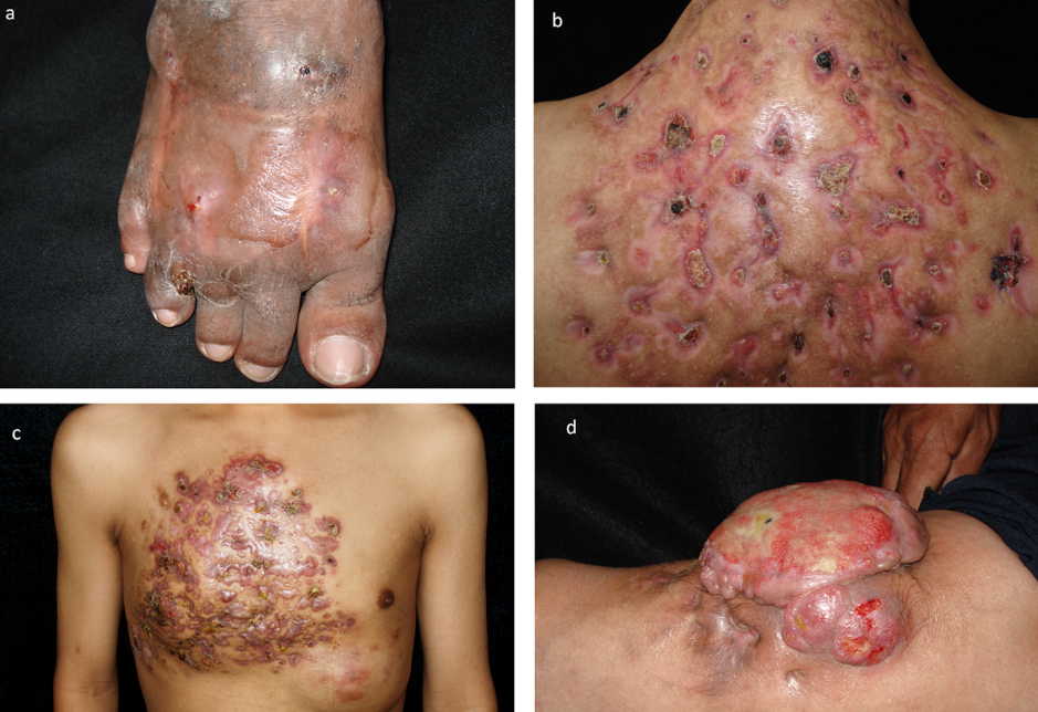 Figure: Image source : DOI:https://doi.org/10.1371/journal.pntd.0003102.g003a)*A. madurae* causing foot mycetomab) N. brasiliensis causing back mycetoma with multiple sinusesc) N. brasiliensis causing extensive mycetoma d)Fusarium solanicomplex causing tumoral mycetoma
Mycetoma possesses occupational risk primarily to field workers and farmers. The clinical feature depends on the site of infection, lesion stage, shape, size, and color of the grains. Three important clinical features of mycetoma are:
- Tumefaction: Tumor-like swelling
- Formation of multiple discharging/draining sinuses (hollow space or cavity within the bone or tissues)
- Grains/granules discharging from sinuses
In rare cases, grains are not formed in which diagnosis is based on direct and cultural findings.
Actinomadura madurai produces large-grain whereas Nocardia spp. produces small-grain mycetoma.
Laboratory diagnosis of Mycetoma
Taking the patient’s history of occupation, trauma, and geographical areas aids in the diagnosis of mycetoma.
Samples: Grains or granules are the samples of choice. Pus exudates can also be collected.
Clean the lesion with antiseptics and collect the grains by pressing the sinus from the periphery. It enhances to pass the discharge in which granules are present. Collect it in the sterile wire gauze.
Direct examination
Wash the grains in sterile saline. Crush the grains using the sterile glass rod and culture them on the suitable media.
KOH wet mount
KOH wet mount helps in observing the characteristics of the fungal structure directly
Procedure of KOH mount
Take a glass slide and add a few drops of KOH to the grains. Place the coverslip and, by applying gentle pressure, crush the grains. Use a glass rod or handle of a loop over the coverslip. Take a petri dish, put the moist filter paper and keep the slide. Leave it for a few hours. Then observe the KOH mount under the microscope.
Actinomycotic grains: It shows thin filaments with a diameter of 0.5 to 1 μm with coccoid or bacillary forms
Eumycotic grains: It shows thick 2-6 μm wide hyphae. Cells are large and swollen up to 15 μm at the margin. Chlamydospores may or may not be present in it.
To diagnose actinomycetoma, direct microscopic observation by Gram staining or acid-fast staining is done.
Gram staining shows the Gram-positive branching filamentous bacteria.
Modified Ziehl-Neelsen/ acid-fast staining (Kinyoun’s method) with 1% sulfuric acid shows Nocardia spp. i.e., reddish-pink-colored filamentous bacteria, but other actinomycetes are not acid-fast.
If septate hyphae are seen in the wet mount smear, then special staining like Hematoxylin and eosin (H &E) staining,Periodic acid-Schiff (PAS) staining, and Grocott’s Methenamine Silver (GMS) staining are done for confirmation.
Culture
After the direct observation of the crushed grains, actinomycetoma or eumycetoma is suspected. When actinomycetoma is suspected, grains samples are washed in normal saline without antibiotics and inoculated in culture media:
- Sabouraud dextrose agar without antibiotics
- Blood agar
- Lowenstein-Jensen media
- Brain-heart infusion agar
When eumycetoma is suspected, grains samples are washed in normal saline with antibiotics-streptopenicillin and penicillin. Then it is inoculated in Sabouraud dextrose agar with antibiotics like chloramphenicol and gentamicin. Modified Sabouraud dextrose agar, i.e., Emmons’ modification, is preferred.
Grains are inoculated in different plates, which increases the chances of isolation of the possible organisms and incubated at different temperatures, i.e., 25℃, 37℃, and 44℃. Then based on the cultural characteristics, causative agents are identified.
Radiodiagnosis of Mycetoma
Radiodiagnosis helps to determine the extent of the disease and study the involved tissue. X-rays, USG, CT scans, and MRI, is done. Mycetoma shows necrosis, generalized osteoporosis, and fusion of smaller bones. USG helps to differentiate eumycetoma, actinomycetoma, and non-mycetomatous lesions. MRI shows the “dot-in-circle,” which is highly specific for mycetoma.
Treatment
Antifungal drugs like oral ketoconazole and itraconazole are used for eumycetoma. Antibiotics like sulfonamides (cotrimoxazole), tetracyclines, streptomycin, amoxycillin, clavulanate, and amikacin are used for actinomycetoma.
References
- Chander, J. (2018). Textbook of Medical Mycology (Fourth edition). Jaypee Brothers Medical Publishers Ltd.

Tankeshwar Acharya, MSc (Medical Microbiology)
Tankeshwar Acharya is an Assistant Professor in the Department of Microbiology at Patan Academy of Health Sciences (PAHS), Nepal, where he has been teaching and practicing clinical microbiology for over 14 years. He is the founder of Microbe Online, one of the leading free microbiology education resources on the web, covering bacteriology, mycology, parasitology, immunology, and clinical laboratory diagnostics written from direct experience in both the classroom and the diagnostic laboratory.