
Mumps Virus: Structure, Pathogenesis, Clinical Findings, and Lab Diagnosis
Mumps virus causes parotitis and can spread to the testes, ovaries, and brain. Learn its structure, how it causes disease step by step, and how it is diagnosed.

On this page
Mumps is usually remembered as the illness that gives children swollen, hamster-like cheeks. In a child it is often mild and self-limiting. The same virus in a teenage boy or an adult is a different problem: it can inflame the testes, the ovaries, the pancreas, or the lining of the brain. One virus, one entry point in the nose, and yet it reaches organs all over the body. Understanding how it travels from the upper airway to such distant places is the key to understanding every complication of mumps.
Mumps virus causes a nonsuppurative (non-pus-forming) swelling of one or both salivary glands, most often the parotid glands. It is classically a mild childhood disease, but in adolescents and adults it more often causes serious complications such as meningitis and orchitis. About one-third of all mumps infections cause no symptoms at all, yet these silent infections can still spread the virus to others.
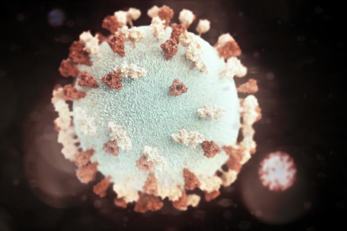 Figure: 3D graphic representation of a spherical-shaped mumps virus particle
Structure and morphology of mumps virus
Mumps virus belongs to the family Paramyxoviridae and is a typical member of that group. Knowing its structure explains both how it is identified in the laboratory and how it infects cells.
It is an enveloped virus with a single-stranded, negative-sense RNA genome that is not segmented. Inside the envelope, the RNA is wound together with protein into a helical nucleocapsid (the ribonucleoprotein, or RNP). Under the electron microscope, this nucleocapsid often unravels from damaged virus particles and appears as a distinctive herringbone or zipper-like pattern. This appearance is characteristic of paramyxoviruses and is a useful identifying feature.
Two glycoproteins stud the surface of the envelope and are central to how the virus works and how the body responds to it:
- HN (hemagglutinin-neuraminidase): the attachment protein. It binds the virus to receptors on the host cell and also carries neuraminidase activity. Because it can bind red blood cells, it is the basis of the hemagglutination and hemadsorption tests used in the laboratory. It is also called the V antigen (viral antigen).
- F (fusion) protein: fuses the viral envelope with the host cell membrane, allowing the genome to enter, and later fuses infected cells with neighbors to form multinucleated giant cells.
Beneath these, the internal nucleocapsid protein is known as the S antigen (soluble antigen). The distinction between the S antigen and V antigen matters for serology, as explained in the immunity section below.
Mumps virus is fragile outside the body. It is rapidly inactivated by heat, formalin, ether, and ultraviolet light, which is why fresh samples must reach the laboratory quickly.
There is only one antigenic type (serotype) of mumps virus, and it shows no significant antigenic variation. This single fact explains why one infection, or a complete vaccination, gives lasting protection.
Pathogenesis of mumps
Humans are the only natural host of mumps virus. The whole disease can be followed as a journey the virus makes from the nose to organs throughout the body.

Entry and primary replication. The virus enters through the upper respiratory tract in droplets of saliva or respiratory secretions. It first multiplies in the epithelial cells lining the nose and upper airway and in the nearby (regional) lymph nodes. At this stage the person has no symptoms.
Primary viremia and spread. From these first sites, the virus enters the bloodstream. This spread through the blood is called viremia, and it is what turns a local respiratory infection into a systemic disease. The blood carries the virus to its preferred targets: the salivary glands (especially the parotids), the testes, the ovaries, the pancreas, the kidneys, and the central nervous system. Mumps virus has a particular tropism, meaning a preference, for glandular tissue and nervous tissue.
Replication in target organs and secondary viremia. Once the virus reaches these organs, it replicates again. This second round of replication seeds a further wave of virus into the blood (a secondary viremia) before the immune response brings the infection under control. This is why several different organs can become inflamed in sequence rather than all at once.
Why the glands swell. In the parotid gland, the virus infects the cells lining the ducts. The resulting inflammation and swelling block the flow of saliva, which causes the pain and the visible swelling. Importantly, this swelling is nonsuppurative: it is inflammation, not a bacterial abscess, so there is no pus.
Incubation and shedding. The incubation period, from infection to the first symptoms, is usually 16 to 18 days, with a range of about 2 to 4 weeks. This long and variable incubation is one reason mumps is hard to contain: an infected person can spread the virus before anyone knows they are ill. The virus is present in saliva from a few days before the swelling begins until about 5 days after, which is the period of greatest infectiousness. It is also shed in urine, and can be detected there for up to about two weeks after symptoms begin.
Clinical Findings
Mumps is often described as an "iceberg" disease, because for every visible case there are many hidden ones. About one-third of infections cause no symptoms, and many more are mild.
In a symptomatic case, the illness begins after an incubation period of about 16 to 18 days with a short prodrome of flu-like illness: fever, malaise, and loss of appetite. This is followed by pain in the parotid glands, which then swell rapidly as inflammation blocks the flow of saliva through the duct. The swollen gland is tender, and anything that stimulates saliva, such as sour or acidic food, sharply increases the pain. Parotitis usually resolves within about a week.
Central nervous system. CNS involvement is common. The virus can be detected in the cerebrospinal fluid in a large share of infections, though most cause no obvious symptoms. Clinically apparent aseptic meningitis occurs in a minority of cases, and encephalitis is rare. Mumps is a classic cause of aseptic (viral) meningitis.
Orchitis. In postpubertal males, roughly one-quarter to one-half develop orchitis, inflammation of the testis, usually on one side and typically about a week after the parotitis. It is intensely painful, and the reason is anatomical: the testis is wrapped in a tough, non-stretchy capsule called the tunica albuginea, so when the inflamed testis swells against this fixed capsule, the pressure causes severe pain. Some testicular atrophy may follow, but sterility is rare, especially since orchitis is usually one-sided.
Oophoritis. Inflammation of the ovaries occurs in about 5 percent of postpubertal females, usually appearing after the parotitis has settled. It does not impair fertility.
Immunity
Immunity to mumps is lifelong after a single infection, and even a symptomless infection produces lasting protection. This is possible because there is only one antigenic type of the virus with no significant variation, so the immune system never has to face a meaningfully different version of it.
After natural infection, the body makes antibodies against the surface HN glycoprotein (the V antigen), the F glycoprotein, and the internal nucleocapsid protein (the S antigen). A cell-mediated immune response develops as well. The two antibody types follow different timelines, which is clinically useful:
- Antibody to the S antigen appears early, within about 3 to 7 days after symptoms begin, but fades within around six months. Its presence therefore signals a recent or current infection.
- Antibody to the V antigen appears later, around four weeks after onset, but persists for years. It is the marker of past infection and lasting immunity.
Infants born to immune mothers are protected by maternal antibody for about the first six months of life.
Laboratory Diagnosis
Lab studies are not required to establish the diagnosis of typical cases. However, they may sometimes be confused with enlargement of parotids due to suppuration, drug sensitivity, tumors, etc.
Isolation and identification of mumps virus
- Suitable samples are saliva, cerebrospinal fluid (CSF), and urine, collected within the first few days after symptoms begin.
- Monkey kidney cell lines are preferred for growing the virus, and samples should be inoculated soon after collection because the virus is fragile.
- For a faster result, immunofluorescence (IFT) can detect viral antigen in a 2 to 3 day cell culture.
- Mumps virus produces little visible cytopathic effect (CPE), mainly cell rounding and the formation of multinucleated giant cells.
- The hemadsorption test detects the virus indirectly: infected cells display HN protein on their surface, which makes added red blood cells stick to them.
Serology
- A rise in antibody level can be shown using paired serum samples (one early, one later). ELISA or the hemagglutination inhibition (HI) test are commonly used.
- Mumps IgM appears early in the illness and lasts about 60 days, making it a reliable marker of recent infection. Antibodies produced by parainfluenza virus infection, a related virus, do not cross-react in the mumps IgM ELISA, so the test stays specific.
- The complement fixation test (CFT), using S and V antigens separately, can also be used and helps date the infection.
Epidemiology
Mumps occurs worldwide, with the highest incidence historically in children aged 5 to 9 years. Humans are the only natural host. It spreads by direct contact, by airborne respiratory droplets, and through fomites (objects contaminated with infected saliva). Because symptomless cases can still transmit the virus, and because the incubation period is long and variable, mumps is difficult to contain without vaccination.
Treatment, Prevention, and Control
There is no specific antiviral treatment for mumps. Care is supportive: rest, fluids, and pain relief. The disease is controlled through prevention.
A live attenuated mumps vaccine provides effective, long-lasting protection, generally for at least 10 years. It produces a mild, non-communicable infection that primes the immune system. It is given as a single subcutaneous injection, either on its own or, far more commonly, combined with the measles and rubella vaccines as the MMR vaccine. The attenuated virus in the vaccine is usually the Jeryl Lynn or Urabe strain.
Because it is a live vaccine, it should not be given during pregnancy or to people with significant immunodeficiency, and it is avoided in those with severe hypersensitivity to neomycin or egg protein. Passive immunization (giving preformed antibody) is not a reliable way to prevent mumps.
How to Remember
The journey ("nose to glands to gonads to brain"): mumps starts in the nose, rides the blood (viremia), and lands in glands and nervous tissue. If you can recite the route, the complications are just stops along it: parotid, testis, ovary, pancreas, meninges.
Why orchitis hurts so much ("no room to swell"): the testis is trapped inside the tough tunica albuginea. Inflamed tissue tries to swell, the capsule will not give, so the pressure becomes severe pain. The anatomy explains the symptom.
S antigen versus V antigen timing ("S for Soon and Short, V for laVe and long"): the S antigen antibody appears soon and lasts a short time (recent infection). The V antigen antibody appears later and lasts for years (past infection and immunity). Match "recent" with S, "remembered" with V.
One serotype, one shot: there is only one antigenic type of mumps virus, so one infection or one complete vaccination protects for life. No serotype variation means no reinfection worries.
Key exam facts
| Fact | Detail |
|---|---|
| Family | Paramyxoviridae |
| Genome | Enveloped, non-segmented, negative-sense single-stranded RNA |
| Nucleocapsid appearance | Herringbone / zipper-like under EM |
| Surface proteins | HN (attachment, = V antigen) and F (fusion) |
| Antigenic types | Only one serotype; no significant variation |
| Natural host | Humans only |
| Transmission | Respiratory droplets, direct contact, fomites |
| Incubation period | 16 to 18 days (range about 2 to 4 weeks) |
| Core mechanism | Respiratory entry → viremia → glandular and neural tissue |
| Hallmark sign | Nonsuppurative parotitis (no pus) |
| Asymptomatic share | About one-third of infections |
| Orchitis | ~25 to 50% of postpubertal males; painful due to tunica albuginea; sterility rare |
| Oophoritis | ~5% of postpubertal females; fertility preserved |
| CNS | Classic cause of aseptic meningitis; encephalitis rare |
| Early serology marker | IgM (appears early, lasts ~60 days) |
| Recent vs past infection | S antigen antibody = recent; V antigen antibody = past/lasting |
| Treatment | Supportive only; no specific antiviral |
| Vaccine | Live attenuated (Jeryl Lynn / Urabe); usually as MMR |
Where Students Get Confused
"Is the parotid swelling an abscess or infection with pus?" No. Mumps parotitis is nonsuppurative, meaning there is no pus. It is viral inflammation that blocks saliva flow, not a bacterial abscess. This is a key way to distinguish it from bacterial parotitis, which does produce pus.
"Does mumps orchitis cause sterility?" Rarely. Orchitis is common and very painful, and some testicular atrophy can follow, but permanent sterility is uncommon because the inflammation is usually one-sided and the other testis is spared. The exam trap is to assume orchitis equals infertility. It usually does not.
"What is the difference between the S antigen and the V antigen?" The S (soluble) antigen is the internal nucleocapsid protein; antibody to it appears early and fades fast, marking recent infection. The V (viral) antigen is the surface HN protein; antibody to it appears later and lasts for years, marking past infection and immunity. Same virus, two markers, two different timelines.
"Why is mumps hard to control if it is usually mild?" Because a large share of infections are symptomless, and even symptomatic people are contagious before the swelling appears. You cannot isolate people you cannot identify. The long, variable incubation period makes this worse. This is why vaccination, not isolation, is the main control tool.
"Is mumps just a childhood disease?" It is mild in children but more dangerous after puberty, when complications like orchitis, oophoritis, and meningitis become more likely. The same virus causes very different severity depending on the age of the patient.
References
- Hviid, A., Rubin, S., & Mühlemann, K. (2008). Mumps. Lancet (London, England), 371(9616), 932–944. https://doi.org/10.1016/S0140-6736(08)60419-5
- Kubota, M., & Hashiguchi, T. (2021). Unique tropism and entry mechanism of mumps virus. Viruses, 13(9), 1746. https://doi.org/10.3390/v13091746
- Mourez, T., & Dina, J. (2018). Le virus des oreillons [Mumps virus: a comprehensive review]. Virologie (Montrouge, France), 22(4), 199–214. https://doi.org/10.1684/vir.2018.0744
- Davison, P., & Morris, J. (2023). Mumps. In StatPearls. StatPearls Publishing.
Frequently Asked Questions
What causes mumps?
What causes mumps?
Mumps is caused by the mumps virus, a member of the Paramyxoviridae family. It is an enveloped RNA virus with only one antigenic type.
How does mumps spread?
How does mumps spread?
Through respiratory droplets, direct contact with infected saliva, and contaminated objects. Humans are the only host. Symptomless carriers can still spread it.
How long is the incubation period of mumps?
How long is the incubation period of mumps?
Usually 16 to 18 days, with a range of about 2 to 4 weeks. This long, variable period makes mumps hard to contain.
Why do the cheeks swell in mumps?
Why do the cheeks swell in mumps?
The virus infects the ducts of the parotid salivary glands. Inflammation blocks saliva flow, and the gland swells and becomes painful. There is no pus, because it is viral inflammation, not a bacterial abscess.
What are the complications of mumps?
What are the complications of mumps?
In adolescents and adults, mumps can cause orchitis (testis inflammation), oophoritis (ovary inflammation), aseptic meningitis, and less commonly pancreatitis or encephalitis. Complications are more common and more serious after puberty.
Does mumps cause infertility?
Does mumps cause infertility?
Rarely. Orchitis is painful and can cause some testicular shrinkage, but permanent sterility is uncommon because it usually affects only one testis.
How is mumps diagnosed in the laboratory?
How is mumps diagnosed in the laboratory?
Most typical cases need no laboratory test. When needed, the virus can be grown from saliva, urine, or CSF, or detected by antibody tests. Mumps IgM is an early marker of recent infection and lasts about 60 days.
Is there a treatment or vaccine for mumps?
Is there a treatment or vaccine for mumps?
There is no specific antiviral treatment; care is supportive. Prevention is through the live attenuated mumps vaccine, usually given as part of the MMR vaccine, which gives long-lasting protection.

Tankeshwar Acharya, MSc (Medical Microbiology)
Tankeshwar Acharya is an Assistant Professor in the Department of Microbiology at Patan Academy of Health Sciences (PAHS), Nepal, where he has been teaching and practicing clinical microbiology for over 14 years. He is the founder of Microbe Online, one of the leading free microbiology education resources on the web, covering bacteriology, mycology, parasitology, immunology, and clinical laboratory diagnostics written from direct experience in both the classroom and the diagnostic laboratory.
Comments
No comments yet. Be the first to share your thoughts.
Leave a comment
All comments are reviewed before they appear.