
Klebsiella oxytoca: How It Differs from K. pneumoniae, and Antibiotic-Associated Colitis
How Klebsiella oxytoca differs from K. pneumoniae (indole positive), the toxin-driven hemorrhagic colitis that makes it distinct, and how the laboratory identifies it.

On this page
A patient started on antibiotics for a minor infection develops sudden bloody diarrhea and cramping a few days later. The usual suspect for antibiotic-associated diarrhea, Clostridioides difficile, tests negative. The cause turns out to be a different organism, one usually thought of as a quiet relative of the pneumonia bacterium: Klebsiella oxytoca.
K. oxytoca is the second most common Klebsiella species, and in the laboratory it looks almost identical to its well-known sister, Klebsiella pneumoniae. But it has one trick its sister lacks: it can produce a toxin that damages the lining of the gut. This page is about what makes K. oxytoca distinct, how it differs from K. pneumoniae, and how the laboratory tells the two apart.
Introduction
Klebsiella oxytoca is a Gram-negative rod of the genus Klebsiella, in the family Enterobacteriaceae. It is the second most common Klebsiella species after Klebsiella pneumoniae, and it shares most of that organism's biology: it is a capsulated, non-motile, lactose-fermenting gut and environmental organism that causes opportunistic, often hospital-acquired, infections.
Because the two species are so similar, the useful way to learn K. oxytoca is by how it differs from K. pneumoniae. There are two differences worth knowing. First, in the laboratory, K. oxytoca is indole-positive while K. pneumoniae is indole-negative; this single test separates them. Second, and more distinctive, K. oxytoca can cause a specific toxin-driven disease, antibiotic-associated hemorrhagic colitis, that K. pneumoniae does not. For the shared genus biology and the full picture of the more common species, see the Klebsiella pneumoniae article.
General properties
K. oxytoca shares the core features of the genus. It is a Gram-negative, non-spore-forming rod, a facultative anaerobe, catalase-positive, oxidase-negative, capsulated, and non-motile.
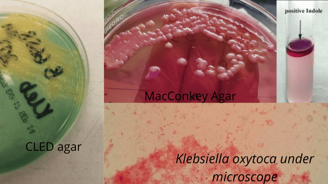 Figure: Some properties of Klebsiella oxytoca
Like K. pneumoniae, it ferments lactose and forms mucoid colonies because of its thick capsule. These shared features, and what they mean for identification, are covered in full on the K. pneumoniae page. What follows here is what makes K. oxytoca different.
Virulence factors and how K. oxytoca causes disease
K. oxytoca carries the same general virulence factors as the rest of the genus, and one extra weapon that makes it distinct.
The shared factors (same as K. pneumoniae). A polysaccharide capsule blocks phagocytosis and is the reason the colonies are mucoid. Pili (fimbriae) let it attach to host cells and to catheters and tubes. Lipopolysaccharide (LPS) drives inflammation and contributes to sepsis. Siderophores scavenge iron so it can grow in the host. These work the same way here as in K. pneumoniae.
The distinctive factor: the tilivalline and tilimycin toxins. This is what sets K. oxytoca apart. Many strains produce small-molecule toxins called tilivalline and tilimycin, which are directly toxic to the cells lining the colon. They damage and kill these cells, causing bleeding and inflammation of the gut wall. K. pneumoniae does not make these toxins, and this is why K. oxytoca causes a disease its sister does not.
How antibiotic-associated hemorrhagic colitis develops
The distinctive disease follows a clear sequence, and the role of antibiotics is the key to it. Normally, K. oxytoca is kept in check by the other bacteria of the gut. When a person takes antibiotics (classically penicillins such as amoxicillin), the normal gut flora is suppressed, which lets K. oxytoca overgrow. The overgrown organism produces tilivalline and tilimycin, which damage the lining of the colon, causing the sudden bloody diarrhea and cramping of hemorrhagic colitis. Importantly, this is a different mechanism and a different organism from the more familiar antibiotic-associated colitis caused by Clostridioides difficile, though the trigger (recent antibiotics) is similar. When the antibiotic is stopped, the colitis usually settles.
The other, more common infections
Apart from the colitis, K. oxytoca causes the same opportunistic, hospital-acquired infections as K. pneumoniae: pneumonia, urinary tract infection, wound and soft-tissue infection, and bloodstream infection that can progress to septic shock. These occur mainly in vulnerable patients, and spread in hospitals mostly on the hands of staff and via contaminated equipment.
Identifying K. oxytoca in the laboratory
Specimen. Urine, blood, sputum, bronchoalveolar lavage, wound swab, or CSF, depending on the infection.
On the plate. K. oxytoca is identical to K. pneumoniae in colony appearance, mucoid and lactose-positive on every medium, so colony morphology tells you it is a Klebsiella but not which species (indole does that). The appearance by medium:
| Medium | Appearance of K. oxytoca |
|---|---|
| MacConkey agar | Mucoid, pink (lactose-fermenting), glistening colonies. |
| Blood agar (agar sangre) | Mucoid, non-hemolytic, cream to greyish-white colonies. |
| Chocolate agar | Mucoid greyish colonies; no hemolysis read here. |
| CLED agar | Mucoid, lactose-fermenting, yellowish colonies (useful for urine). |
You cannot tell the two species apart by colony appearance; both look mucoid and lactose-positive. The one test that separates it from K. pneumoniae: indole.
K. oxytoca is indole-positive; K. pneumoniae is indole-negative. This is the deciding test. Everything else in the two species' biochemical profiles is largely the same, so when a laboratory reports "indole-positive Klebsiella," it is pointing at K. oxytoca.
The identification panel.
| Test | K. oxytoca result | What it tells you |
|---|---|---|
| Oxidase | Negative | Places it in the Enterobacteriaceae. |
| Catalase | Positive | Consistent. |
| Indole | Positive | The key split from K. pneumoniae (indole-negative). |
| Motility | Non-motile | Like the rest of the genus; no H antigen. |
| Voges-Proskauer (VP) | Positive | Genus pattern (neutral fermentation). |
| Citrate | Positive | Genus pattern. |
| Urease | Positive (weak) | Weakly positive, like K. pneumoniae. |
| ONPG | Positive | Lactose fermenter. |
| Lysine decarboxylase | Positive | Genus pattern. |
| Ornithine decarboxylase | Negative | Helps distinguish from some other genera. |
| TSI | A/A, gas positive, no H₂S | Ferments both sugars, strong gas, no H₂S. |
Resistance and treatment
Like K. pneumoniae, K. oxytoca is an important carrier of antibiotic resistance. Many strains produce extended-spectrum beta-lactamases (ESBLs), and some produce carbapenemases, so it can be multidrug-resistant. K. oxytoca also carries a chromosomal beta-lactamase that makes it naturally resistant to some penicillins.
For serious infections, treatment depends on susceptibility testing, and a carbapenem is often used for ESBL-producing strains. The detail of ESBL and carbapenem resistance is covered in the beta-lactamase article. For the toxin-associated hemorrhagic colitis, the main step is usually stopping the triggering antibiotic, after which the colitis typically resolves; this is a clinical decision. Specific drug doses and durations are not covered here.
How to remember
Indole is the whole difference in the lab. K. oxytoca and K. pneumoniae look identical, mucoid and pink, and behave almost identically. The one test that splits them is indole: oxytoca is indole-positive. Link the "o" of oxytoca to the positive result if it helps. If a lab reports an indole-positive mucoid Klebsiella, it is oxytoca.
The toxin sister. K. pneumoniae is the lung one; K. oxytoca is the one with a gut toxin. Picture oxytoca carrying an extra weapon its sister lacks: tilivalline, which cuts the colon lining and causes bloody diarrhea after antibiotics. That toxin is why this organism has its own disease and its own page.
Antibiotics unleash it. The colitis needs a trigger: antibiotics wipe out the competing gut flora, oxytoca overgrows, and its toxin damages the colon. It looks like C. difficile colitis (recent antibiotics, bloody diarrhea) but is a different organism. When the antibiotic stops, it usually settles.
Key exam facts
| Fact | Detail |
|---|---|
| Organism / family | Klebsiella oxytoca, genus Klebsiella, family Enterobacteriaceae |
| Rank | Second most common Klebsiella species (after K. pneumoniae) |
| Colony | Mucoid, pink (lactose fermenter) on MacConkey, like K. pneumoniae |
| Motility | Non-motile |
| Key lab difference from K. pneumoniae | Indole-positive (K. pneumoniae is indole-negative) |
| VP / citrate / urease | VP +, citrate +, urease weak + |
| TSI | A/A, gas positive, no H₂S |
| Distinctive virulence factor | Tilivalline and tilimycin toxins |
| Distinctive disease | Antibiotic-associated hemorrhagic colitis |
| Common infections | Pneumonia, UTI, wound infection, bloodstream infection |
| Resistance | ESBL and carbapenemase producer; chromosomal beta-lactamase |
| Colitis management | Stop the triggering antibiotic |
Where students get confused
K. oxytoca vs K. pneumoniae. They look identical on the plate and share almost all biochemistry. The single reliable separator is indole: K. oxytoca positive, K. pneumoniae negative.
The colitis is not C. difficile. K. oxytoca hemorrhagic colitis follows antibiotics and causes bloody diarrhea, which sounds like C. difficile, but it is a different organism with a different toxin (tilivalline, not the C. difficile toxins). A C. difficile-negative antibiotic-associated bloody colitis should raise the possibility of K. oxytoca.
Antibiotics cause it, and stopping them treats it. It seems backward that an infection is triggered by antibiotics and treated by stopping them. The reason is that antibiotics remove the competing gut flora and let K. oxytoca overgrow; removing the trigger lets the flora recover.
"Oxytoca" is not about oxygen or oxytocin. The name confuses students. It refers to rapid lactose fermentation (Greek for "quick-producing"), not oxygen, not the hormone oxytocin.
It is still a serious hospital pathogen. The colitis story can make K. oxytoca sound like a niche gut organism, but like K. pneumoniae it causes pneumonia, UTI, and bloodstream infection in vulnerable patients and carries serious resistance.
References
- Podschun, R., & Ullmann, U. (1998). Klebsiella spp. as nosocomial pathogens: epidemiology, taxonomy, typing methods, and pathogenicity factors. Clinical Microbiology Reviews, 11(4), 589–603. https://doi.org/10.1128/CMR.11.4.589
- Tille, P. M. (2022). Bailey & Scott's Diagnostic Microbiology (15th ed.). Elsevier.
- Madigan, M. T., Bender, K. S., Buckley, D. H., Sattley, W. M., & Stahl, D. A. (2021). Brock Biology of Microorganisms (16th ed.). Pearson.
- Högenauer, C., Langner, C., Beubler, E., et al. (2006). Klebsiella oxytoca as a causative organism of antibiotic-associated hemorrhagic colitis. New England Journal of Medicine, 355(23), 2418–2426. https://doi.org/10.1056/NEJMoa054765
Frequently Asked Questions
How do you tell Klebsiella oxytoca from Klebsiella pneumoniae?
How do you tell Klebsiella oxytoca from Klebsiella pneumoniae?
By the indole test. K. oxytoca is indole-positive and K. pneumoniae is indole-negative. The two species look identical on culture (both mucoid and lactose-fermenting) and share almost all other biochemistry, so indole is the deciding test.
What disease does Klebsiella oxytoca cause that K. pneumoniae does not?
What disease does Klebsiella oxytoca cause that K. pneumoniae does not?
Antibiotic-associated hemorrhagic colitis. K. oxytoca can produce toxins (tilivalline and tilimycin) that damage the lining of the colon, causing bloody diarrhea after antibiotic use. K. pneumoniae does not make these toxins.
Is Klebsiella oxytoca indole positive or negative?
Is Klebsiella oxytoca indole positive or negative?
Positive. This is its main laboratory difference from K. pneumoniae, which is indole-negative.
How is K. oxytoca colitis different from C. difficile colitis?
How is K. oxytoca colitis different from C. difficile colitis?
Both follow antibiotic use and cause diarrhea, but they are different organisms with different toxins. K. oxytoca colitis is caused by tilivalline and typically causes bloody diarrhea, and it usually settles when the triggering antibiotic is stopped. A C. difficile-negative antibiotic-associated bloody colitis can be due to K. oxytoca.
What infections does Klebsiella oxytoca cause?
What infections does Klebsiella oxytoca cause?
Besides the antibiotic-associated colitis, it causes the same opportunistic hospital infections as K. pneumoniae: pneumonia, urinary tract infection, wound and soft-tissue infection, and bloodstream infection, mainly in vulnerable patients.
Is Klebsiella oxytoca antibiotic resistant?
Is Klebsiella oxytoca antibiotic resistant?
Often, yes. Many strains produce extended-spectrum beta-lactamases (ESBLs), and some produce carbapenemases, so infections can be multidrug-resistant. Treatment is guided by susceptibility testing.
What does the name "oxytoca" mean?
What does the name "oxytoca" mean?
It refers to rapid production, describing the organism's quick lactose fermentation. It is not related to oxygen or to the hormone oxytocin.

Tankeshwar Acharya, MSc (Medical Microbiology)
Tankeshwar Acharya is an Assistant Professor in the Department of Microbiology at Patan Academy of Health Sciences (PAHS), Nepal, where he has been teaching and practicing clinical microbiology for over 14 years. He is the founder of Microbe Online, one of the leading free microbiology education resources on the web, covering bacteriology, mycology, parasitology, immunology, and clinical laboratory diagnostics written from direct experience in both the classroom and the diagnostic laboratory.
Comments
No comments yet. Be the first to share your thoughts.
Leave a comment
All comments are reviewed before they appear.