
Indole Test: Principle, Procedure, Interpretation & Indole-Positive Organisms

On this page
Indole test is used to determine the ability of an organism to split the amino acid tryptophan to form the compound indole. It's one of the four IMViC tests used to differentiate members of the family Enterobacteriaceae and it's usually the fastest of the four to read.
Why It Matters
Picture a urine culture growing a lactose-fermenting Gram-negative rod on MacConkey agar. Full identification and sensitivity will take another 24–48 hours but a clinician needs an empiric antibiotic choice today. A 20-second spot indole test, combined with the colony's oxidase result, can presumptively call it E. coli right then: indole-positive, oxidase-negative, pink lactose-fermenter on MacConkey. That presumptive call is often enough to start a sensible empiric regimen while the formal culture finishes.
This is also why indole is one of the oldest tests in clinical bacteriology. It doesn't need expensive reagents or incubators beyond what every lab already has, and it still separates indole-positive E. coli from indole-negative Enterobacter and Klebsiella pneumoniae faster than almost any other single test.
Principle
Tryptophan is hydrolyzed by the enzyme tryptophanase into three end products: indole, pyruvate, and ammonia. Indole, when present, combines with the aldehyde in the test reagent to form a colored compound:
- Pink to red-violet (quinoidal compound): if a benzaldehyde reagent (Kovac's or Ehrlich's) is used
- Blue to green: if a cinnamaldehyde reagent (DMACA) is used
No tryptophanase → no indole → no color change (indole-negative).
 Figure: Indole Test Reaction
Procedure of Indole Test
There are two ways to run this test, and which one you choose matters.
1. Spot (Rapid) Indole Test
Best for: quick presumptive ID directly from an isolated colony.
- Saturate a filter paper with reagent, then rub a colony sample onto it or
- Swab the colony and add a drop of reagent to the swab or
- Add reagent directly onto the colony on the agar plate
Read the color change within 20 seconds.
2. Conventional Tube Method
Best for: confirming a negative spot test, or testing weak indole producers that the rapid method misses.
- Inoculate tryptophan broth with the test organism.
- Incubate at 37°C for 24–48 hours.
- Kovac's method: Add 0.5 mL Kovac's reagent down the side of the tube; read the color at the meniscus.
- Ehrlich's method (for anaerobes/fastidious organisms): Add 0.5 mL xylene, mix and let settle, then add 6 drops of Ehrlich's reagent down the side; read the color below the xylene layer.
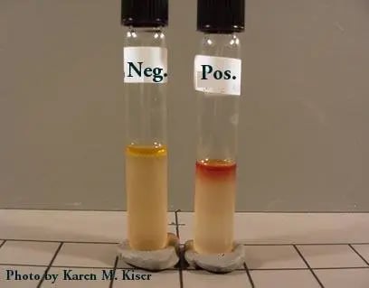 Figure: Indole Test Results: Positive (Right Tube)-development of Red-ring
Where students actually get confused
- Reagent choice isn't optional. For weak producers, anaerobes, and fastidious organisms like Cardiobacterium hominis, Kovac's reagent can miss the reaction. Ehrlich's reagent with a xylene extraction step is more sensitive and is preferred in the tube method for this group. For rapid spot testing of anaerobes, the cinnamaldehyde reagent (DMACA) is the validated choice.
- Don't inoculate from MacConkey or EMB agar. The dyes in these media interfere with color interpretation. Use a non-selective medium for the colony you're testing.
- Don't use Mueller-Hinton agar colonies either. Tryptophan is destroyed during the acid hydrolysis used to make the medium, which can produce a false negative.
- A negative spot test isn't the final answer. If it's negative, run the tube method as some organisms are weak producers that only the more sensitive tube test will catch.
- Watch pigmented colonies on a spot test. A naturally pigmented organism (for example the blue-green of Pseudomonas aeruginosa) can tint the filter paper and be misread. Judge the color of the reagent reaction, not the colony's own color, and confirm in a tube if unsure.
- Avoid plates with a nitrate disk nearby. Nitrate can cause a false-negative spot indole reaction.
Result Interpretation
| Reaction | Reagent | Result |
|---|---|---|
| Positive | Benzaldehyde (Kovac's/Ehrlich's) | Brown-red to purple-red color within 20 seconds |
| Positive | Cinnamaldehyde (DMACA) | Blue to blue-green color |
| Negative | Either | Colorless or pale yellow |
Reporting:
- E. coli (the classic indole-positive member), Vibrio, Aeromonas, Plesiomonas, and Pasteurella species are indole positive. Indole positivity varies across the Enterobacteriaceae, which is exactly what makes it useful for differentiation.
- Most strains of P. vulgaris, M. morganii, and Providencia: indole positive
- Fastidious Gram-negatives like Cardiobacterium hominis and Bergeyella zoohelcum: indole positive
- Cutibacterium acnes: indole positive
The "OK VIP" Trick
Three closely related genus-pairs trip students up every exam season because one species is indole-positive and its close relative is indole-negative:
| Genus | Indole-positive species | Indole-negative species |
|---|---|---|
| Klebsiella | K. oxytoca | K. pneumoniae |
| Citrobacter | C. koseri | C. freundii |
| Proteus | P. vulgaris | P. mirabilis |
Remember it as "OK VIP": Oxytoca (Klebsiella), Koseri (Citrobacter), Vulgaris (Proteus) → Indole Positive. Everything else in that trio defaults to negative.
The Klebsiella split is worth more than an exam mark. K. oxytoca, the indole-positive one, is the species behind antibiotic-associated hemorrhagic colitis: it produces a toxin (tilivalline) that damages colonic mucosa, and it classically emerges when penicillins suppress competing flora. So "the indole-positive Klebsiella" is not just a practical-exam distinction; it names the Klebsiella you think of when a patient develops bloody diarrhea after starting an antibiotic. A mnemonic you can attach to a real clinical picture is one you keep.
Multitest Media That Include Indole
In diagnostic labs, indole is rarely tested alone. It's usually read off a combination medium:
| Medium | Tests for |
|---|---|
| SIM | Indole + Motility + H₂S |
| MIU | Indole + Motility + Urease |
| MIO | Indole + Motility + Ornithine decarboxylase |
Quality Control
- Discard benzaldehyde reagents (Kovac's/Ehrlich's) if the color isn't pale yellow before use.
- QC every new reagent lot before use; QC in-house reagents weekly — they degrade, especially if not kept at 4°C.
| Organism | Expected result |
|---|---|
| E. coli ATCC 25922 | Positive |
| Pseudomonas aeruginosa ATCC 27853 | Negative |
| Porphyromonas asaccharolytica ATCC 25260 (Ehrlich's, anaerobes) | Positive |
| Bacteroides fragilis ATCC 25285 (Ehrlich's, anaerobes) | Negative |
Limitations
- The growth medium must contain adequate tryptophan so avoid colonies from Mueller-Hinton agar.
- Avoid plates with a nitrate disk nearby (false negatives).
- Only cinnamaldehyde reagent is validated for spot-testing anaerobe. It's more sensitive but less stable than benzaldehyde reagents.
- A negative spot test should be confirmed with the tube method before reporting indole-negative.
- Fastidious Gram-negative rods (e.g., C. hominis) may need a heavier inoculum and extraction step.
Key exam facts in one table
| Question | Answer |
|---|---|
| What enzyme does the indole test detect? | Tryptophanase |
| Three products of tryptophan hydrolysis | Indole, pyruvate, ammonia |
| Kovac's/Ehrlich's positive color | Pink to red-violet (benzaldehyde reagents) |
| DMACA positive color | Blue to blue-green (cinnamaldehyde reagent) |
| Classic indole-positive organism | E. coli |
| The three OK VIP positives | K. oxytoca, C. koseri, P. vulgaris |
| Why not test from MacConkey/EMB/MHA? | Dyes interfere; MHA lacks tryptophan (false negatives) |
| Most sensitive reagent for anaerobes (spot) | Cinnamaldehyde (DMACA) |
References and further readings
- MacWilliams MP. Indole test protocol. American Society for Microbiology, 2009.
- Leber AL, editor. Clinical Microbiology Procedures Handbook. 4th ed. Washington, DC: ASM Press; 2016. doi:10.1128/9781555818814
- Tille PM. Bailey and Scott's Diagnostic Microbiology. 15th ed. St. Louis: Elsevier; 2022.
Frequently Asked Questions
What's the difference between Kovac's and Ehrlich's reagent?
Why is my indole test negative when I expected E. coli to be positive?
Is the indole test enough to identify E. coli on its own?
No. It's a presumptive test. Combine it with oxidase and colony morphology on MacConkey for a same-day presumptive call, but confirm with full biochemical/serological identification before finalizing a report.
Why does the Klebsiella oxytoca versus Klebsiella pneumoniae indole result matter clinically?
Why does the Klebsiella oxytoca versus Klebsiella pneumoniae indole result matter clinically?
Because K. oxytoca, the indole-positive species, is the cause of antibiotic-associated hemorrhagic colitis. It produces the toxin tilivalline and tends to emerge when antibiotics suppress competing gut flora, presenting as bloody diarrhea after starting an antibiotic. K. pneumoniae, the indole-negative species, is not associated with this syndrome. So the indole split that students learn as OK VIP also separates two clinically different organisms.

Tankeshwar Acharya, MSc (Medical Microbiology)
Tankeshwar Acharya is an Assistant Professor in the Department of Microbiology at Patan Academy of Health Sciences (PAHS), Nepal, where he has been teaching and practicing clinical microbiology for over 14 years. He is the founder of Microbe Online, one of the leading free microbiology education resources on the web, covering bacteriology, mycology, parasitology, immunology, and clinical laboratory diagnostics written from direct experience in both the classroom and the diagnostic laboratory.
Comments
No comments yet. Be the first to share your thoughts.
Leave a comment
All comments are reviewed before they appear.