
Preparation of Gram Stain Reagents: Formulas, Storage, and Quality Control

On this page
A microbiology laboratory reports an unusual result: three consecutive blood culture smears have shown all organisms as gram-negative, including a known Staphylococcus aureus positive control. The attending physician delays anti-staphylococcal therapy. The investigation reveals the iodine solution in use had degraded — it had turned yellow instead of remaining brown, losing its mordant function. Every smear stained with that batch had produced false gram-negative results.
Reagent quality directly determines diagnostic accuracy in Gram staining. A correctly prepared and stored reagent batch produces reliable results for months; a degraded or improperly prepared batch generates misleading results that can affect patient management. Understanding how each reagent is prepared, why it must be stored correctly, and how to verify its quality before clinical use is not a minor procedural detail — it is a patient safety issue.
Gram staining requires the use of different chemical reagents which can be purchased in ready-to-use forms from commercial suppliers or can be prepared at the laboratory by mixing different chemicals in an appropriate amount.
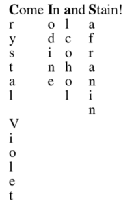 Gram staining technique requires simultaneous use of chemical reagents for a fixed period followed by washing; Primary stain (crystal violet), Mordant (iodine), Decolorizer (ethanol or acid-alcohol), and Counterstain****(safranin or dilute carbol-fuchsin).
Stained slide is air-dried and observed under oil immersion (100x) using a bright field microscope. Gram-positive bacteria appear blue/purple while Gram-negative appear as pink/red.
Crystal violet
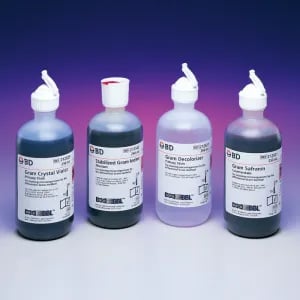 Figure: Gram stain reagent
- Dissolve 2.0 g certified crystal violet into 20.0 ml of 95% ethyl alcohol.
- Dissolve 0.8 g ammonium oxalate into 80.0 ml distilled water.
- Mix the two solutions together and allow them to stand overnight at room temperature (25°C).
- Filter through coarse filter paper before use.
- Store at room temperature (25°C).
Why crystal violet is the primary stain: Crystal violet (CV) is a basic (cationic) dye with high affinity for negatively charged bacterial cell components. When prepared correctly, it should appear as a deep purple solution. A pale or faded crystal violet solution — indicating degradation or excessive dilution — will produce weak or absent primary staining on all organisms, causing false-negative results.
Gram’s iodine
- Grind 1.0 g iodine (crystalline) and 2.0 g potassium iodide in a mortar. Small additions of distilled water may be helpful in preparing the solution.
- Add to 300.0 ml distilled water.
- Store at room temperature (25°C) in a foil-covered bottle (to protect solution from light).
Storage critical point: Gram's iodine must be stored in a brown or amber glass bottle away from light. A freshly prepared or good-quality iodine solution is dark brown. As it degrades, it turns yellow — at this point it has lost most of its mordant activity and must be discarded immediately. Using yellow iodine is the most common single cause of false gram-negative results in clinical laboratories. Check the colour of the iodine bottle before every staining session.
Decolorizer
Some workers prefer to use acetone by itself, ethanol 95% v/v, or ethanol-iodine as the decolorizing solution. A mixture of acetone-alcohol is recommended because it decolorizes more rapidly than ethanol 95% v/v, and is less likely to over-decolorize smears than acetone without alcohol added.
The most critical step: Decolourisation is where Gram staining succeeds or fails. The decoloriser (acetone-alcohol) must be applied for exactly the right duration — typically 10–15 seconds, added drop by drop until the effluent runs clear. Under-decolourisation leaves gram-negative organisms appearing gram-positive; over-decolourisation strips the crystal violet from gram-positive organisms, making them appear gram-negative. Reagent quality alone does not guarantee correct results — technique matters equally.
Acetone-alcohol
decolorizer
To make 1 liter :
- Acetone………..500 ml
- Ethanol or methanol, absolute* …………..475 ml
- Distilled water…………25 ml
*Technical grade is adequate.
- Mix the distilled water with absolute ethanol (ethyl alcohol) or methanol (methyl alcohol). Transfer the solution to a screw-cap bottle of 1 liter capacity. Caution: Ethanol and methanol are highly flammable, therefore use well away from an open flame.
- Measure the acetone, and add immediately to the alcohol solution. Mix well. Caution: Acetone is a highly flammable chemical that vaporizes rapidly, therefore use it well away from an open flame.
- Label the bottle, and mark it Highly Flammable. Store in a safe place at room temperature. The reagent is stable indefinitely.
- For use: Transfer a small amount of the reagent to a dispensing container that can be closed when not in use.
Counterstain
Safranin and dilute carbol-fuchsin are commonly used counterstain in Gram staining procedure, another being Neutral red (it stains gonococci and meningococci well).
Preparation of Safranin
- Add 2.5 g certified safranin-O to 100.0 ml 95% ethyl alcohol.
- Add 10.0 ml safranin and ethyl alcohol solution made in step 1 to 90.0 ml distilled water.
- Store at room temperature (25°C).
Preparation of dilute carbol-fuchsin
(may be a more effective counterstain than safranin)
- Dissolve 0.3 g basic fuchsin in 10.0 ml 95% ethyl alcohol.
- Add 5.0 ml melted phenol crystals to 95.0 ml distilled water.
- Add the 5% phenol solution to the fuchsin solution and let stand overnight.
- Filter through coarse filter paper.
- Store at room temperature (25°C) in a foil-covered bottle for up to 1 year.
Storage Conditions and Shelf Life
Proper storage is as important as correct preparation. A correctly prepared reagent that is stored incorrectly will degrade and produce unreliable results.
| Reagent | Storage Condition | Shelf Life | Discard When |
|---|---|---|---|
| Crystal violet | Room temperature (15–25°C), tightly capped | 12 months | Colour fades from deep purple to pale violet |
| Gram's iodine | Brown/amber glass, room temperature, away from light | 3–6 months | Solution turns yellow (from dark brown) |
| Decoloriser (acetone-alcohol) | Tightly capped, away from heat and flame | 12 months | Evaporation reduces volume significantly; discard and prepare fresh |
| Safranin | Room temperature, tightly capped | 12 months | Colour fades from deep red to pale pink |
| Dilute carbol-fuchsin | Brown glass, room temperature | 6–12 months | Colour fades or precipitate forms |
| Neutral red | Room temperature | 12 months | Colour fades |
General rules:
- Label every bottle with the preparation date and expiry date
- Transfer working volumes to small dispensing bottles — do not repeatedly open the stock bottle
- Keep dispensing bottles capped when not in use to prevent evaporation (decoloriser) and oxidation (iodine)
- Never top up a dispensing bottle with new reagent — discard the old, clean the bottle, refill with fresh
Quality Control of Gram Stain Reagents
Quality control is not optional in a clinical microbiology laboratory — it is the mechanism by which you know your results are trustworthy before they reach the clinician.
Before first clinical use of any new reagent batch
Prepare a control smear containing both:
- A known gram-positive organism: Staphylococcus aureus (ATCC 25923) or any confirmed gram-positive clinical isolate
- A known gram-negative organism: Escherichia coli (ATCC 25922) or any confirmed gram-negative clinical isolate
Stain the control smear with the new batch. Expected results:
- S. aureus: blue-purple cocci in clusters
- E. coli: pink-red rods
If either control fails — repeat with a fresh smear before investigating the reagent batch.
Daily QC check (recommended)
Many laboratories include a QC smear with each batch of clinical specimens. This does not need to be a named ATCC strain — any confirmed gram-positive and gram-negative organism from a recent culture works.
The neutrophil nuclei check (in-run QC)
On any specimen containing pus cells, the nuclei of neutrophils serve as a built-in QC indicator:
- Nuclei staining red/pink = correct decolourisation
- Nuclei staining blue/purple = under-decolourisation — repeat the slide with longer decoloriser contact time
This check is free, requires no additional reagents, and catches the most common staining error immediately.
Troubleshooting Gram Stain Reagent Problems
| Observation | Likely Reagent Problem | Investigation and Action |
|---|---|---|
| All organisms staining gram-negative (including GP controls) | Iodine degraded (yellow colour) — mordant failure | Check iodine colour; replace if yellow; retest with fresh iodine |
| All organisms staining gram-positive (including GN controls) | Insufficient decoloriser; decoloriser too dilute | Check decoloriser composition; ensure adequate contact time (drop by drop until effluent runs clear) |
| Faint or absent primary staining (pale smears) | Crystal violet degraded or too dilute | Check crystal violet colour (should be deep purple); replace if faded |
| Poor counterstaining (cells colourless after decolorisation) | Safranin too dilute or degraded | Check safranin colour (should be deep red); replace if pale |
| Precipitate in stain deposits on slide | Unfiltered stain; precipitated dye crystals | Filter staining solutions through Whatman No. 1 filter paper before use |
| Background too dark (obscures organisms) | Smear too thick; crystal violet contact time too long | Prepare thinner smear; reduce crystal violet time to 1 minute |
| Inconsistent results between different staff | Variable decoloriser technique | Standardise technique: drop-by-drop until effluent runs clear; time 10–15 seconds maximum |
Why Each Reagent Matters: The Teaching Framework
Understanding what each reagent does — not just how to prepare it — prevents the most common errors:
| Reagent | Role | What Happens If It Fails |
|---|---|---|
| Crystal violet | Primary stain — colours all cells purple | Degraded CV → pale or absent staining on everything |
| Gram's iodine | Mordant — forms CV-I complex within the cell | Degraded iodine → CV washes out of GP and GN alike → everything appears GN |
| Decoloriser | Distinguishes GP from GN — the critical step | Too weak/short → under-decolourisation → GN appears GP; Too strong/long → over-decolourisation → GP appears GN |
| Safranin | Counterstain — stains decolourised GN cells pink/red | Degraded safranin → GN cells remain colourless, appearing absent |
Every Gram staining error — false GP, false GN, or no staining — can be traced back to failure of one of these four reagents or failure of the decolourisation technique.
How to Remember
The iodine colour rule — "Brown is good, yellow is gone": Gram's iodine should always be dark brown. The moment it turns yellow, it has lost its mordant activity. Check the bottle colour before every session — this takes two seconds and prevents the most common cause of false gram-negative results.
The decoloriser mantra — "10-15 seconds, drop by drop, until clear": These three parameters — time, technique, and endpoint — are what distinguish a reliable decolourisation from one that produces false results. No reagent preparation, however perfect, compensates for poor decolourisation technique.
The "CIDS" reagent sequence: Crystal violet → Iodine → Decoloriser → Safranin — same as "ComeInAndStain" from the main Gram staining article.
Key Exam Facts in One Table
| Reagent | Role | Key Preparation Point | Storage | Failure Sign |
|---|---|---|---|---|
| Crystal violet | Primary stain | 2% in 20% ethanol (Hucker's) | Room temp, tightly capped | Faded purple → replace |
| Gram's iodine | Mordant | Iodine + KI in water (Gram's formula) | Brown glass, away from light | Turns yellow → discard immediately |
| Acetone-alcohol | Decoloriser | 1:1 acetone:95% ethanol | Tightly capped, away from heat | Evaporates → prepare fresh |
| Safranin | Counterstain | 0.25% in water | Room temp, tightly capped | Faded pink → replace |
| Dilute carbol-fuchsin | Alternative counterstain | 0.3% basic fuchsin in phenol-water | Brown glass, room temp | Faded → replace |
| Neutral red | Alternative counterstain | 0.1% in distilled water | Room temp | Faded → replace |
References
- Tripathi N, Sapra A. Gram Staining. [Updated 2023 Aug 14]. In: StatPearls [Internet]. Treasure Island (FL): StatPearls Publishing; 2023 Jan-. Available from: https://www.ncbi.nlm.nih.gov/books/NBK562156/
- Davies, J. A., Anderson, G. K., Beveridge, T. J., & Clark, H. C. (1983). Chemical mechanism of the Gram stain and synthesis of a new electron-opaque marker for electron microscopy which replaces the iodine mordant of the stain. Journal of bacteriology, 156(2), 837–845. https://doi.org/10.1128/jb.156.2.837-845.1983
- Forbes BA, Sahm DF, Weissfeld AS. Bailey & Scott's Diagnostic Microbiology. 14th ed. Elsevier; 2023.
- Garcia LS. Clinical Microbiology Procedures Handbook. 4th ed. ASM Press; 2016.
- Murray PR, Rosenthal KS, Pfaller MA. Medical Microbiology. 9th ed. Elsevier; 2020.
Frequently Asked Questions
How do I know if Gram's iodine solution has degraded and needs replacing?
What quality control should be done on a new batch of Gram stain reagents?
What is the shelf life of Gram stain reagents and how should they be stored?

Tankeshwar Acharya, MSc (Medical Microbiology)
Tankeshwar Acharya is an Assistant Professor in the Department of Microbiology at Patan Academy of Health Sciences (PAHS), Nepal, where he has been teaching and practicing clinical microbiology for over 14 years. He is the founder of Microbe Online, one of the leading free microbiology education resources on the web, covering bacteriology, mycology, parasitology, immunology, and clinical laboratory diagnostics written from direct experience in both the classroom and the diagnostic laboratory.
Comments
No comments yet. Be the first to share your thoughts.
Leave a comment
All comments are reviewed before they appear.